Procedures in Dermatology
Dr Sami Fatehi MBBS,MSc,MD, PhD
Overview
- Shave biopsy
- Punch biopsy
- Incision & Drainage (I&D)
- Excisions
- Electrodessication & Curettage (ED&C)
- Cryotherapy (viral warts, SK, Cherry angiomas)
What is a skin biopsy?
- A skin biopsy is a diagnostic procedure in which a portion of skin (and/or subcutis) is submitted to the pathology lab.
- This specimen is fixed, sectioned and placed on slides for histologic analysis
- Special stains can be used to detect fungus, bacteria, immune complexes, lymphocytes, inflammatory mediators, arthropods.
- The aim is that the pathologist can provide more information to aid in diagnosing the disease.
Why do skin biopsy?
- Skin biopsies usually provide diagnostic information that adds to the clinical picture already at hand.
- Many skin diseases have characteristic findings on routine histology that are highly diagnostic
- Ascertain benign vs. malignant, infectious vs. autoimmune, exogenous vs. endogenous process.
When you take skin biopsy it should:
- Provides an adequate specimen for the pathologist to review
- Using the utmost care and knowledge of anatomy to minimize the potential morbidity of the procedure.
- Post-biopsy wound care patient education.
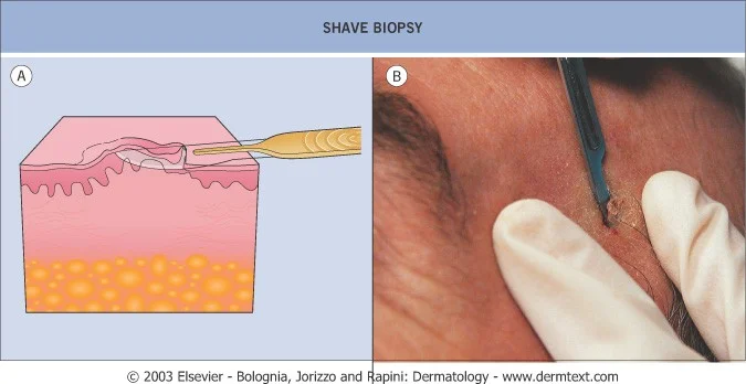
1- Biopsy by shave technique
- Removal of representative piece of skin by oblique incision with a blade.
- Can use scalpel or Dermoblade
- Idea is to sample both lesional and normal-appearing perilesional skin
- Depth needs to get down to at least superficial upper dermis - biopsies of epidermis only usually unsatisfactory.
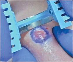
Dermablade
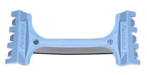
Shave biopsy using dermablade
When to do a shave
- In sensitive anatomic locations
- The highly active patient: Shave biopsy wounds have no limitation on activity.
- The patient who can’t/doesn’t want to come back for suture removal from punch biopsy.
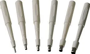
2-Biopsy by punch technique
- Removal of a representative piece of skin and subcutis with a punch
- Best way to look at it is like a little cylindrical cookie-cutter which punches all the way through the skin
- Usually a more involved procedure than shave needing, extra time for anesthesia, hemostasis and would closure
Disposable tool bas… lesion
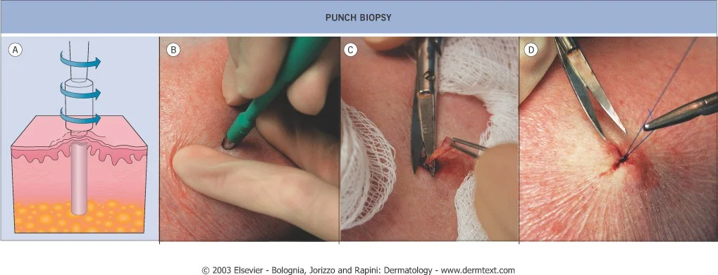
When to do a punch
- Punch superior for any skin diseases where a picture of the deep dermis/subcutis is diagnostic.
- Tends to provide more information for inflammatory skin disorders, as they tend to involve greater depth of dermis
- Better choice for deeply-seated lesions in dermis and subcutis.
3- Excision and Encisional biopsy
- Procedure whereby a full thickness specimen of skin is removed either for therapeutic or diagnostic purposes.
- Excisions usually in elliptical shape oriented along skin tension lines
- Suture the wound
- Procedure learned by seeing/doing.
Why do an excision?
Usually done to completely remove a lesion for therapeutic reasons:
- Skin cancer
- Dysplastic nevus (abnormal mole)
- Epidermal inclusion cyst
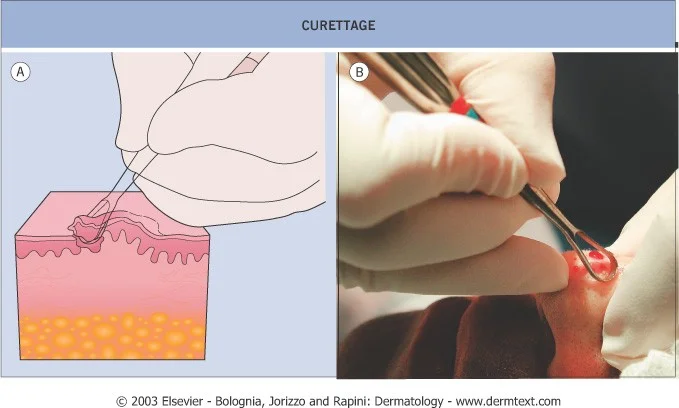
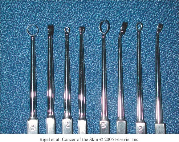
4- Electrodessication and Curettage (ED&C)
- Essentially a process whereby superficial cancerous (SCC) and pre-cancerous growths are removed from the skin by repeated scraping and burning.
- An effective, safe, expedient means of treating certain skin cancers in certain locations.
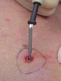
Curretage
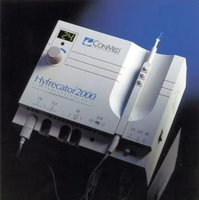
Hyfrecator
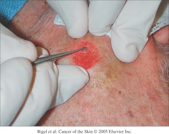
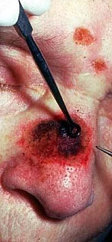
burning
burn and scrap
ED&C indications
-
Indicated for SCC in situ, superficial and selected nodular BCC.
-
Benign growths like warts
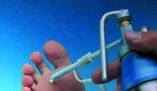
Cryotherapy
- The destruction of skin lesions
- using a cold substance
- Most commonly liquid
- nitrogen
- Destruction is selective,
- affecting tissue only
Cryotherapy - Indications
- Benign lesions
- Premalignant lesions
- Malignant lesions
Table 1.
Some of the common conditions responsive to cryosurgery.
| Benign lesions | Pre-malignant lesions | Malignant Lesions |
|---|---|---|
| Viral Warts | Actinic/solar keratoses | Superficial basal cell carcinomas |
| Skin tags | Bowens disease (Intra-epithelial carcinoma) | |
| Seborrhoeic keratoses | Actinic cheilitis | |
| Sebaceous hyperplasia | Cherry angiomas | |
| Molluscum contagiosum | ||
| Milia |
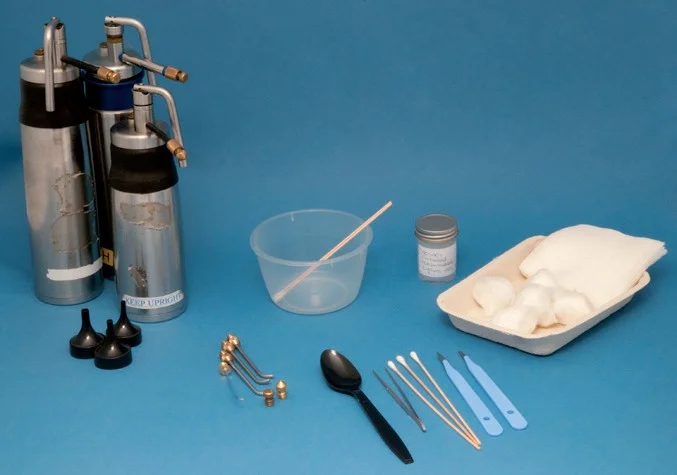
Cryotherapy - Equipment
The equipment required depends on the method and technique used
Methods: depending on lesion site’s
- Open spray - - 40 °C - best outcome
- Cotton bud - - 20 °C - eyelid
- Metal forceps - - 15 °C - neck