BASICS OF ECG
Table of Contents
-
- What is an ECG?
- With ECGs WE CAN IDENTIFY
- Impulse Conduction & the ECG
- NORMAL ECG
- The PQRST
- The PR Interval
- The ECG Paper
- ECG LEADS
- Bipolar Leads
- Unipolar Leads
- The Standard ECG
- Standard Limb Leads
- Standard Limb Leads (Angles)
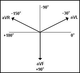
- Augmented Limb Leads
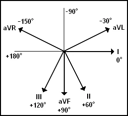
- All Limb Leads
- Precordial Leads
- Left Ventricular Hypertrophy
- LEFT VENTRICULAR HYPERTROPHY
- LEFT VENTRICULAR HYPERTROPHY
- LEFT VENTRICULAR HYPERTROPHY
- Views of the Heart
- ST Elevation
- ST Elevation (Cont.)
- Anterior View of the Heart
- Anterior Myocardial Infarction
- Most Likely Occluded Coronary Artery
- ECG 4
- ECG 5
- ECG 6
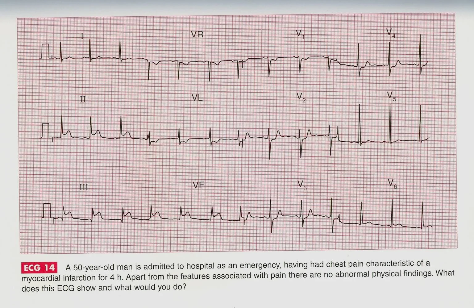
- ECG 14
- Anterior MI
- Putting It All Together
- Interpretation
- Other MI Locations
- Other MI Locations
- Anterolateral MI
BASICS OF ECG
What is an ECG?
- The electrocardiogram (ECG) is a representation of the electrical events of the cardiac cycle.
- Each event has a distinctive waveform.
- The study of waveforms can lead to greater insight into a patient’s cardiac pathophysiology.
With ECGs WE CAN IDENTIFY
- Arrhythmias
- Myocardial ischemia and infarction
- Pericarditis
- Chamber hypertrophy
- Electrolyte disturbances (i.e. hyperkalemia, hypokalemia)
- Drug toxicity (i.e. digoxin and drugs which prolong the QT interval)
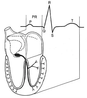
Impulse Conduction & the ECG
-
Sinoatrial node
- AV node
- Bundle of His
- Bundle Branches
- Purkinje fibers
- Bundle Branches
- Bundle of His
- AV node
-
PR
- P
- Q
- R
- S
- T
- S
- R
- Q
- P
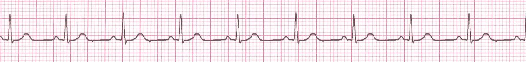
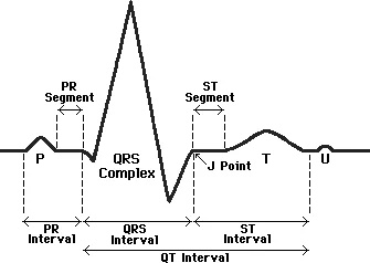
NORMAL ECG
- P
- QRS Complex
- Q
- R
- S
- PR Interval
- PR Segment
- ST Segment
- T
- QT Interval
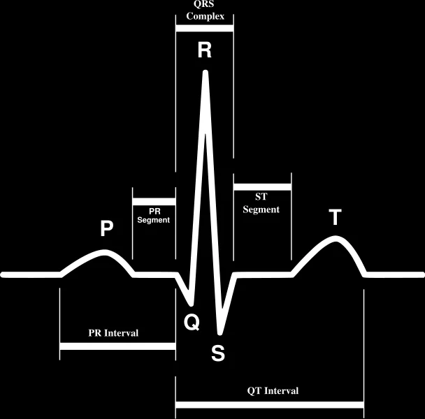
The PQRST
- P wave - Atrial depolarization
- QRS - Ventricular depolarization
- T wave - Ventricular repolarization
The PR Interval
-
Atrial depolarization
- +
- delay in AV junction
- (AV node/Bundle of His)
-
(delay allows time for the atria to contract before the ventricles contract)
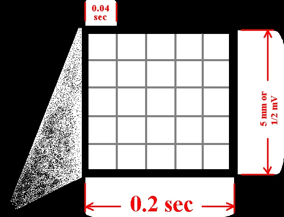
The ECG Paper
-
Horizontally
- One small box - 0.04 s
- One large box - 0.20 s
-
Vertically
- One large box - 0.5 mV
-
5 mm = 12 mV

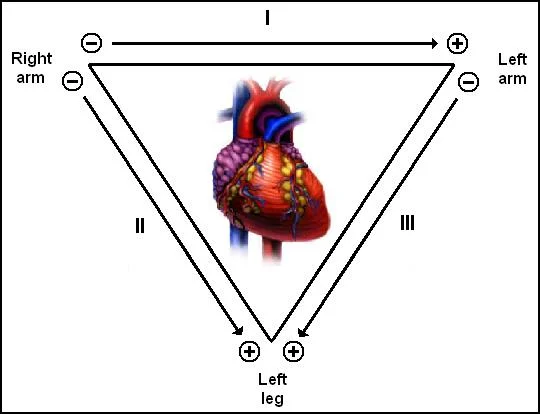
ECG LEADS
Leads measure the difference in electrical potential between two points.
Bipolar Leads
- Two different points on the body
Unipolar Leads
- One point on the body and a virtual reference point with zero electrical potential, located in the center of the heart
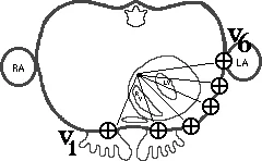
The Standard ECG
The standard ECG has 12 leads:
- 3 Standard Limb Leads
- 3 Augmented Limb Leads
- 6 Precordial Leads
Standard Limb Leads
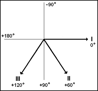
Standard Limb Leads (Angles)
Augmented Limb Leads
All Limb Leads
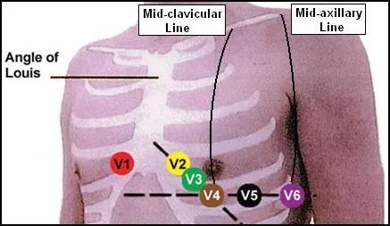
Precordial Leads
- Angle of Louis
- Mid-clavicular Line
- Mid-axillary Line
- V1
- V2
- V3
- V4
- V5
- V6
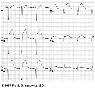
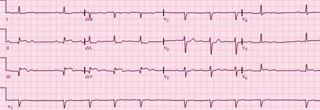
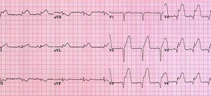
Left Ventricular Hypertrophy
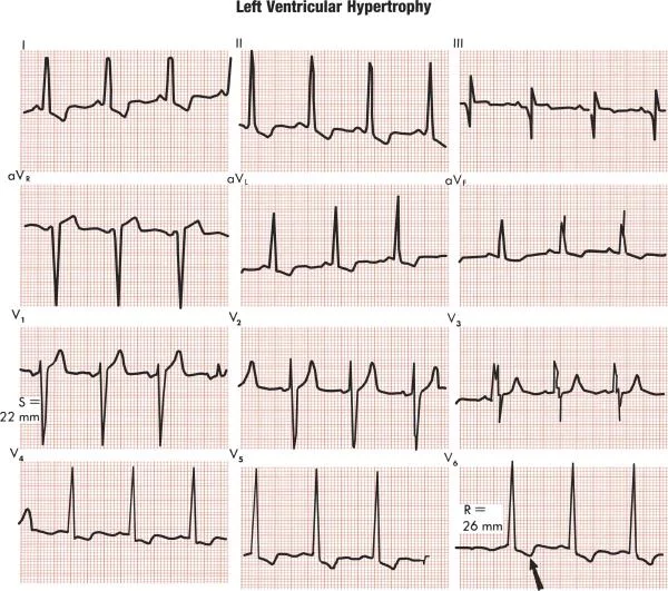
LEFT VENTRICULAR HYPERTROPHY
Compare these two 12-lead ECGs. What stands out as different with the second one?
Normal Left Ventricular Hypertrophy
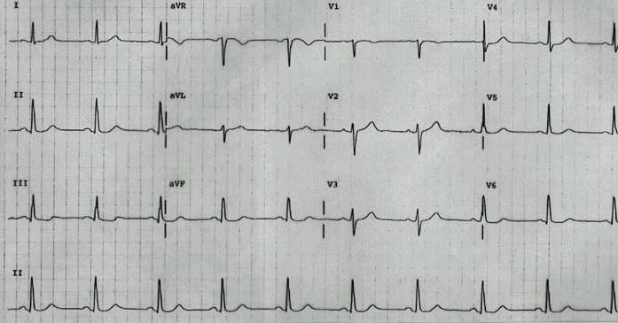
Answer: The QRS complexes are very tall (increased voltage) 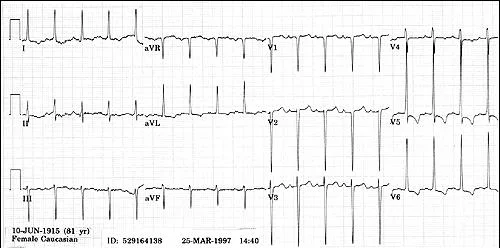
LEFT VENTRICULAR HYPERTROPHY
-
Criteria exists to diagnose LVH using a 12-lead ECG.
- For example:
- The R wave in V5 or V6 plus the S wave in V1 or V2 exceeds 35 mm.
- For example:
-
However, for now, all you need to know is that the QRS voltage increases with LVH.
Q3. A 58-year-old man is a known case of diabetes and hypertension came for routine follow up. His ECG is shown.
Views of the Heart
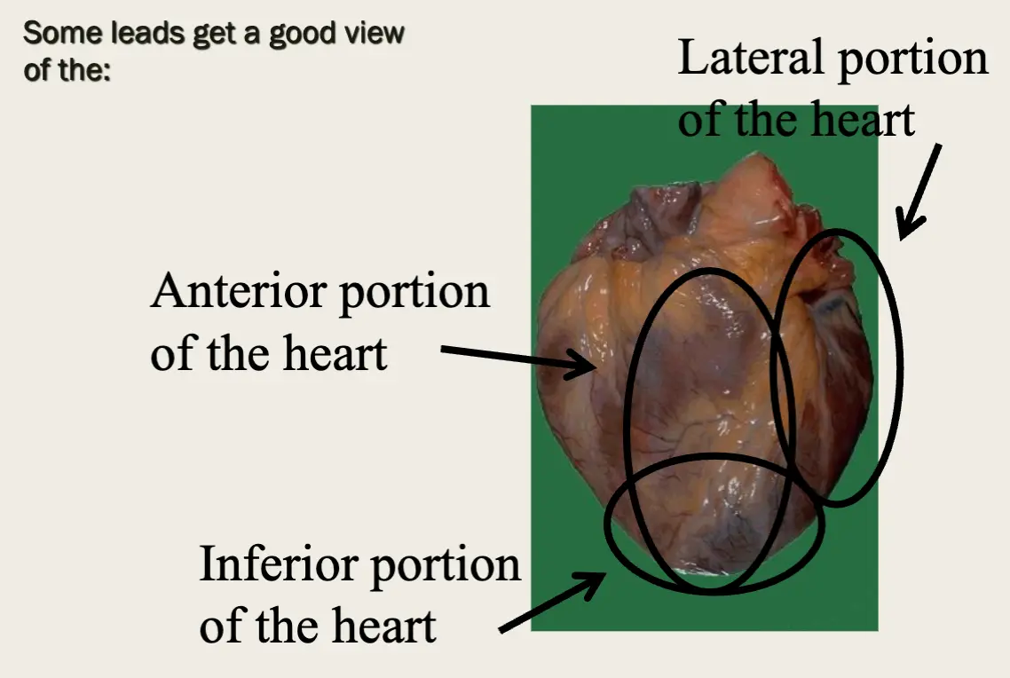
- Some leads get a good view of the:
- Anterior portion of the heart
- Lateral portion of the heart
- Inferior portion of the heart
ST Elevation
One way to diagnose an acute MI is to look for elevation of the ST segment.
ST Elevation (Cont.)
- Elevation of the ST segment (greater than 1 small box) in 2 leads is consistent with a myocardial infarction.
Anterior View of the Heart
The anterior portion of the heart is best viewed using leads V1-V4.
Anterior Myocardial Infarction
If you see changes in leads V1 - V4 that are consistent with a myocardial infarction, you can conclude that it is an anterior wall myocardial infarction.
Most Likely Occluded Coronary Artery
- Left Coronary Artery (Acute Anterior MI) ST elevation in V1 to V6
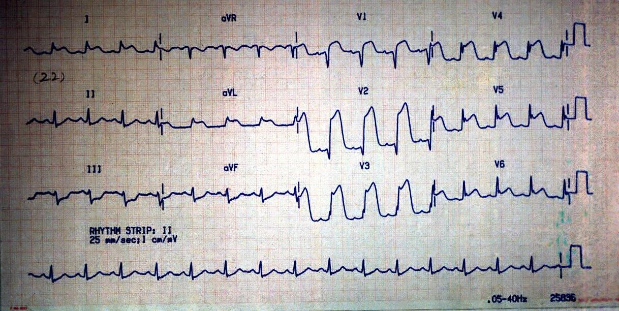
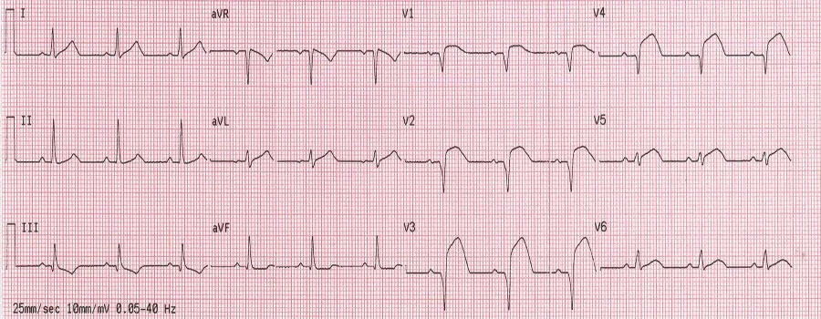
ECG 4
A 50-year-old man is seen in the A & E department with severe central chest pain which has been present for 18 h. What does this ECG show?
Acute anterior MI
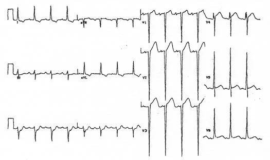
ECG 5
A 56-year-old man presents with 2 hours H/O epigastric pain.
- AF (Irregular irregularity of heart beats in lead aV1)
ECG 6
A 74 year old man presents to emergency room with chest pain of 2 hours duration.
What is your diagnosis?
- A. Acute anterior MI
- B. Acute inferior MI
- C. Acute pericarditis
- D. Acute posterior MI
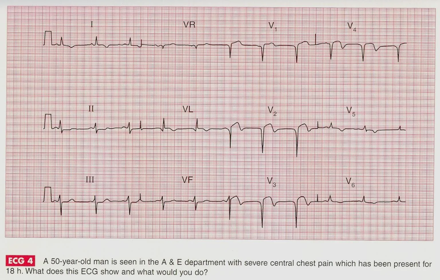
ECG 14
A 50-year-old man is admitted to hospital as an emergency, having had chest pain characteristic of a myocardial infarction for 4 h. Apart from the features associated with pain there are no abnormal physical findings. What does this ECG show?
Acute inferior MI
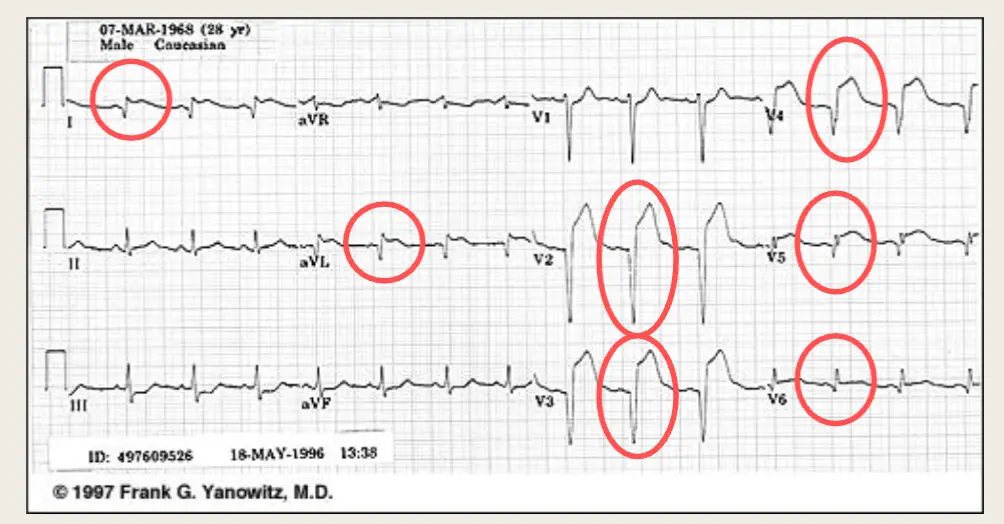
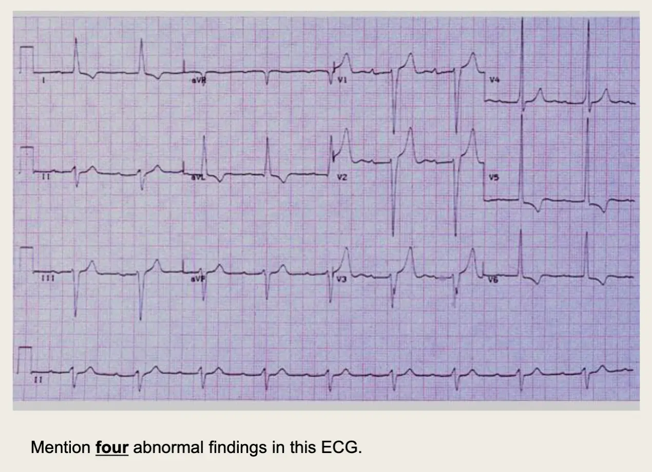
Anterior MI
Putting It All Together
Do you think this person is having a myocardial infarction. If so, where?
 Yes, this person is having an acute anterior wall myocardial infarction.
Other MI Locations
Now that you know where to look for an anterior wall myocardial infarction let’s look at how you would determine if the MI involves the lateral wall or the inferior wall of the heart.
Other MI Locations
-
First, take a look again at this picture of the heart.
- Anterior portion of the heart
- Inferior portion of the heart
- Lateral portion of the heart
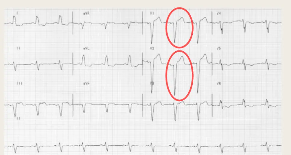
Anterolateral MI
This person’s MI involves both the anterior wall (V2-V4) and the lateral wall (V5-V6, I, and AVL) !