Proptosis
Case Presentation
A 66-year-old Caucasian female presented with gradually increasing protrusion of her right eye over a one-year period. She complained of increased tearing and foreign body sensation. She denied any change in vision, pain with eye movement, double vision, headache, or weight loss. Her past medical history was significant for hypertension, benign breast lump removal, and skin nodule removal. She had no previous eye surgery or trauma. Her family history was negative for malignancy or endocrine disorders.
Physical Examination
The physical examination revealed a visual acuity of 20/20 both eyes, full color vision, full visual fields, with motility examination of the right eye restricted in lateral gaze. Pupils were normal, with no afferent pupillary defect noted. On external examination, her right eye was grossly proptotic. Slit lamp examination detected conjunctival injection of her right eye. Dilated fundus examination showed normal optic discs with no evidence of disc edema. No lymphadenopathy was detected.
Retinoblastoma never cause proptosis in children. In children, proptosis can be due to rhabdomyosarcoma (malignant), benign orbital cellulitis. In adults, causes include thyroid eye disease and cavernous hemangioma. Z
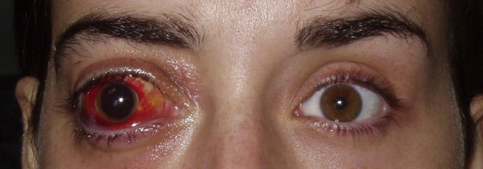 Right Proptosis
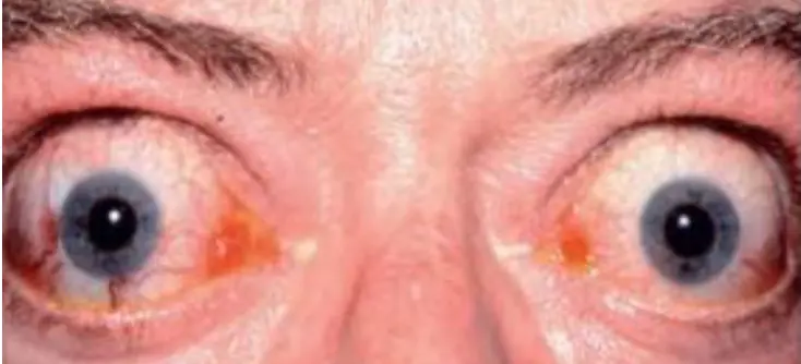 Bilateral thyroid eye disease (proptosis, exophthalmos)
1- What is diagnosis?
Proptosis or Proptosis due to thyroid disease (according to the case )
2- Differential diagnosis
-
Trauma (retrobulbar hemorrhage)
-
Orbital inflammatory pseudotumor
-
Orbital infectious cellulitis
-
Orbital tumors (benign or malignant)
-
Lacrimal gland tumors
-
Carotid-cavernous fistula
-
Orbital varix
-
Orbital pseudotumor, orbital cellulitis, cavernous sinus thrombosis, intraorbital neoplasm
3- Clinical Examination & Investigations
Clinical Exam
- ophthalmic examination
- Visual Acuity –
- visual field -
- Slit lamp –
- Retena and
- direct ophthalmoscope /Fundoscopy
- Pupil exam - * Pupil reaction How is Proptosis Diagnosed?
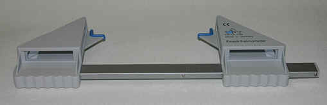
- Globes from above: measured with an exophthalmometer
- The distance between the lateral orbital rim and the corneal apex is used as a measure for proptosis. This distance is normally 18 mm.
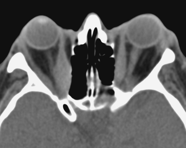
Investigations Which Neuroimaging Test is Best to Evaluate the Etiology of Proptosis?
- CT scans are superior in most cases.
- MRI may be desirable in certain cases when optic nerve dysfunction is present.
- MRI is sensitive in identifying extraocular muscle oedema and other soft tissue.
- TFT
4- Treatments
Treatment of the cause. * Systemic steroids * Radiotherapy * Surgical decompression
NB: if the case is due hyperthyroidism :
- Control hyperthyrodism
- Proptosis by Systemic Steroid and Radiotherapy and Surgical decompression
- Soft tissue involvement by Anti inflammatory + Lubricant
- Eye lid Retraction by lid Surgery
- Myopathy by muscle surgery