Basic Airway
ABDULLAH ALSAKKA
Objectives
Review airway anatomy
Review basic airway maneuvers
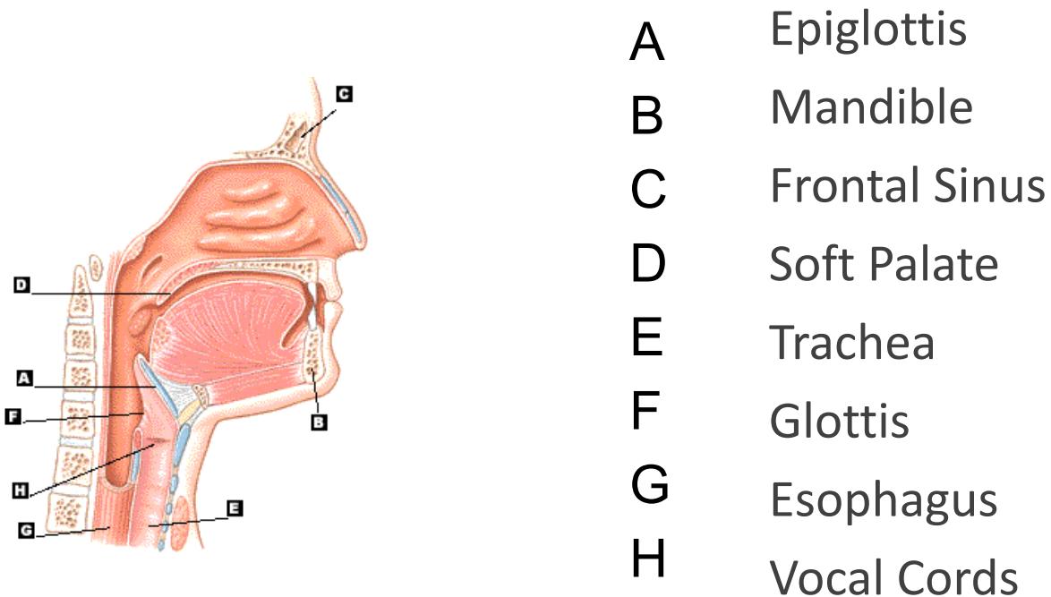
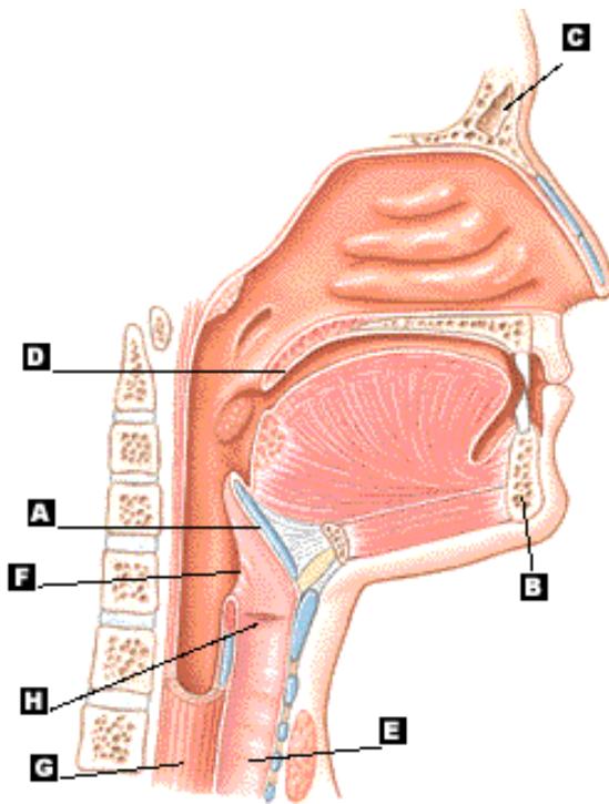
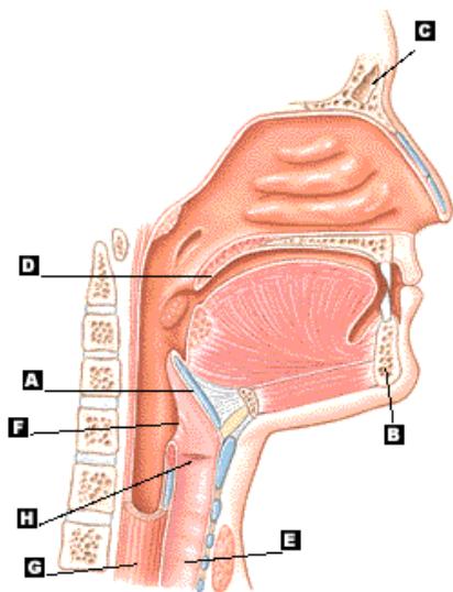
The Upper Airway
Diagram of the upper airway showing labeled structures:
| A | Epiglottis |
|---|---|
| B | Mandible |
| C | Frontal Sinus |
| D | Soft Palate |
| E | Trachea |
| F | Glottis |
| G | Esophagus |
| H | Vocal Cords |
The Upper Airway
Other Structures
- Nasopharynx
- Oropharynx
- Hypopharynx
- Larynx
Functions
Functions of the Upper Airway
Passageway for air
Warm
Filter
Humidify
Protection
- Gag Reflex
- Cough
Speech
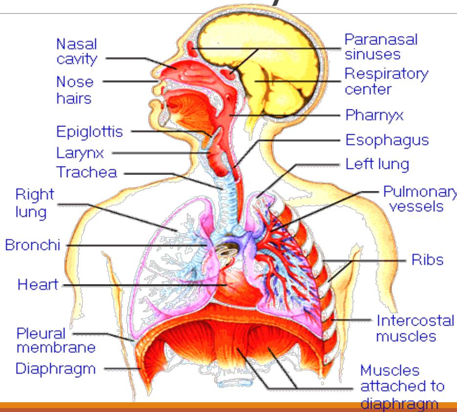
Upper and Lower Airways
Diagram illustrating the upper and lower airways and associated structures.
- Nasal cavity
- Nose hairs
- Epiglottis
- Larynx
- Trachea
- Right lung
- Bronchi
- Heart
- Pleural membrane
- Diaphragm
- Paranasal sinuses
- Respiratory center
- Pharynx
- Esophagus
- Left lung
- Pulmonary vessels
- Ribs
- Intercostal muscles
- Muscles attached to diaphragm
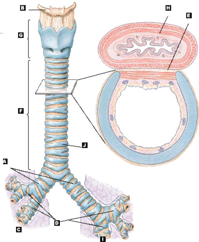
The Lower Airway
A Primary Bronchi
B Hyoid Bone
C Right Lung
D Secondary Bronchi
E Tracheal Ligament
F Trachea
G Larynx
H Esophagus
I Left Lung
J Trachea
J
Airway Anatomy
Upper Airway
- Pharynx
- Epiglottis
- Glottis
- Vocal cords
- Larynx
Lower Airway
- Trachea
- Bronchi
- Alveoli
- Lung tissue, consisting of lobes and lobules (3 on the right and 2 on the left)
- Pleura
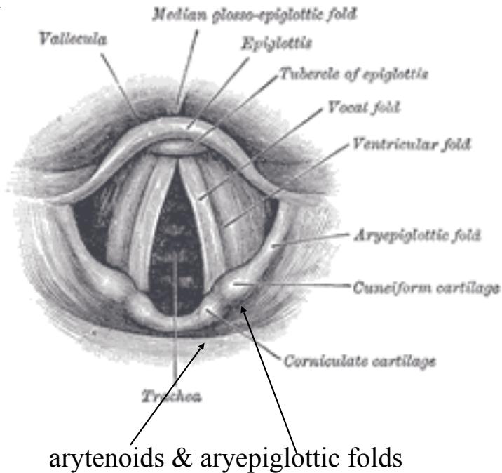
Airway Anatomy
Diagram illustrating the anatomy of the larynx and upper trachea. Labeled structures include:
- Vallecula
- Median glosso-epiglottic fold
- Epiglottis
- Tubercle of epiglottis
- Vocal fold
- Ventricular fold
- Aryepiglottic fold
- Cuneiform cartilage
- Corniculate cartilage
- Trachea
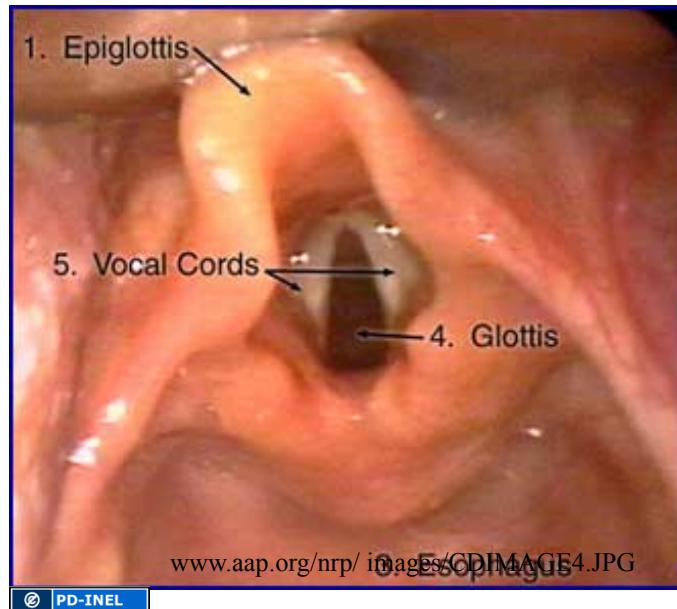
Endoscopic view of the larynx showing the following labeled structures:
- Epiglottis
- Vocal Cords
- Glottis
Source attribution: www.aap.org/nrp/images/CDIMAGE4.JPG
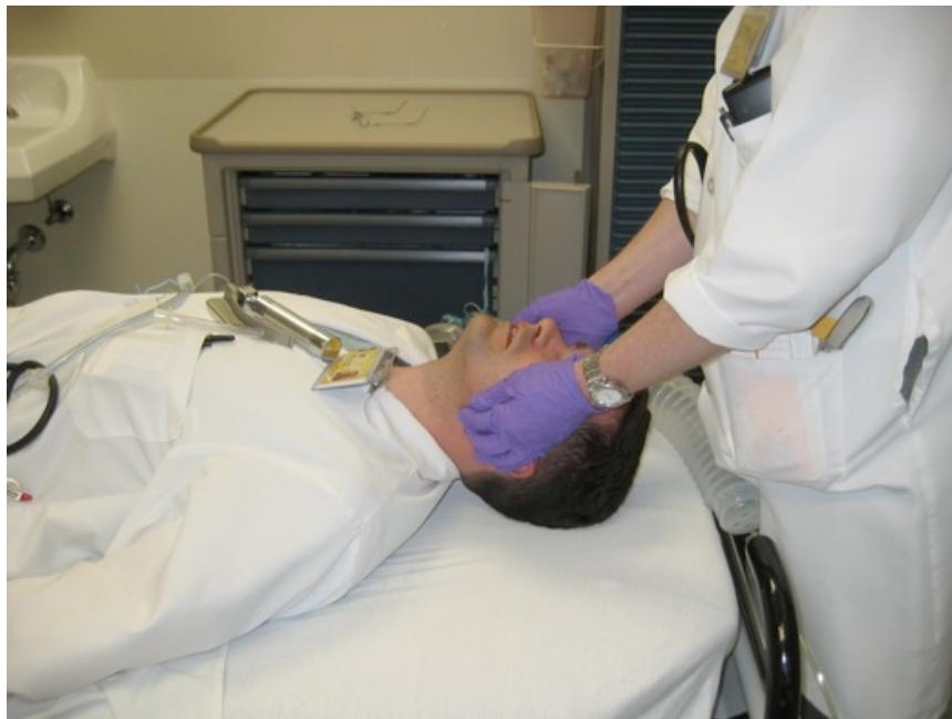
Basic Airway Maneuvers
ALWAYS REMEMBER THE BASICS
These skills should be used prior to initiating any advanced airway technique
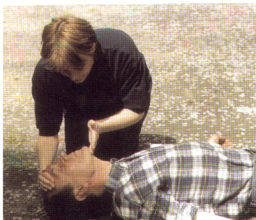
- Head-tilt/chin lift
- Jaw thrust
- Modified jaw thrust (for trauma patients)
- Sellick’s maneuver
Airway Management Techniques Patency Maneuvers
Head tilt with chin lift, or jaw-thrust maneuver
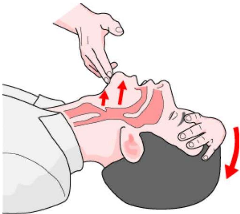
Opening the airway
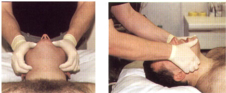
Jaw thrust technique may be needed if C-spine injury
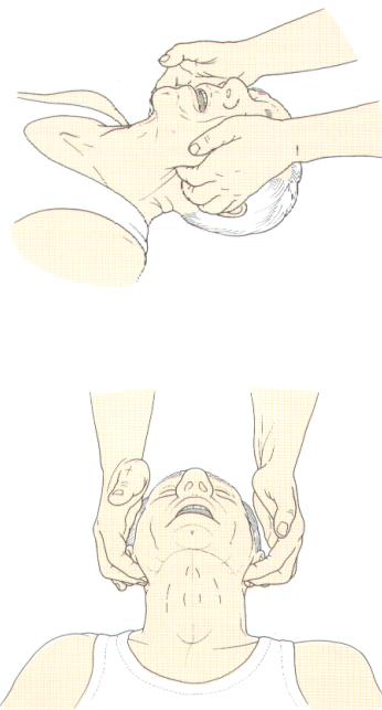
Figure 5.1 Jaw thrust
1.Oropharyngeal Airway
Size is measured from the corner of the mouth to the angle of the jaw
Sizes range from 0-6
It holds the tongue away from the posterior pharynx, but does not isolate the trachea
Simple airway adjuncts
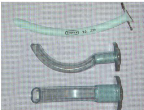
Figure 5.2 Nasopharyngeal and oropharyngeal airways
Oral Airway continued
The oral airway is inserted with the curve towards the side of the mouth
Then rotated so that the curve of the airway matches the curve of the tongue
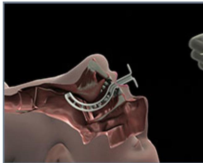
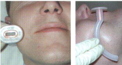
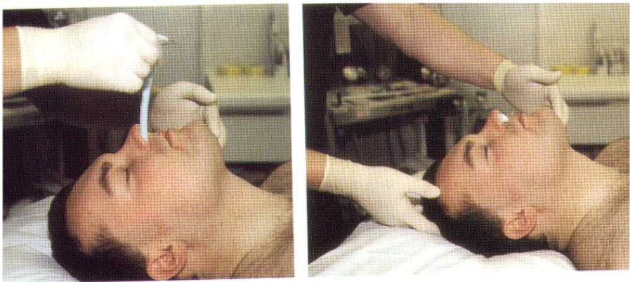
Figure 5.3 Sizing of an oropharyngeal airway
Oropharyngeal airway insertion
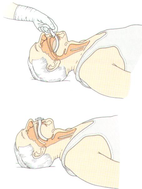
Figure 5.4 Insertion of an oropharyngeal airway
2.Nasopharyngeal Airway
Soft plastic or rubber tube that is designed to pass just inferior to the base of the tongue
Passed through one of the nares and can be used in patients with an intact gag reflex
CONTRAINDICATED in cases of suspected or possible basilar skull fracture
Sizes range from 17-26 cm in length and 6-9 mm internal diameter
Measured from tip of the nose to the corner of the patients ear
The nasal airway is lubricated with a water soluble lubricant
The beveled tip is inserted directed towards the septum, with the airway directed perpendicular to the face
If resistance is met, rotating the airway may help or the other nare may be used
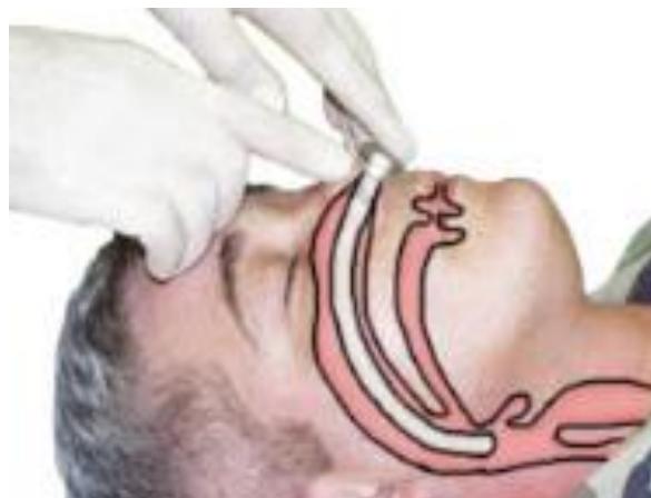
Nasopharyngeal airway insertion
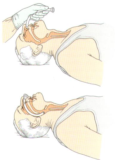
Figure 5.5 Insertion of an nasopharyngeal airway
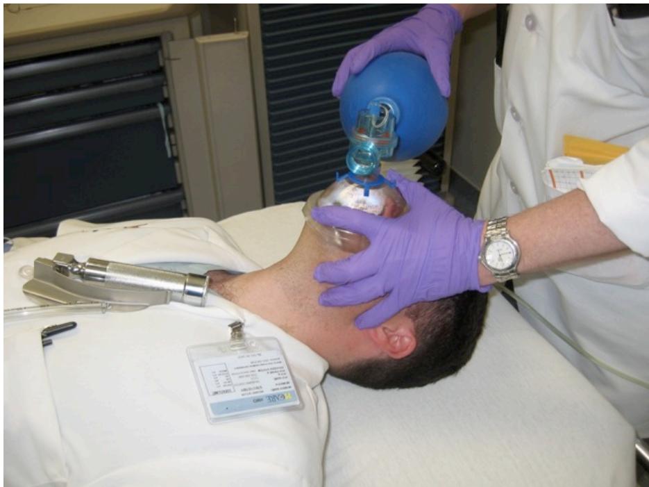
Bag-Valve-Mask Ventilation
- Very important skill to know
- May provide temporary or definitive airway management.
- One person - importance of a good seal.
- Two person technique more effective.
- In EMS setting may be as useful as endotracheal intubation.
Bag-Valve-Mask Ventilation