Central venous access
Dr Abdulaziz Alrabiah, MD
Definition:
- tip of the catheter is placed in the great veins or the right atrium
- it can be single lumen or multiple lumen
sites of insertion
- ✓ internal jugular vein
- ✓ subclavian vein
- ✓ femoral vein
Indications
- No peripheral IV or IO access
- CVP monitoring
Central Venous Pressure - drug administration ( interpose , chemotherapy , TPN …etc) interposes
Cardiogenic shock - renal replacement therapy
- transvenous pacing
Contraindications
- coagulopathy
- respiratory failure
- raised ICP (use femoral vein )
- obstructed vein (thrombus or tumour )
- overlying skin infection
- burn
- uncooperative patient
complications
immediate
- Pneumothorax (Subclavian > IJV)
- failure to locate vein
- arterial puncture
- haemothorax
- haematoma
- arrhythmia
- thoracic duct injury
- guide wire embolus
- air embolus
— migration of wire
needs interventional
radiology
Early
- haemopericardium → cardiac tamponade
- pneumothorax
- blockage
- cylothorax
- kinking of catheter
late infection (2.5/1000 catheters)
- catheter fracture
- vascular erosion
- vessels stenosis
- thrombosis
- osteomyelitis of clavicle
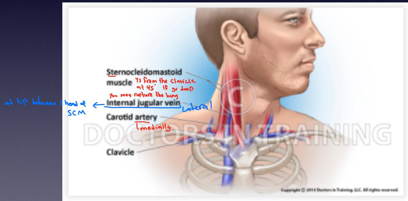
Anatomy of IJV and Subclavian vein
CC verify
- Sternocleidomastoid muscle (SCM): Located anteriorly, serving as a landmark.
- Internal jugular vein (IJV): Runs posterior to the SCM. relative to the sternocleidomastoid muscle and the clavicle. at tip between 2 head of SCM, 1/3 from the clavicle, at 45° if go deep, You may rupture the lung.
- Carotid artery: Runs anterior to the IJV, medial to the IJV.
- Clavicle: Bony structure at the base of the neck.
anatomy of femoral vein
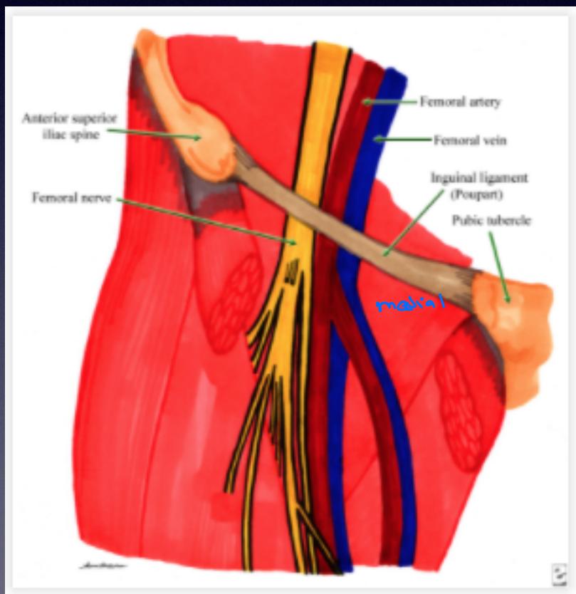
1 cm below
inguinal ligament toward umbilicus
if you go more than 1 cm risk of puncturing
the Artery
method of insertion
- Confirmation of position
- ultrasound visualisation of needle insertion, guidewire placement and CVC
- chest Xray if IJV or subclavian
- pressure measurement
- assess for CVP trace