Forensic Medicine OSPE
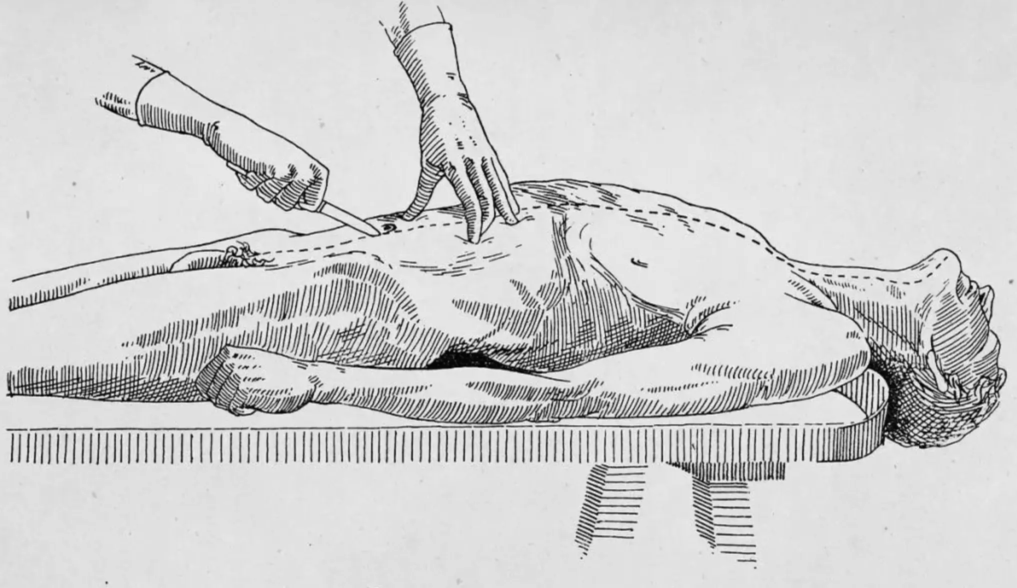
Table of Contents
- General Forensic Data & Organ Weights
- Wounds and Injuries
- Asphyxia and Mechanical Causes
- Firearm Injuries
- Burns and Thermal Injuries
- Head Injuries and Skull Fractures
- Age Determination
- Postmortem Changes
- Death Investigation
- Neonatal and Infant Death
- Electrical Injuries
- Clinical Stations and Autopsy Findings
اللهم يا معلّم موسى علّمني، ويا مفهم سليمان فهّمني، ويا مؤتي لقمان الحكمة وفصل الخطاب آتني الحكمة وفصل الخطاب اللهم اجعل ألستنا عامرة بذكرك، وقلوبنا بخشيتك، وأسرارنا بطاعتك، إنك على كل شيء قدير، حسبنا الله ونعم الوكيل
Updates through https://medatlax.com/Clinical/Level-11/Forensics/Forensics within ospe - will be highlighted yellow
Collected from past batches; Lina Serhan, Leenah Turjoman, Norah Almusallam, Aminah Alessa, Hanadi, Mubarak, Adel, and Khaled AlSafadi
Quick Reference Tables
Wound Classification
| Wound Type | Characteristics | Common Causes |
|---|---|---|
| Abrasion | Superficial, minimal bleeding | Friction, impact, pressure |
| Contusion | Bruising, color changes | Blunt trauma |
| Incised | Clean edges, gaping | Sharp instruments |
| Lacerated | Irregular, tissue bridging | Blunt trauma |
| Stab | Deep, can be penetrating | Pointed instruments |
Antemortem vs Postmortem Wounds
| Feature | Antemortem Wound | Postmortem Wound |
|---|---|---|
| Hemorrhage | Severe, arterial spurting, clots | Slight, oozing, no clots |
| Edges | Gaping, everted | No gaping, no eversion |
| Vital Reactions | Present (redness, swelling) | Absent |
| Microscopy | Cellular infiltration | Absent |
| Chemical | Increased serotonin/histamine | No increase |
Age Determination Summary
| Bone/Joint | Age Indicator | Fusion Age |
|---|---|---|
| Elbow | Trochlea-capitulum union | Below 14 years |
| Knee | Femur/tibia/fibula union | Below 21 years |
| Hand | Metacarpal-phalangeal union | Below 18 years |
| Sternum | Manubrium-body union | 40-60 years |
Asphyxia Types
| Type | Mechanism | Key Findings |
|---|---|---|
| Hanging | Ligature suspension | Oblique ligature mark |
| Strangulation | Neck compression | Horizontal ligature mark |
| Smothering | External airway obstruction | Perioral abrasions |
| Choking | Internal airway obstruction | Foreign material |
Force & Effect of hanging
| Structure | Force (kg) | Effect |
|---|---|---|
| Jugular vein | 2 | Venous congestion |
| Carotid arteries | 5 | Arterial compression |
| Trachea | 15 | Airway obstruction |
| Vertebral arteries | 30 | Complete obstruction |
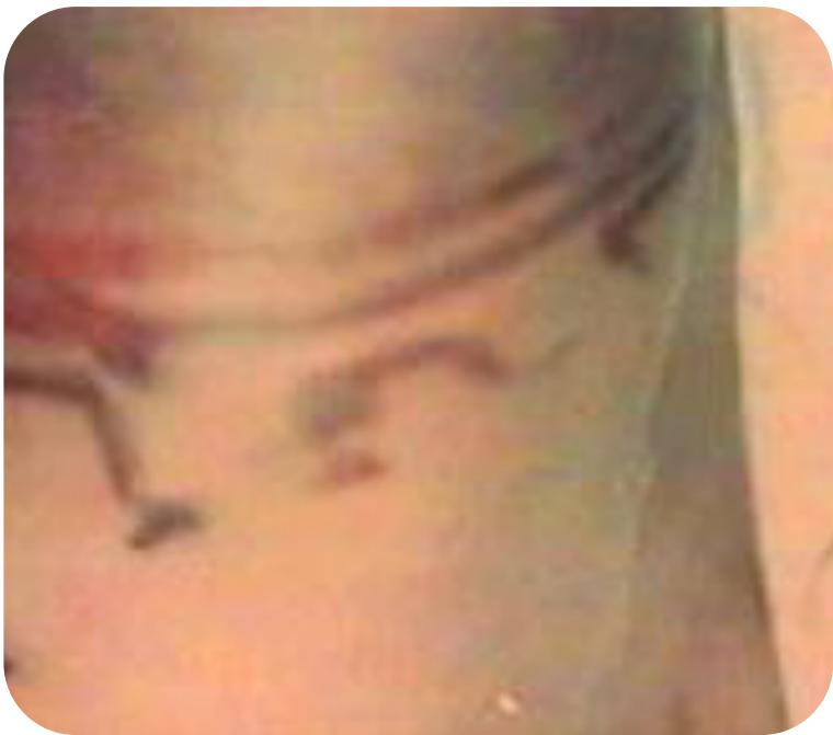
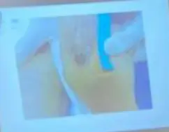
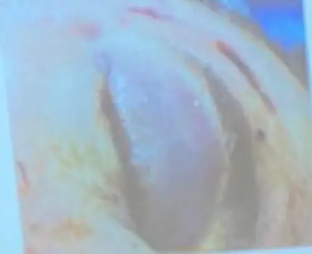
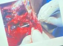
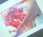
Sexual Assault / Abuse Findings
- Description of Abuse:
- Locate tears (e.g., at 12 o’clock, 11 o’clock).
- Determine if recent or old.
- Check for edema
- Traction Test: Same picture with traction may show fresh blood.
General Forensic Data & Organ Weights
Normal Organ Weights
- Brain: 1300 - 1400 g
- Heart: 280 - 312 g
- Liver: 1500 g
Wounds and Injuries
Types of Wounds
Abrasions
- Impact Abrasion: Caused by perpendicular blunt force, often takes shape of causative object
- Type: Contusion Impact Abrasion.
- Cause: Hitting by car.
- Question: Does this injury alone can cause death? No.
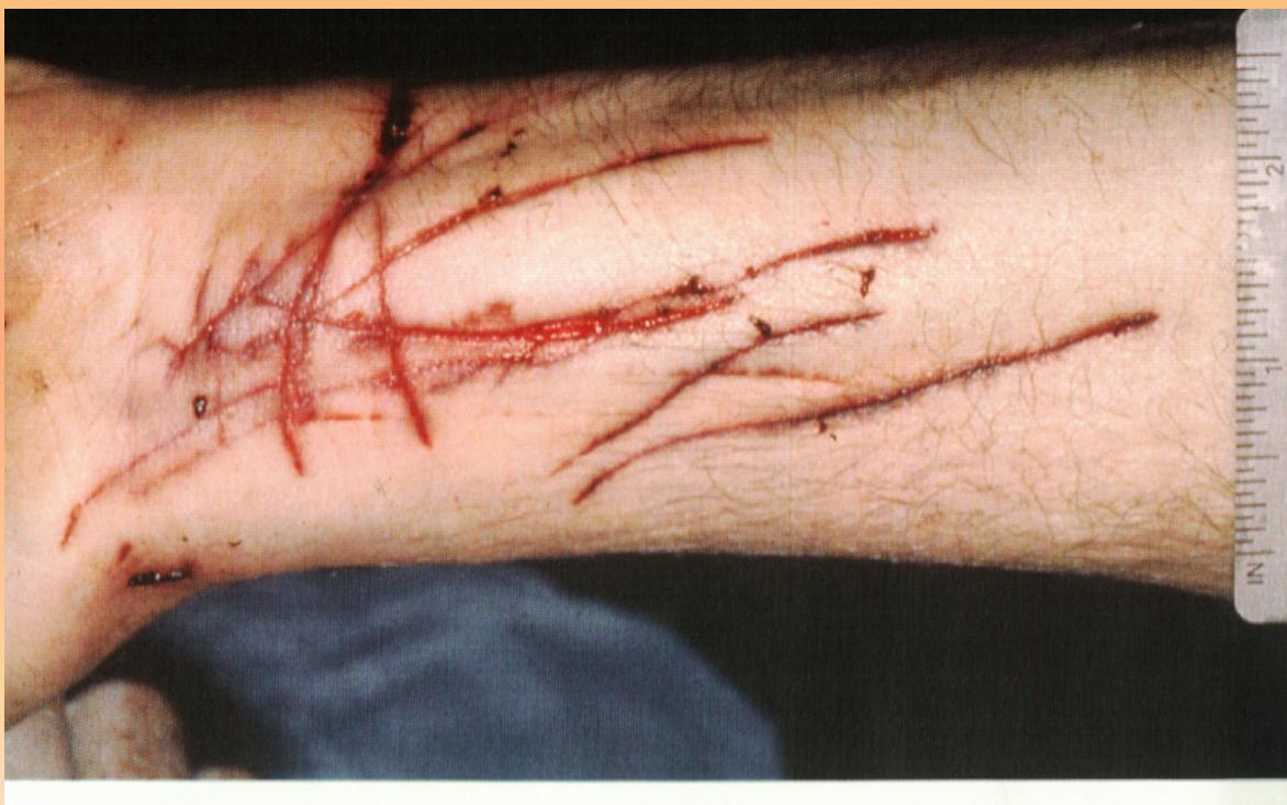
- Friction/Sliding Abrasion (Scratches): Caused by movement between skin and rough object or sharp object (nails)
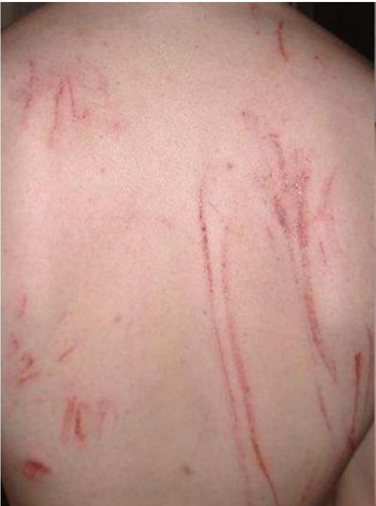
- Question: State the specific type of injury? Linear abrasions (Sliding).
- Type: Grazed abrasion (Dragging) / Scratches (Sliding).
- Characteristics: Shows parallel longitudinal lines.
- Cause: Sharp object passing across skin (e.g., fingernails).
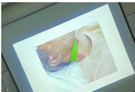
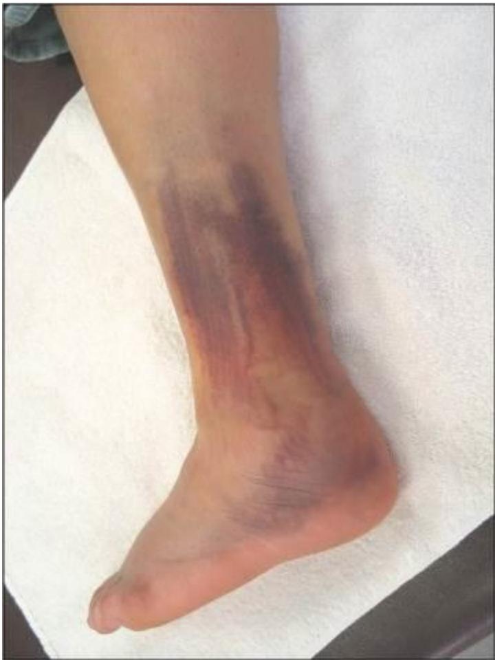
- Pressure Abrasion: Seen in hanging cases
-
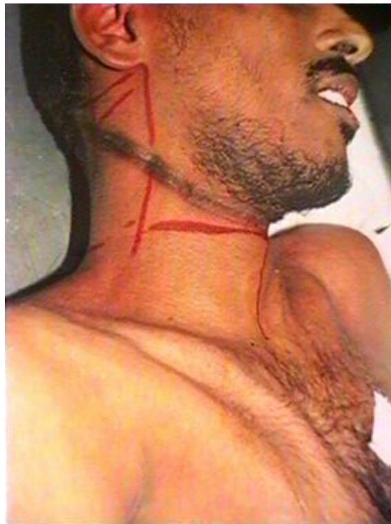
- Type: Friction abrasion (Ligature mark).
- Scenario: Complete typical hanging.
- Cause of death: Asphyxia.
- Note: The red color on pic is not injury, it’s a marker.
-
- Imprint Abrasion: Caused by fingernails
-
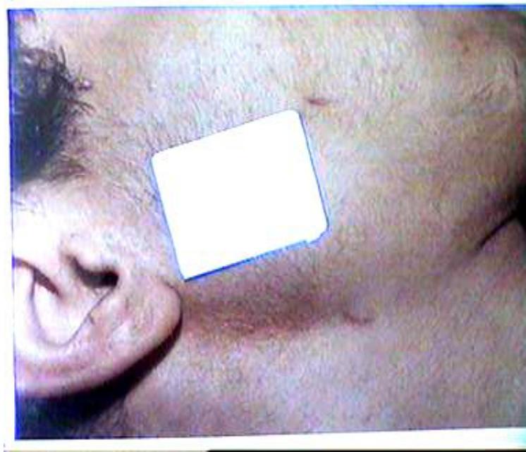
- Question: Cause of death? Undetermined cause of death.
- Type: Impact Abrasion.
- Cause: Finger Nails (Throttling).
- Note: Can identify the position of assailant by the direction of injury.
-
Contusions (Bruises)
- Characteristics: Antemortem (color changes), can cause death via embolism, neurogenic shock, hematoma, septicemia
- Age Determination by Color:
| Color | Age | Mechanism |
|---|---|---|
| Bright red | 0-2 days | |
| Bluish | 2-3 days | Due to reduced HB |
| Green | 4-5 days | |
| Yellow | 7-10 days | Due to Bilirubin |
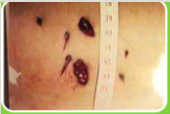
- Questions and Images:
- Question: Which of the following is correct concerning the color changes of that wound? Bluish discoloration is due to reduced HB.
- Question: State the age of injury, state why? 2-3 days due to presence bluish color.
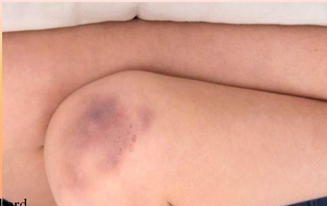
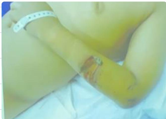
- Characteristics: Antemortem (changing in color).
- Age: 1-3 days (bluish purple).
- Cause of death: Embolism, Neurogenic shock, hematoma, septicemia.
- Instrument: Blunt.
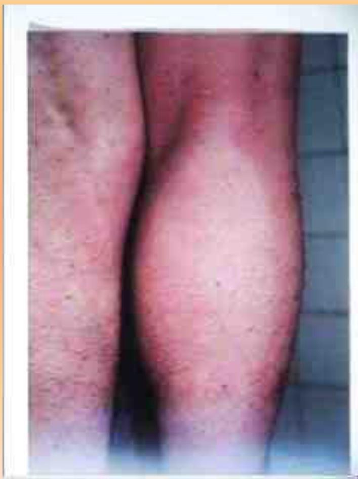
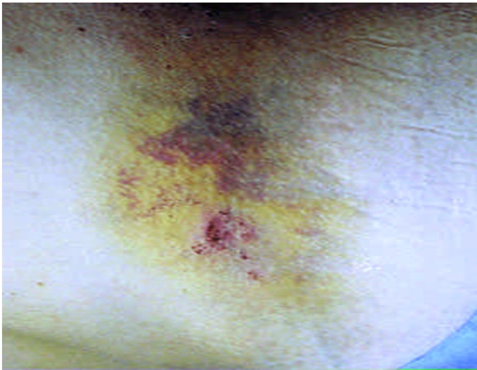 - Age: 7-10 days (yellow color).
 - Type: Localized contusion.
- Example: Hammer.
- Type: Localized contusion.
- Example: Hammer.
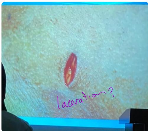
Cut Wounds (Incised Wounds)
- Characteristics: Sharp edges, gaping, severe bleeding, antemortem (swelling edges)
- Causes of death/Dangers: Severe bleeding, air embolism, infection, cutting of nerves and tendons
- Types:
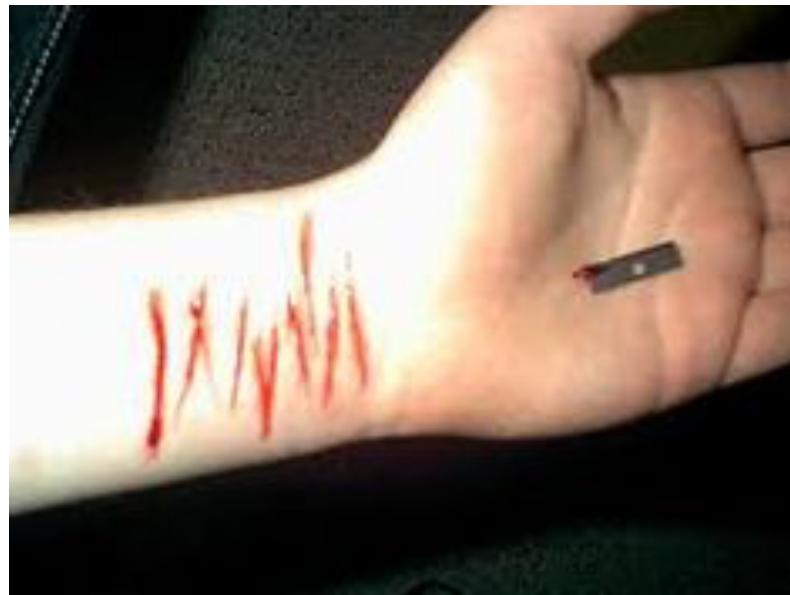
- Multiple cut wounds:
 (Antemortem, Sharp instrument).
- Multiple cut wounds:
- Hesitation wounds:
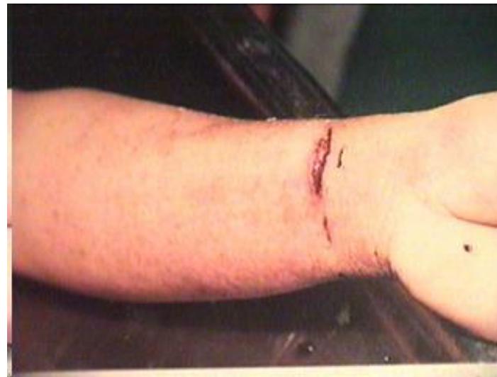
- Instrument: Knife.
- Characteristics: Sharp edges, gaping, severe bleeding.
- Dangers: Air embolism, cutting of nerves and tendons, infection.
-
- Cut wound.
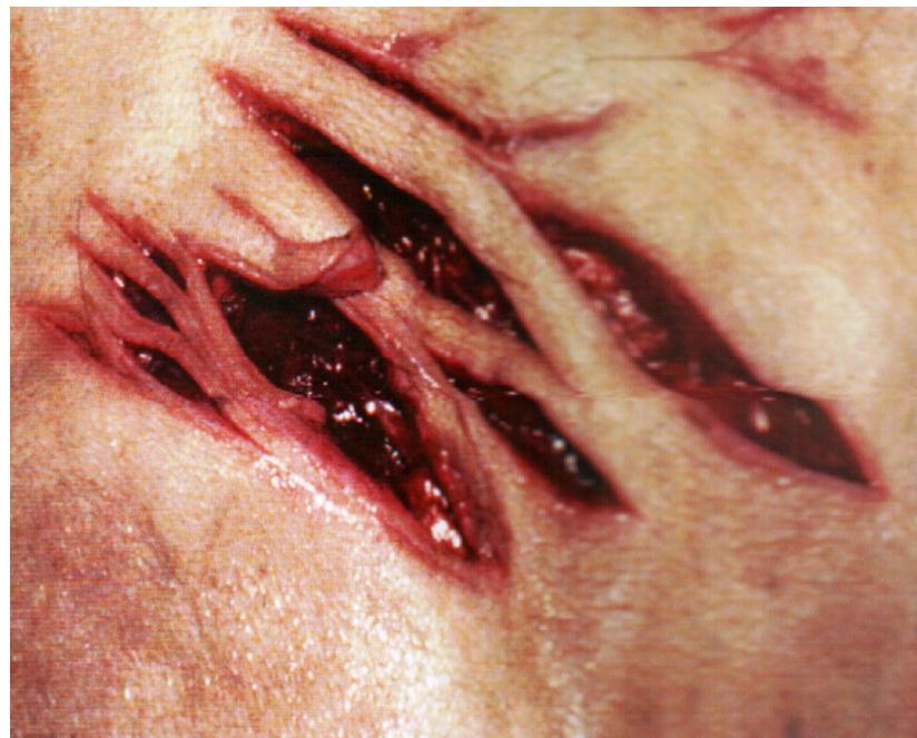
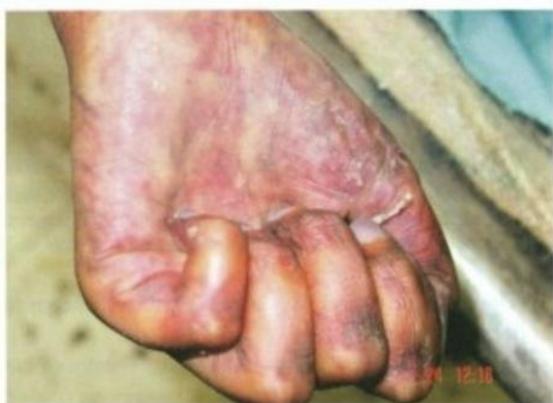
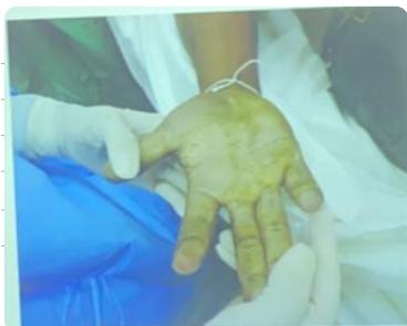
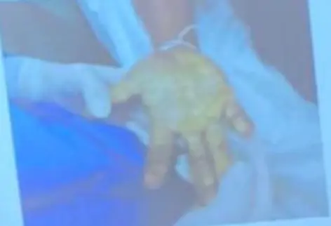
- Defense wounds:
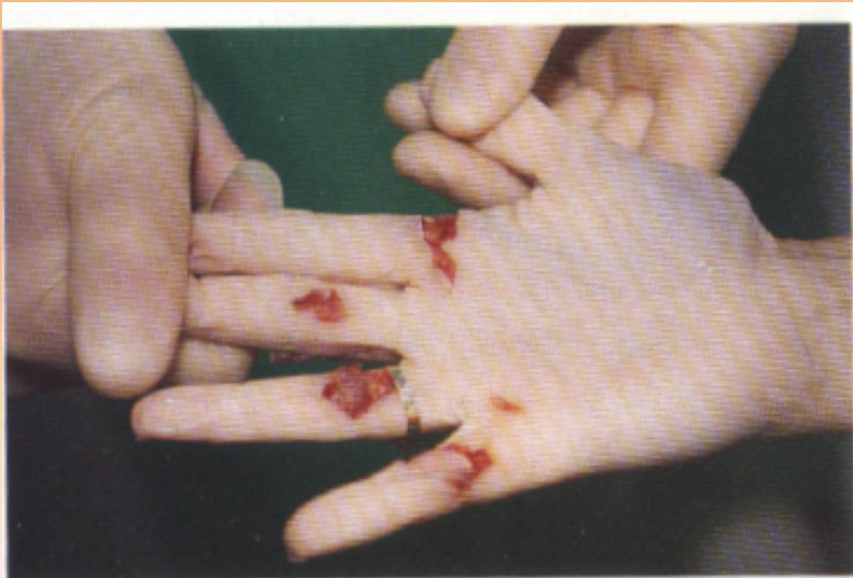
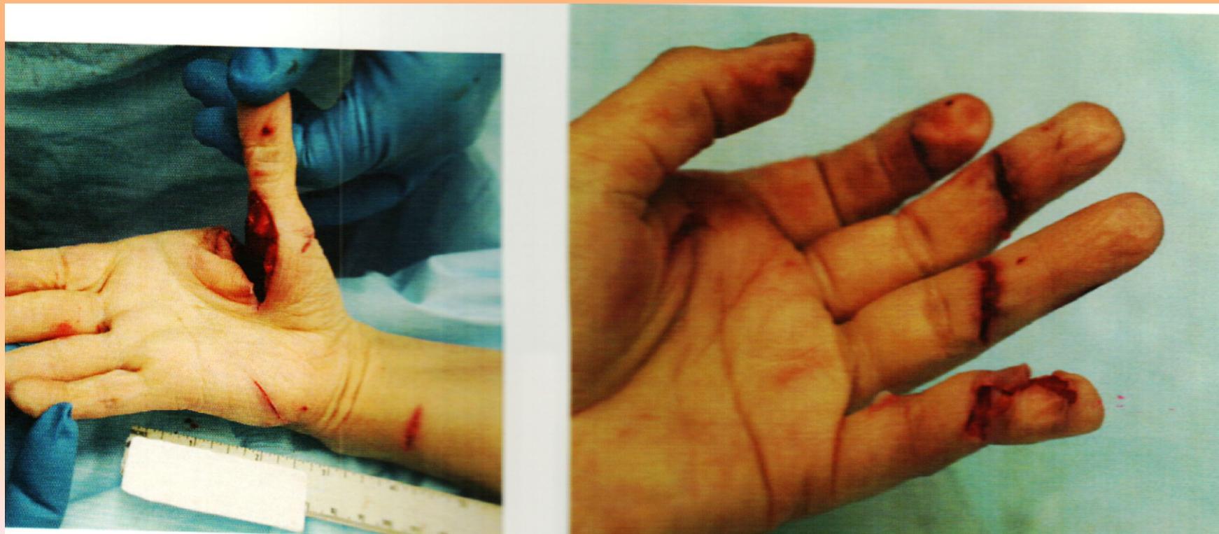
- Instrument: Knife.
- Description: Severe laceration of palmar surface, cuts across flexor surface.
- Cut Throat:
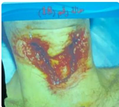
- Description: Transverse injury at ant aspect of neck with irregular border.
- Cause of death: Carotid injury or injury to main vessels.
- Specific Case Questions:
- State type of lesion? Incised wound.
- This lesion classified as: Postmortem. (Finger points to Lividity, not 4th wound).
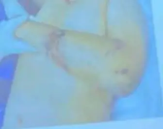
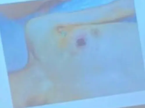
- Description: Clean cut wound of RT shoulder below clavicle 1-2 cm. Clean sharp edge.
- Characteristics: Antemortem (swelling edges, gap).
- Cause of death: Severe bleeding, Air embolism, Infection, hemorhage
- Instrument: Sharp (knife, razor, glass).
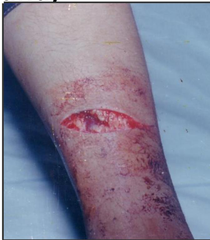
Lacerated Wounds
- Characteristics: Irregular edges, tissue bridging, caused by blunt trauma
- Questions:
- Description: Lacerated wound, linear with bridging tissue (diagnostic).
 - Type: Contused Wound (Specific: Lacerated).
- Cause of death: Hematoma.
- Instrument: Heavy blunt object (stone, fall).
- Hallmark: Presence of tissue bridging.
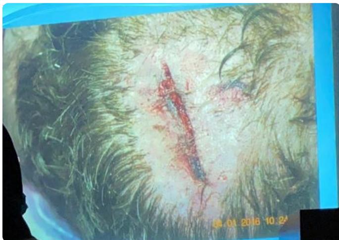
- Description: Edges severely damaged, highly irregular.
- Type: Contused Wound (Specific: Lacerated).
- Cause of death: Hematoma.
- Instrument: Heavy blunt object (stone, fall).
- Hallmark: Presence of tissue bridging.
- Description: Edges severely damaged, highly irregular.
- Type: Contused wound (Edges abraded, contusions around).
- Instrument: Blunt. injury (stick, stone, fall).
- Distinction: Not lacerated because lacerated has more angles and more irregularity.
- Characteristics: Antemortem (bleeding and clotting).
- Cause of death: Subdural and epidural hemorrhage.
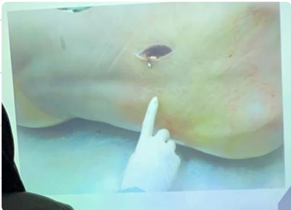
Stab and Puncture Wounds
- Characteristics: Deep, clean cut, can be penetrating or perforating
- Puncture Wounds (Pointed, Blunt Edge):
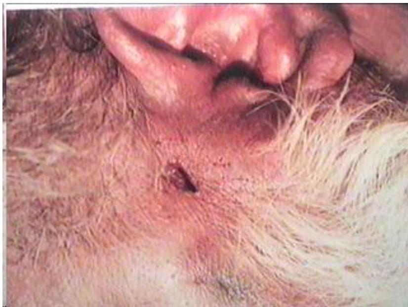
- Type: Puncture penetrating injury / Stab “punctured” wound.
- Instrument: Nail from a nail firing gun / Nail / Screw / Stick.
- Transfixing/Perforating Wounds:
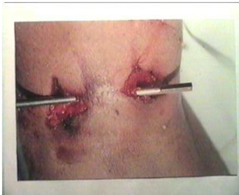
- Type: Transfixing wound (perforating).
- Instrument: Stick, pole.
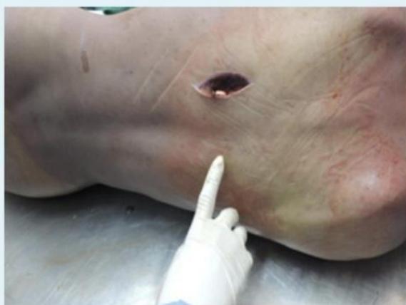
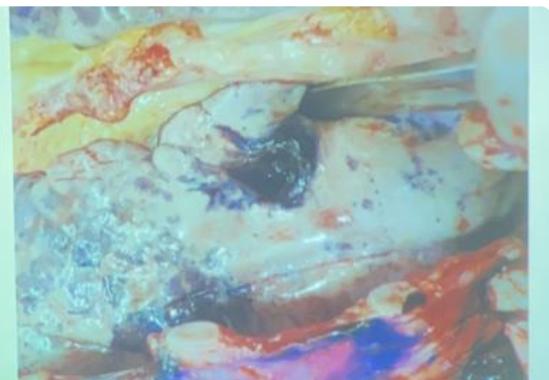
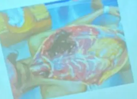
- Penetrating Wounds:
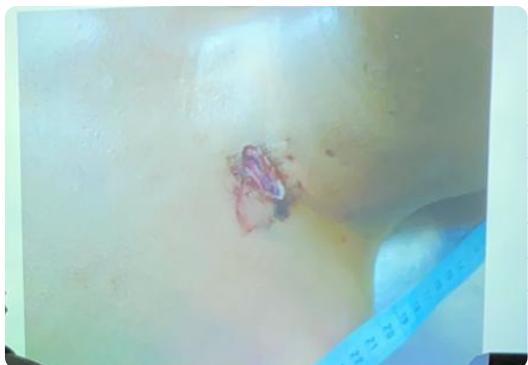
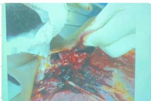
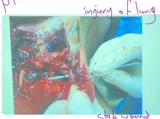
- Description: Cut/Stab to neck. Injury of lung.
- Note: Stab wound has more depth than cut wound.

- Type: Stab penetrating wound to heart.
- Organ: Heart.
- Cause of death: Hemorrhage.
- Characteristics: A.M Wound.
- General Stab Questions:
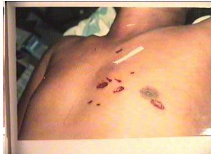
- Type: Multiple stab wounds.
- Characteristics: Antemortem.
- Instrument: Knife.
- Type: Stab wound + Hesitation wound.
- Characteristics: Antemortem.
- Type: Stab wound.
- Characters: More deep than long, clean cut.
- Angles: Two pointed (bi-bladed) or One pointed/one transverse (mono-bladed).
- Dangers: Neurogenic shock, Injury to vital organs, Internal hemorrhage, Sepsis.
Asphyxia and Mechanical Causes
Types of Asphyxia
Hanging
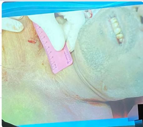
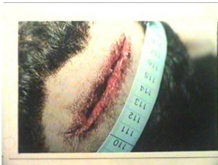
- Typical Hanging: Knot at nape of neck, ligature mark oblique upward
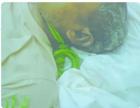
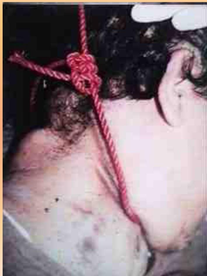
- Scenario: Picture of hanging man: it’s imp. To describe fabric, ligature mark and its location. Ligature mark, contused abrasion located at the upper part of the neck under the RT ear by 8cm located from the chin and just below the hair line and you have to mention if oblique upward or downward.
- Partial Hanging: Weight of part of body - 5kg
- Question: If 5kg wt? Cerebral anemia/Carotid compression.
- Constricting Forces:
- Question 1: A constricting force of can compress the trachea: c. 15 K.G.
- Question 2: In such cases what is the common direction of the ligature mark: b. Oblique.
| Structure | Force (kg) | Effect |
|---|---|---|
| Jugular vein | 2 | Venous congestion |
| Carotid arteries | 5 | Arterial compression |
| Trachea | 15 | Airway obstruction |
| Vertebral arteries | 30 | Complete obstruction |
Clinical Signs
- Petechial Hemorrhage: Sign of asphyxia
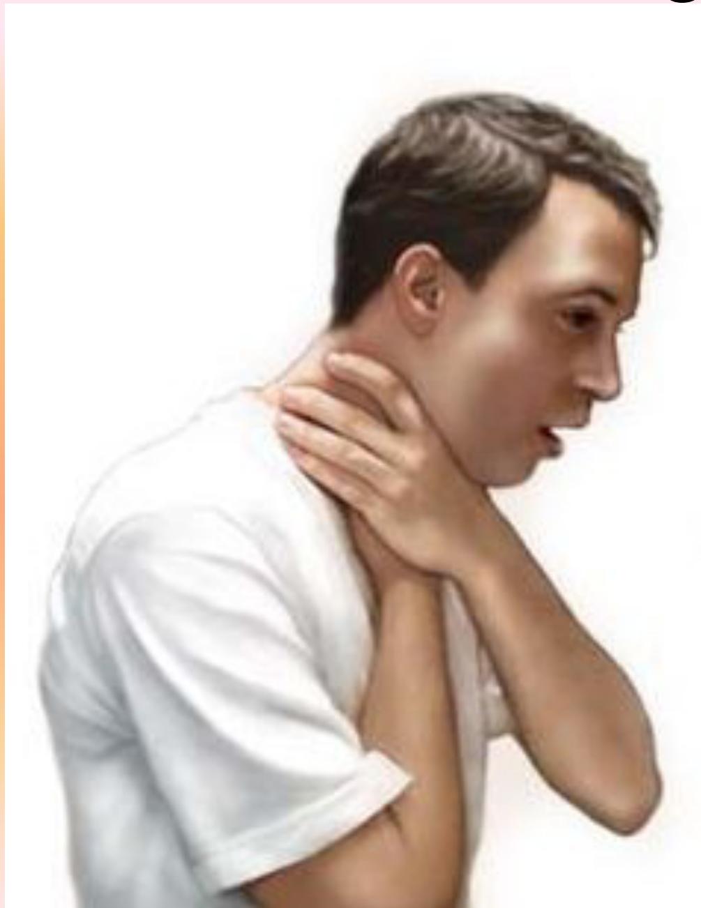
Strangulation
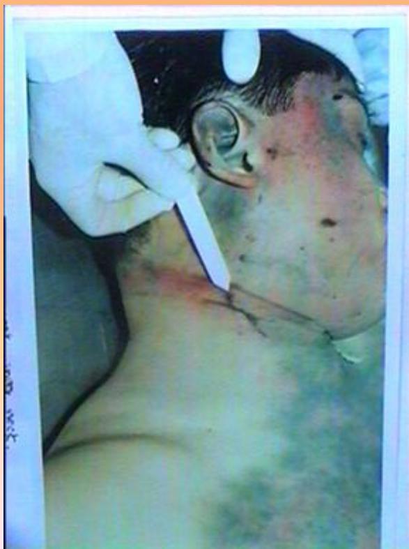
- Throttling: Manual strangulation by hands
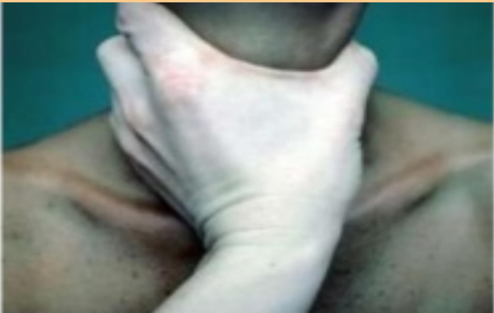
- Cause of death: Combined asphyxia and venous congestion.
- Specific type: Throttling, manual Strangulation.
Smothering
- Characteristics: Abrasions around mouth and nose
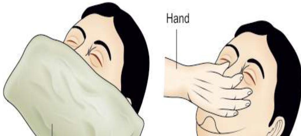
- Cause of death: Asphyxia (suffocation). Cessation of respiration. - Obstructing air passage by hands/pillow/towel.
- Type: Mechanical asphyxia “Suffocation” smothering.
- Description: Abrasions around mouth and nose.
- Cause of death:
- Description: Smothering (asphyxia).
Choking
- Characteristics: Blocking internal air passage by foreign material
- Description: Suffocation; Chocking - Blocking Internal Air Passage.
- Examples: Food particles, fruit seeds in children, tongue in comatose patients.
Firearm Injuries
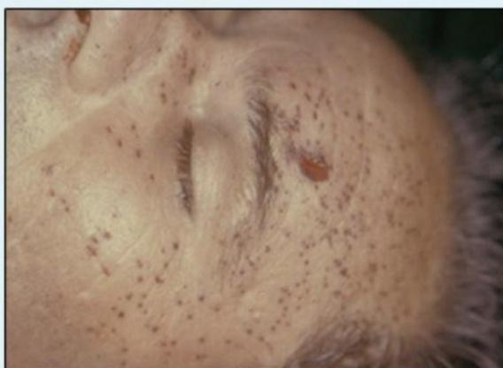
Ballistic Findings
-
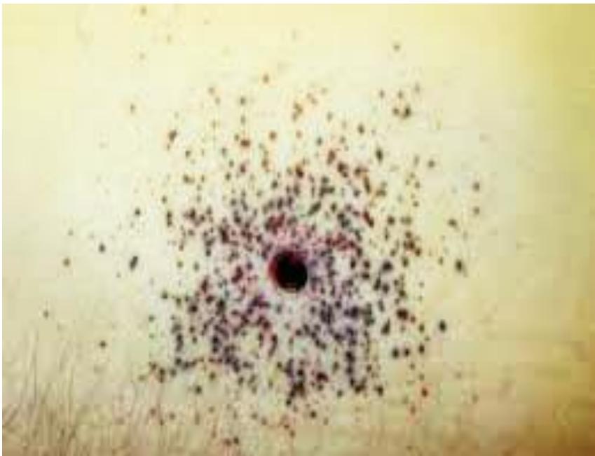
Tattooing: Present up to 2 yards (unburnt gunpowder particles)
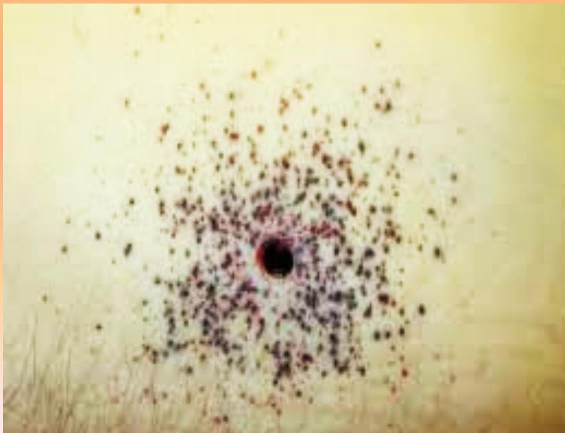
- Description: Unburnt gun powder pierce under superficial skin layers.
-
Blackening: Present up to 1 yard
- In track:
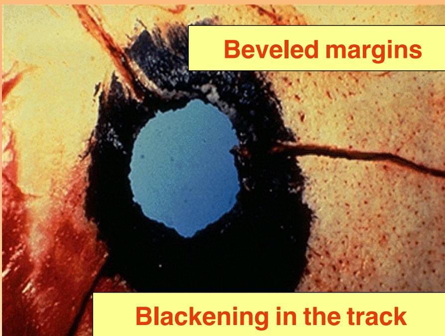
- In track:
-
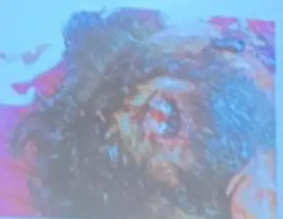
Contact Range: Shows burning and muzzle print
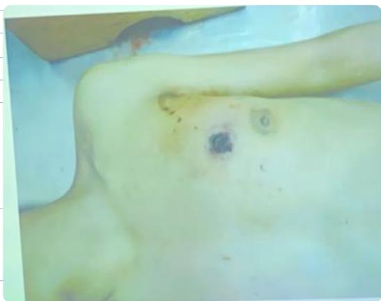
- Description: A picture of male autopsy showed round injury of upper limb. Above LT nipple showed loss of tissue with muzzle print of black color.
- Spot dx: Gunshot.
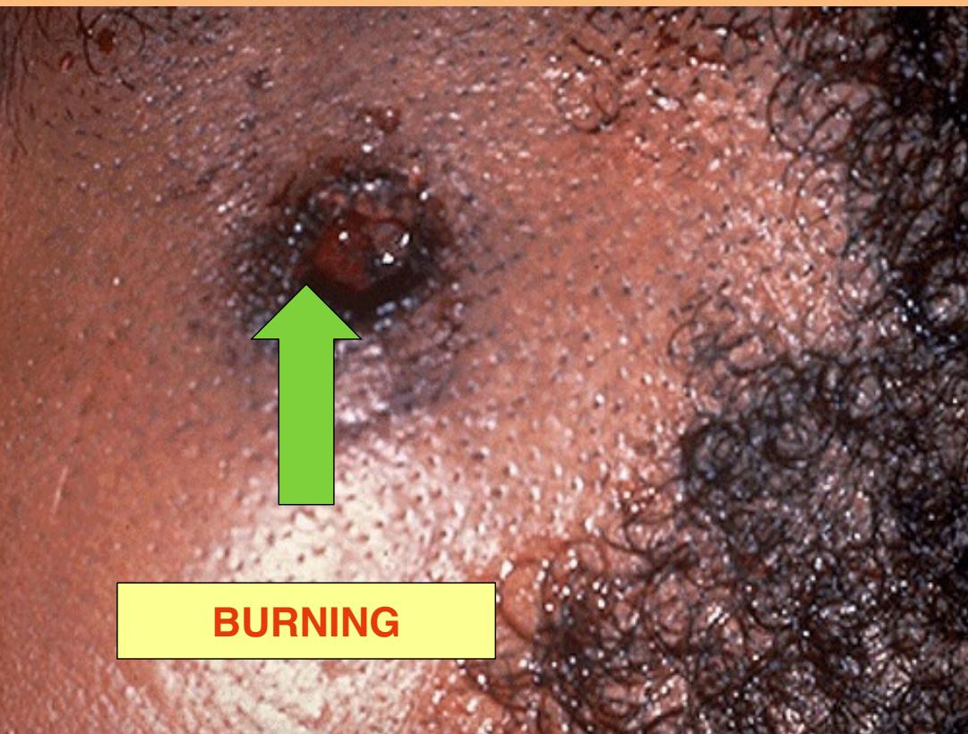
- Description: Burning inlet close range.
- Burning Distance: Few inches in case of revolver & one foot in case of a Shotgun.
Wound Characteristics
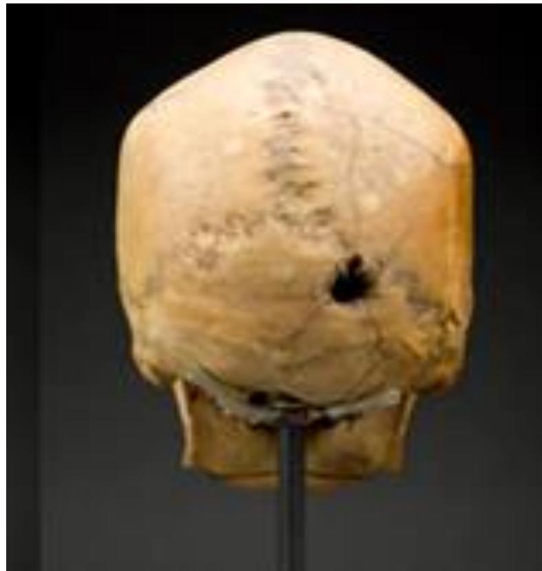
- Entry Wound: Loss of tissue, collar of abrasion
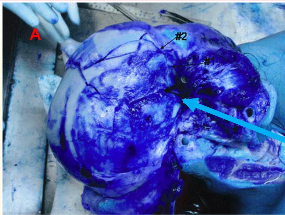
- Description: Inlet with fragments of bone.
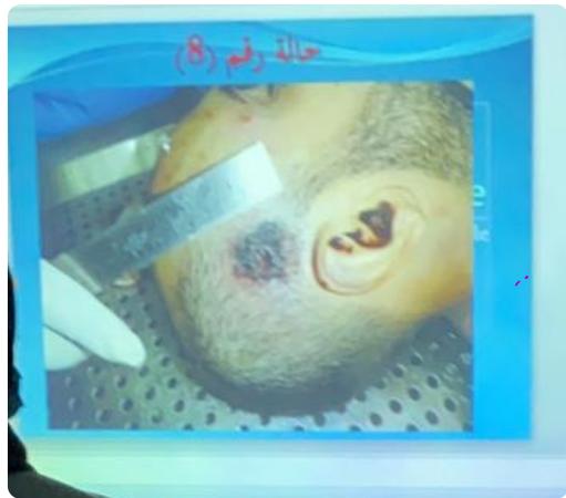
- Description: Entry wound in RT side of head, above RT ear by 5cm, with blood from ear.
- Internal Injuries:
 - Hemothorax/Fluid
 - Lung injury
- Bullet Extraction:
 - Removal after X-ray
- Exit Wound: External beveling
- Type: Exit wound that shows external beveling.
- Mechanism of Beveling:
- The table struck first (entry) is supported from below, resulting in a small circular hole with clean margins.
- The table struck second (exit) has no support, resulting in a bigger irregular hole with beveled margins.
- Importance: Gives direction of fire.
Distance Determination
- Contact: Burning and muzzle print present
- Near range: Less than 2 yards, tattooing present
- Distant: No tattooing or blackening
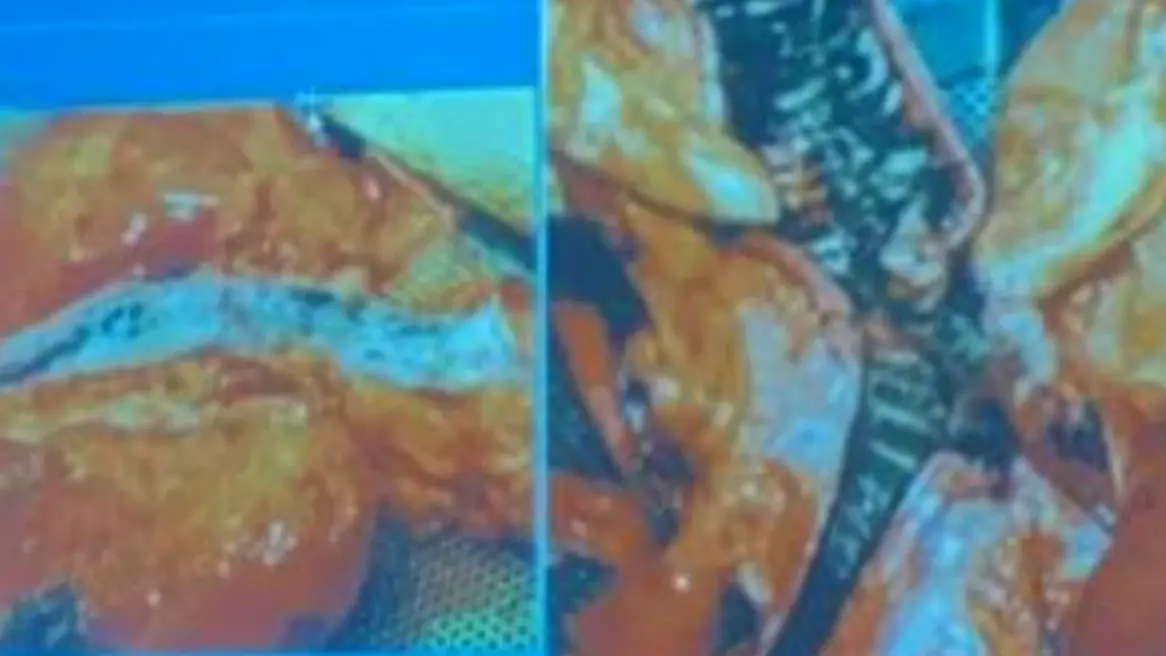
Burns and Thermal Injuries
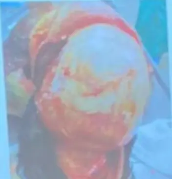
Burn Degrees
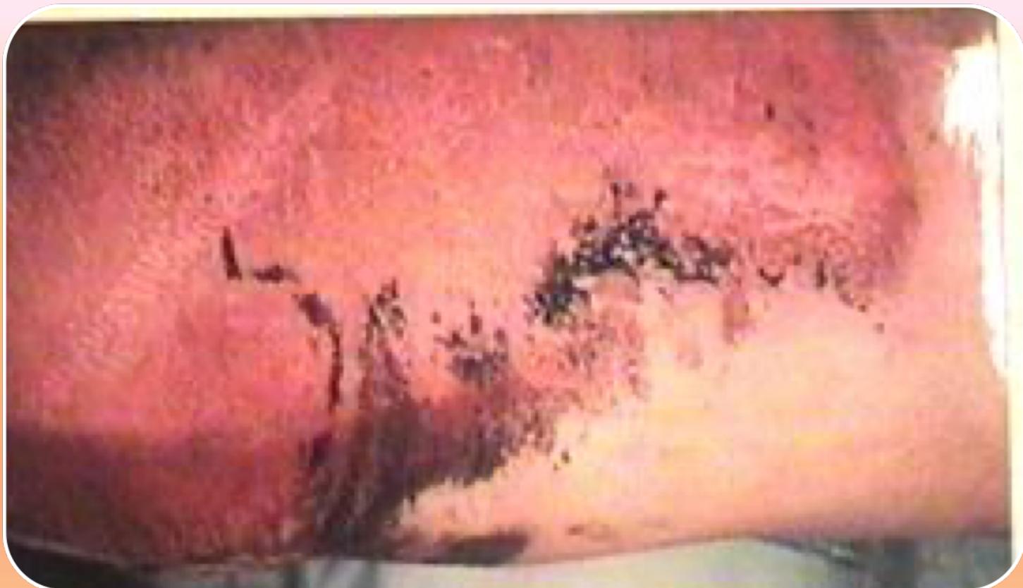
- 1st Degree: Epidermal involvement, redness only
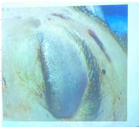
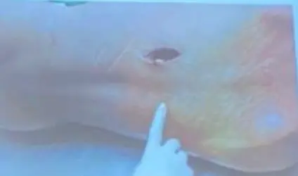
- 2nd Degree: Blister formation, increased vascular permeability
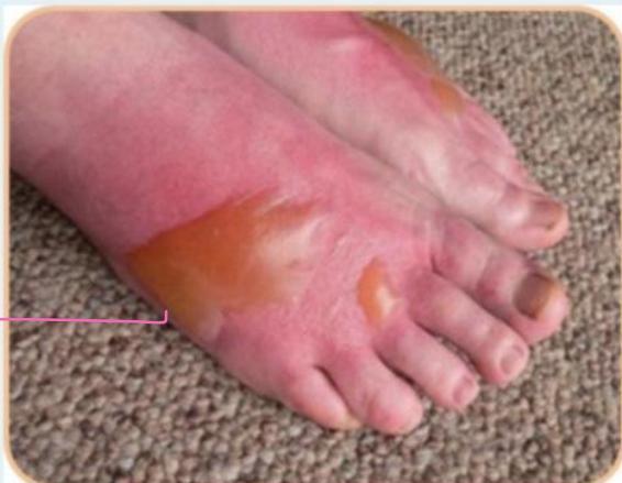
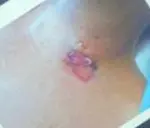
- Question: State type of lesion? Scald: second degree burn.
- Arrow points to?: Blisters.
- Description: Scald, 2nd degree, Blister formation.
- Complications: Hypovolemic shock, electrolyte disturbance, secondary infection.
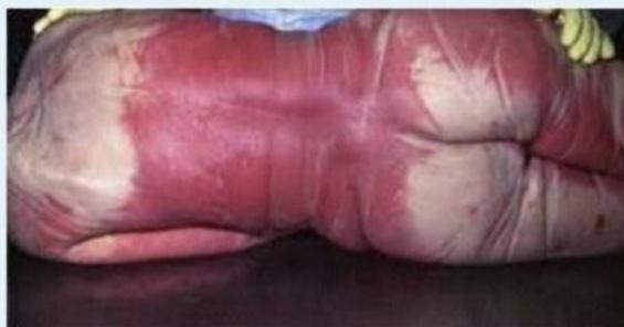
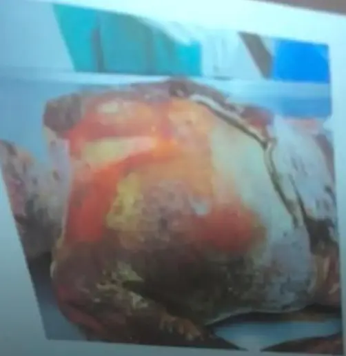
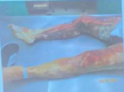
- 3rd Degree: Full-thickness destruction, dryness, deep tissue involvement
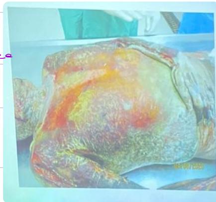
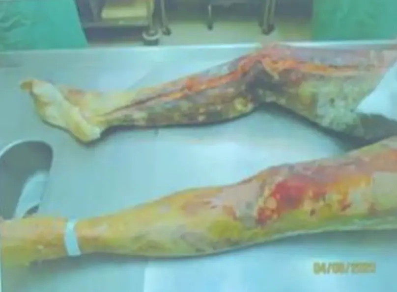
-
- Extensive severe burn (thighs)
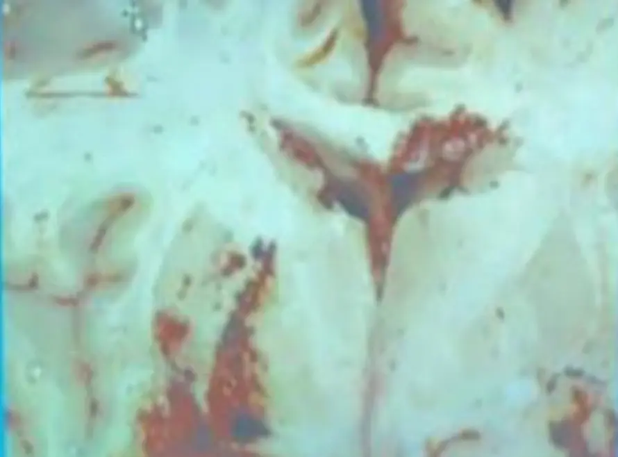
Inhalation Injury
- Findings: Soot in trachea, cherry red lung (CO poisoning)
- Scenario: 35 yo M found in his burned room. Shows soot lining trachea.
Burn Assessment Protocol
- Site: Anatomical location
- Degree: Severity determination
- Extent: Localized vs generalized
Head Injuries and Skull Fractures
Bleeding Patterns
-
a. Epidural hematoma → associated with Temporal bone fracture (Rupture middle meningeal artery)
-
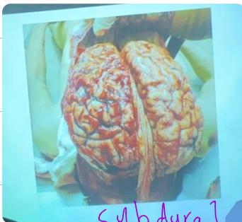
b. Subdural hematoma
-
Subarachnoid: Not washable (stays if washed)
-
Intraventricular Hemorrhage:
-
Dorsal Aspect Bleeding:
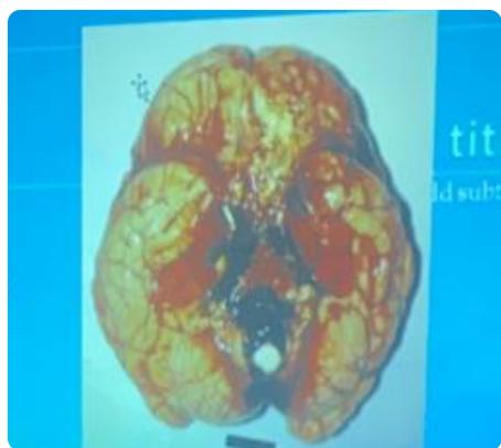
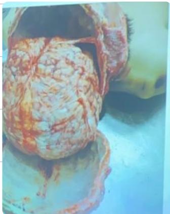
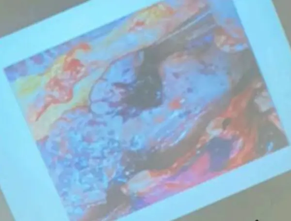
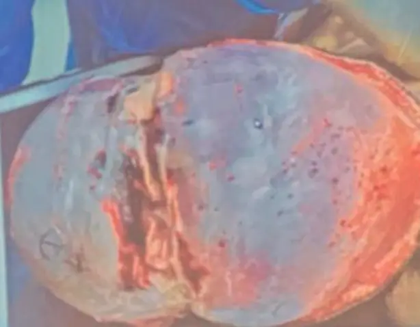
- Brain Contusion:
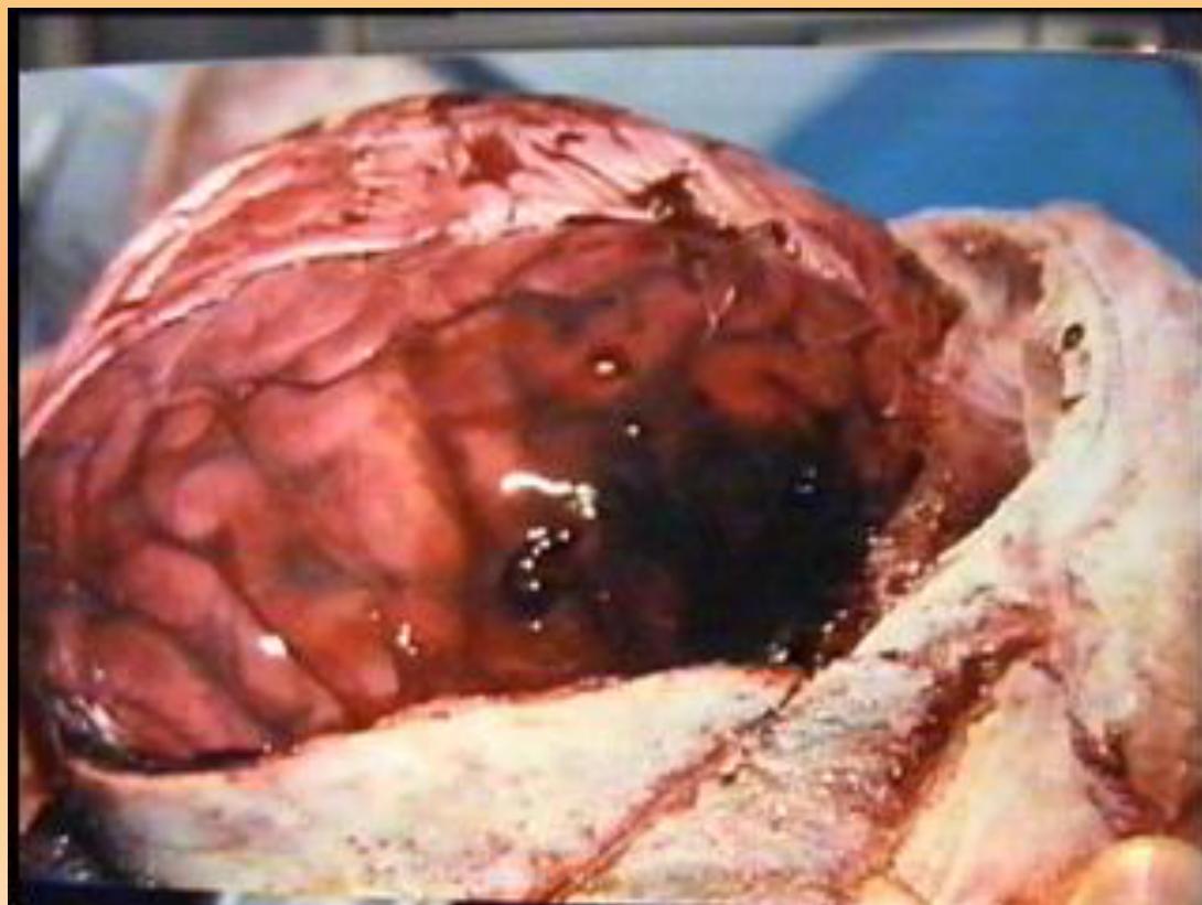
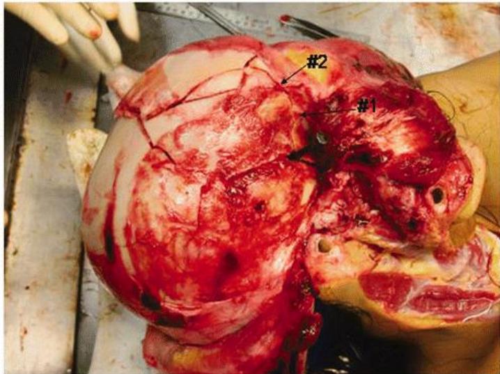
- Description: Contused Wound accompanied with severe skull fracture. Brain Contusion.
Skull Fracture Types
- Linear Fracture: Simple break
- Depressed Fracture: Bone pushed inward
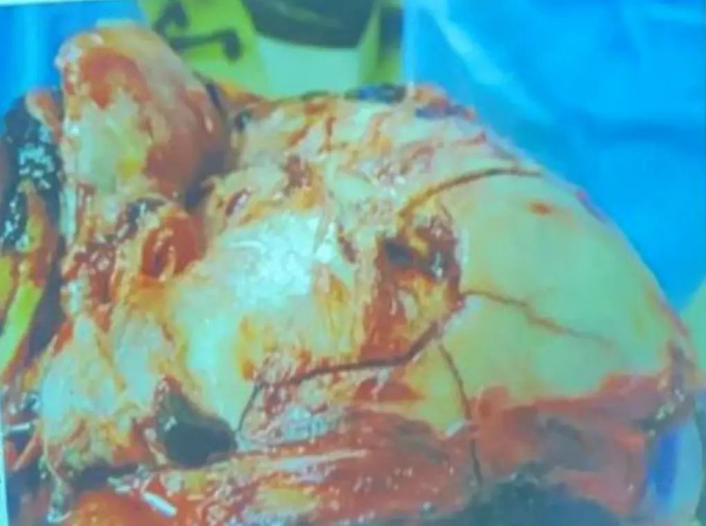
- Mosaic Fracture: Depressed-comminuted with multiple fragments
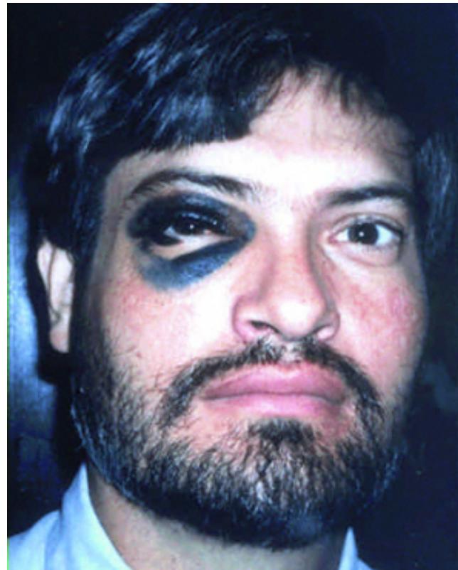
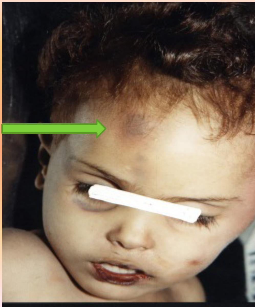
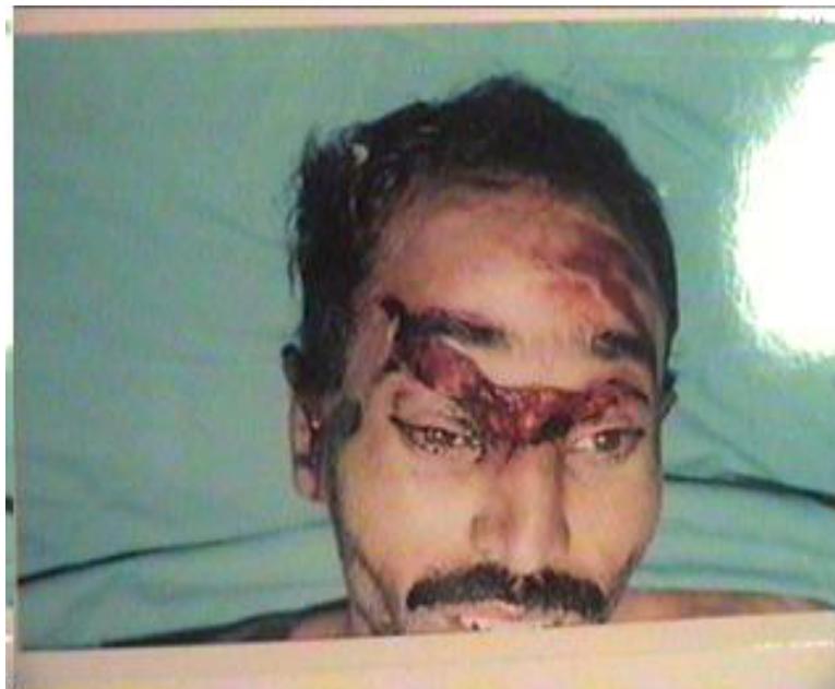
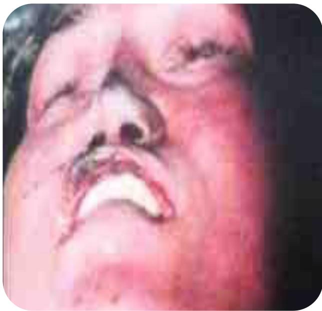
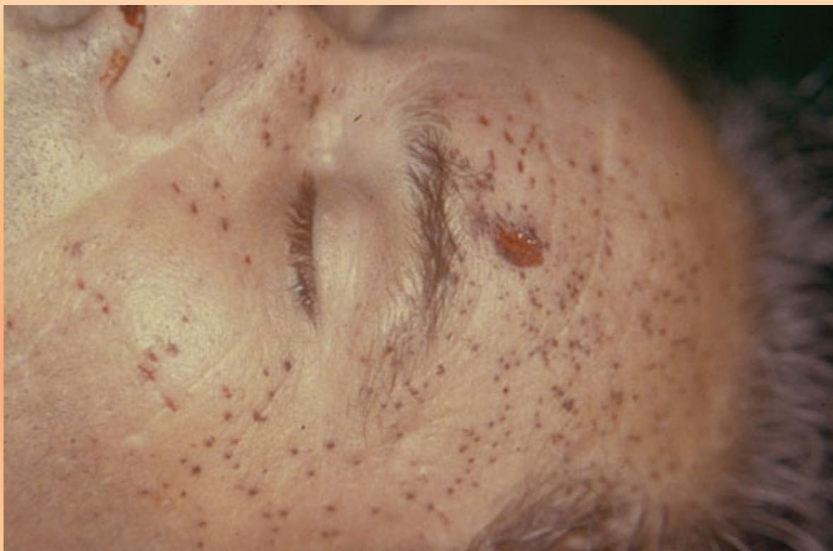
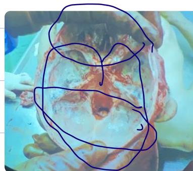
- Finding: Black eyes (Raccoon eye).
- Cause: Blunt trauma to head or face. Direct violence to anterior cranial fossa.
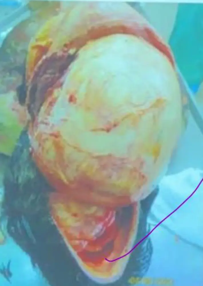
Scalp Examination
- Normal Scalp: Free of fracture upon reflection
Cranial Fossa Boundaries
- Posterior Cranial Fossa:
- Anteriorly: Temporal bone
- Posteriorly: Occipital bone
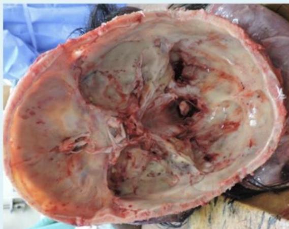
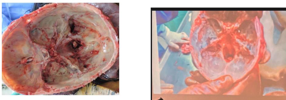
- Question: What is the boundaries of the posterior cranial fossa? Anteriorly temporal bone and posteriorly occipital bone.
- a. Epidural hematoma → associated with Temporal bone fracture
- Epidural: Not washable (lens-shaped bleeding)
Age Determination
Bone Fusion Ages
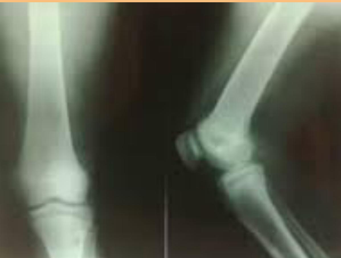
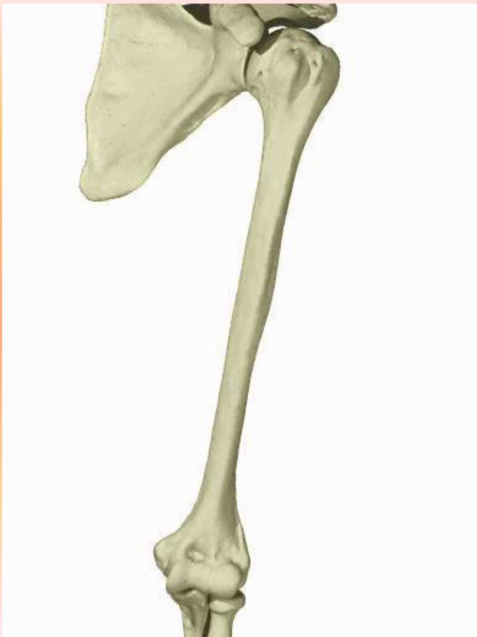
- Elbow Joint (Below 14 years): Trochlea not united with capitulum
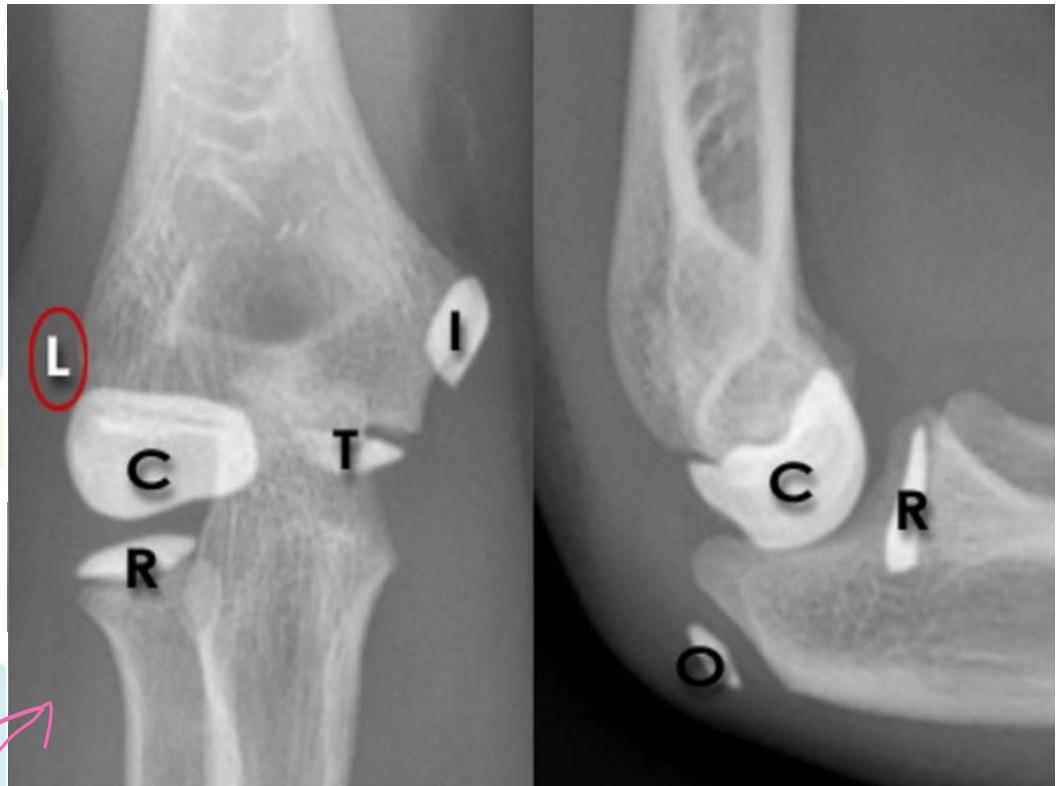
- Identify Age: Below 14 years.
- Reasoning: Trochlea is not united with capitulum.
- Note: 16 Years - Lateral epicondyle with shaft of Humerus union (Male ID Card).
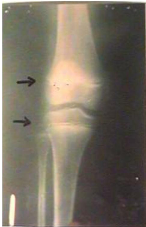
- Knee Joint (Below 21 years):
- Lower end of femur not united with shaft
- Upper ends of tibia and fibula not united with shafts
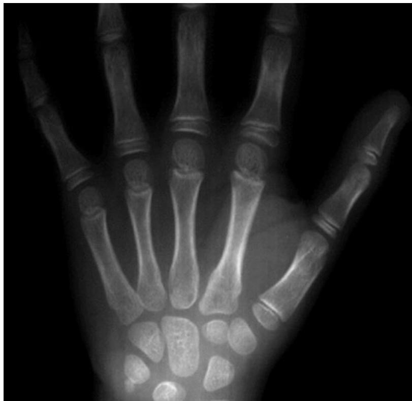
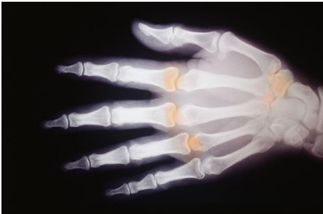
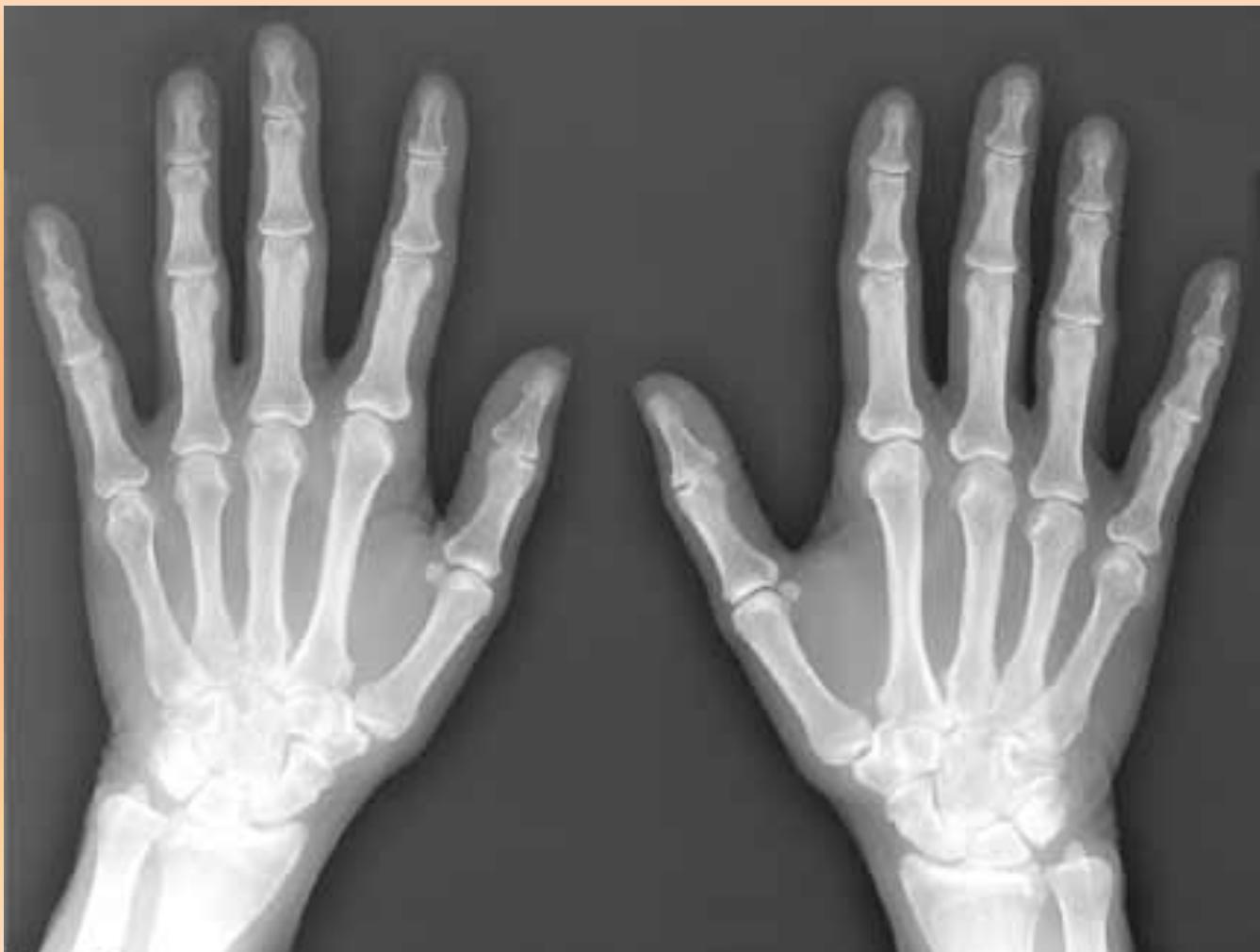
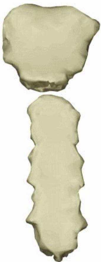
- Hand Joints (Below 18 years):
- Distal ends of metacarpals not united with shafts
- Proximal ends of phalanges not united
- Above 17/18/21 years:
- Above 17: Medial epicondyle/Upper radius united (
- Above 17: Medial epicondyle/Upper radius united (
- Above 18: All hand bones united (
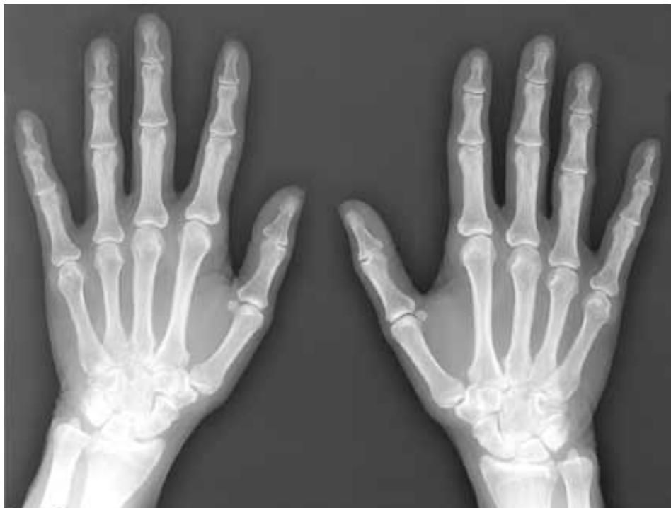
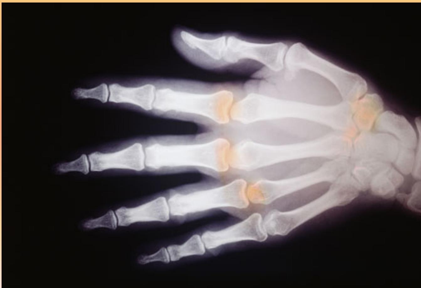 )
) -
- 21 or Above: Lower radius/ulna united (
 )
)
- 21 or Above: Lower radius/ulna united (
- Sternum (40-60 years):
- Manubrium not united with body
- Xiphoid process united with body at 40
Postmortem Changes
Livor Mortis (Hypostasis)
- Definition: Postmortem discoloration due to blood accumulation in dependent parts
- Timeline:
- Starts: 30 minutes - 3 hours
- Fixed: 6-8 hours
- Color Variations:
- Normal: Purplish
- Cherry red: Carbon monoxide poisoning
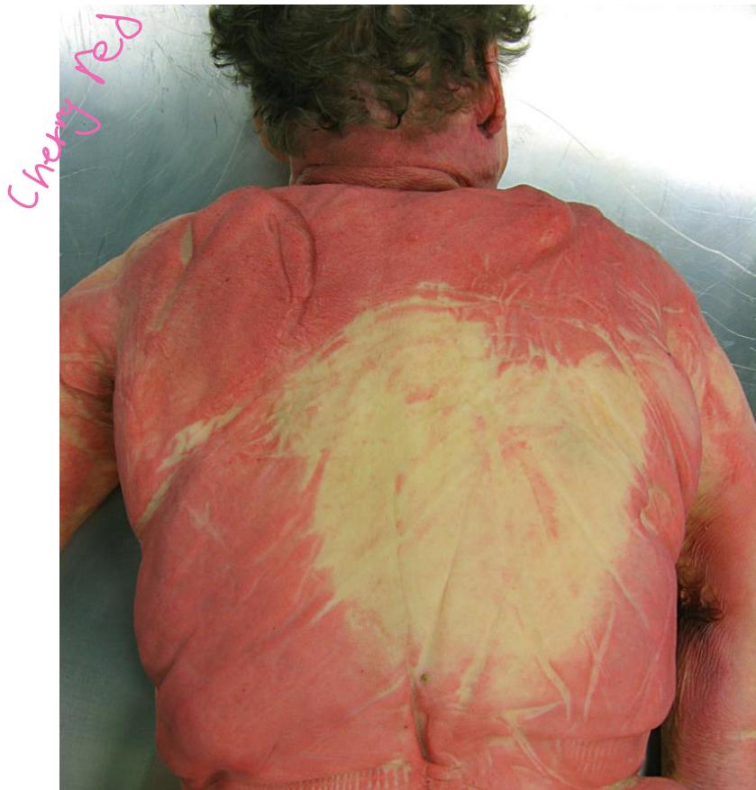
- Question: State the postmortem changes, and determine the cause of death If the color changes in the dead body is cheery red. Livour mortis and death is due to Anemic Anoxia from Carbon Monoxide Poisoning.
- Questions:
- Question: What is the name of this finding? Lividity.
- Question: Fixation time? 6 to 8 hours.
- Question: We can notice this finding in: Anemic anoxia.
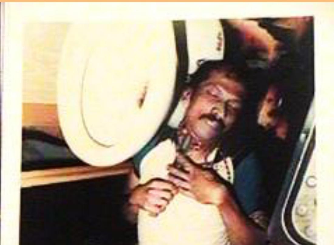
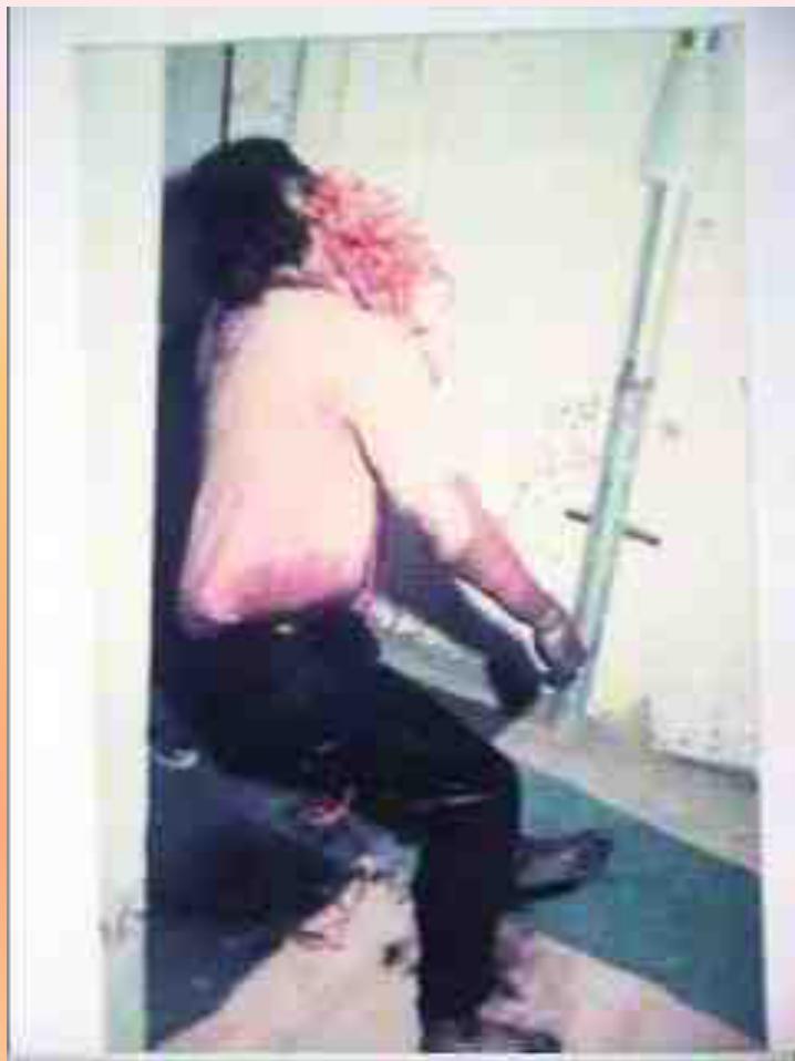
Cadaveric Spasm
- Definition: Instantaneous rigidity at moment of death
- Common in: Drowning victims (grasping objects), Soldier gripping weapon.
- Scenario: A forensic on call doctor receive a call about a drowning case, when arrive to the scene, he notice this finding, choose the correct answer: d. Cadaveric spasm.
Death Investigation
Manner of Death Determination
- Homicide:
- Question: Determine the manner of death: Homicide.
- Question: Determine the distance of injury from the used weapon: Near range firing less than 2 yards.
Neonatal and Infant Death
Types of Birth
-
Dead-born Child (Stillbirth):
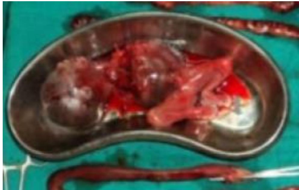
- Question: State the type of Birth? Dead-born Child (Dead Birth).
- Question: If the length of the fetus is 30 cm. Determine its age. Miscarriage for 6 lunar months fetus.
-
Aborted Child:
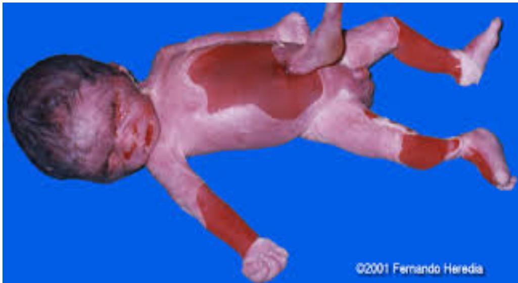
Maceration
- Definition: Aseptic autolysis of dead fetus in uterus
- Characteristics: No bacterial action, unlike putrefaction
- Early signs: 6-12 hours - desquamation, brown-red umbilical cord
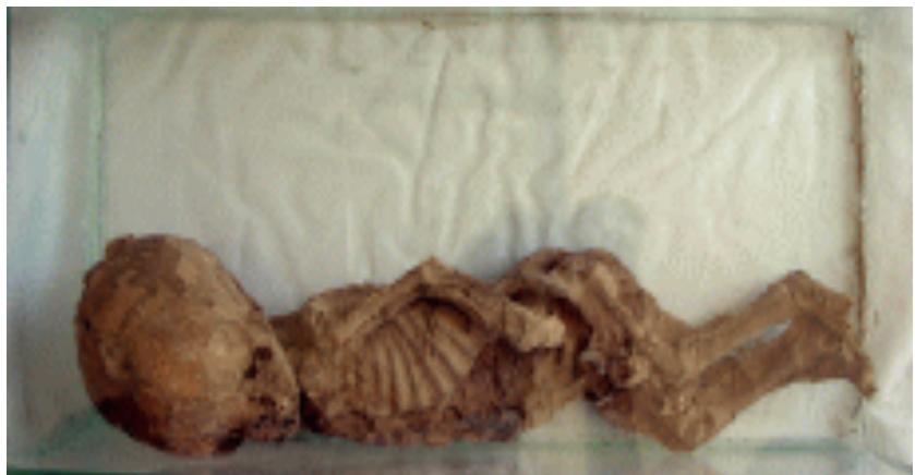
Mummification
- Definition: Dehydration of dead fetus
Electrical Injuries
Characteristics
- Inlet: Depressed center, dark margin, more severe
- Exit: Less destructive, small lesion
- Cause of death: Current passage through vital organs (heart) → Disturbance of pacemaker
- Scenario: With brief hx: male found lying beside his car with electrical wire beside him then it’s electrocution… look for the inlet and exit of the injury… exit not always found. It cause death when the electricity tract pass through vital organ like heart as in case from right arm to left arm disturbance of pacemaker.
- Description: Electrocution with dark margin of (inlet), depressed discolored lesion with irregular borders.
Clinical Stations and Autopsy Findings
Station-Based Questions
Station 1: Burn Patient
- Finding: Coagulative necrosis, soot in trachea if inhaled while alive
Station 2: Postmortem Changes
- Findings: Hypostasis, cadaveric spasm, putrefaction
Station 3: Burn Patient - Fracture
- Finding: Artifact from prolonged flame exposure, not real fracture
Station 4: Autopsy Procedure
- First step: Reflect the scalp
- Fracture type from blunt force: Depressed fracture
Station 5: Electrical Injury
- Finding: Volcano appearance around wound edges
- Protection against current: Rubber
Station 6: Gunshot Wound
- Finding: Collar of abrasion, tissue loss prevents suturing
Station 7: Gunshot - Internal Injuries
- Findings: Penetrating lung injury, potential heart involvement
Station 8: Postmortem Wound
- Finding: Incised wound during autopsy, no gaping (postmortem)
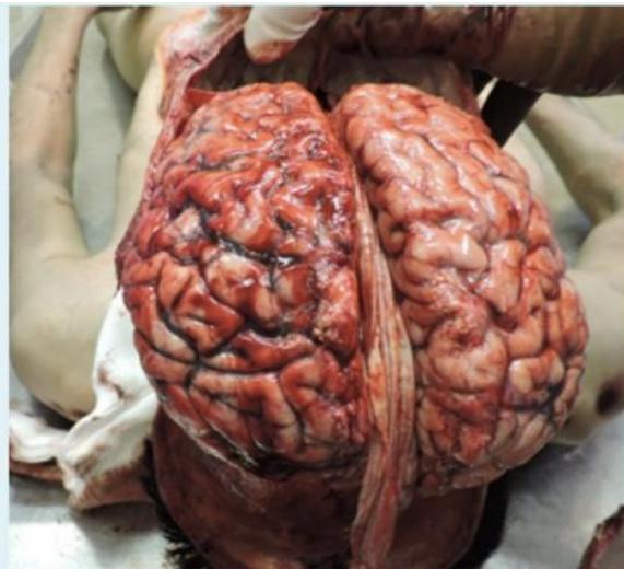
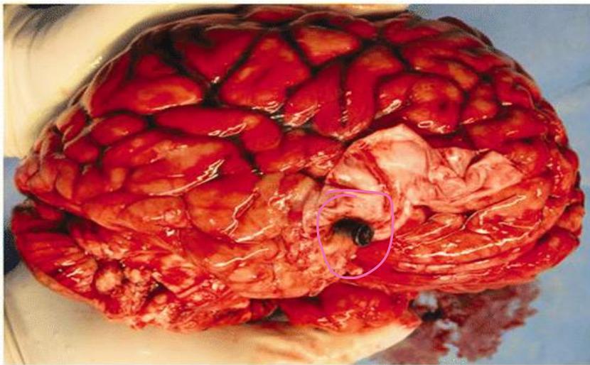
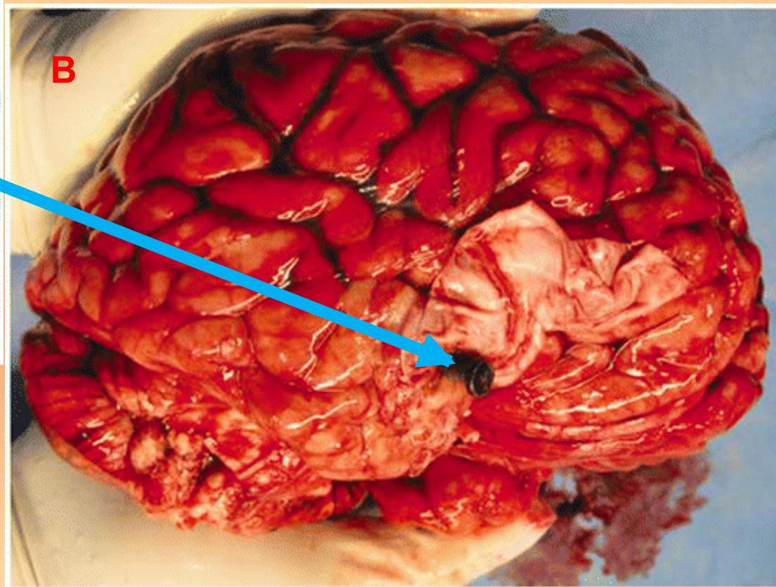
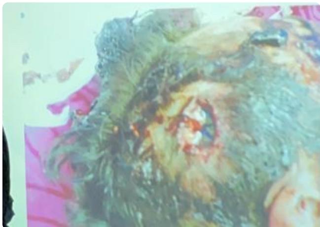
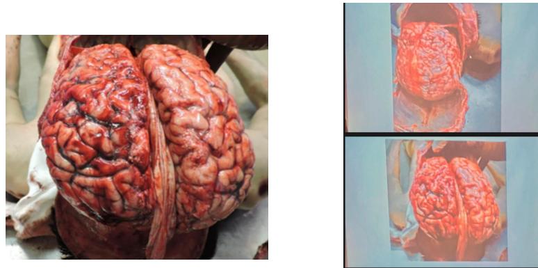
Station 9: Gunshot Head Injury
- Finding: Brain tissue protrusion, characteristic tissue loss
Station 10: Raccoon Eye
- Finding: Ecchymosis upper eyelid, indicates basal skull fracture
Station 11: Mosaic Fracture
- Finding: Depressed-comminuted skull fracture with multiple particles
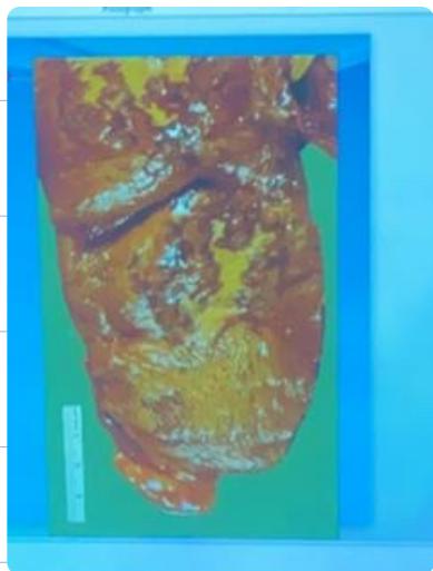
Station 12: Pale Scalp
- Finding: Very pale scalp indicates anemia/bleeding
Station 13: Brain Hemorrhage
- Question: Differentiate subdural vs subarachnoid hemorrhage
- Answer: Subdural is washable, subarachnoid is not (stays even if washed).
Station 14: Skull Base
- Question: Boundaries of posterior cranial fossa
- Answer: See Cranial Fossa Boundaries. (Anteriorly: Temporal bone, Posteriorly: Occipital bone)
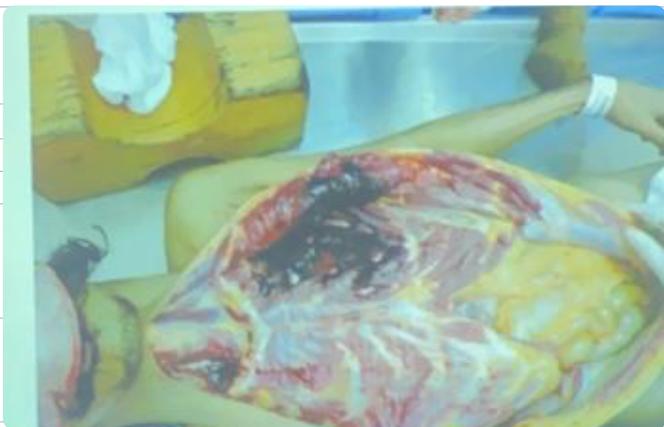
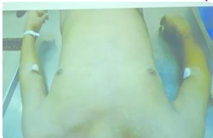
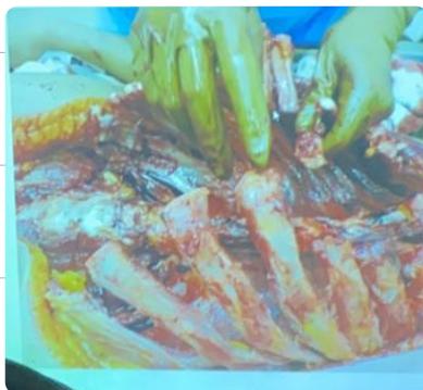
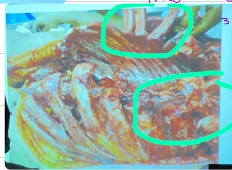
Station 15: Fall from Height
- Findings: No external wounds, bilateral rib fractures, flail chest
- Scenarios:
- Clean body externally, internal severe damage
- Lungs removed
- Rib fractures (Bilateral, 3 ribs)

Station 16: Stab Wound
- Findings: Stab to apical lung, difference between stab and incised wound
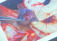
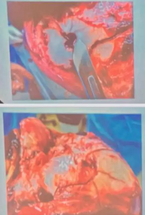
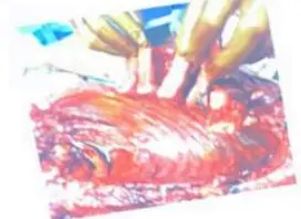
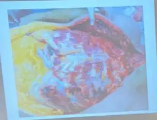
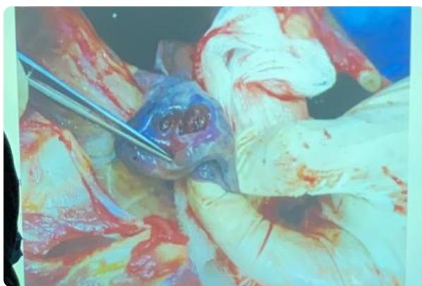
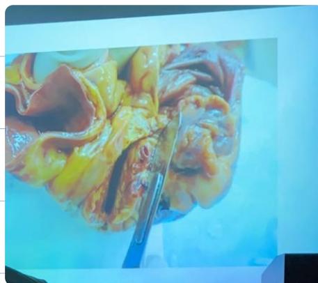
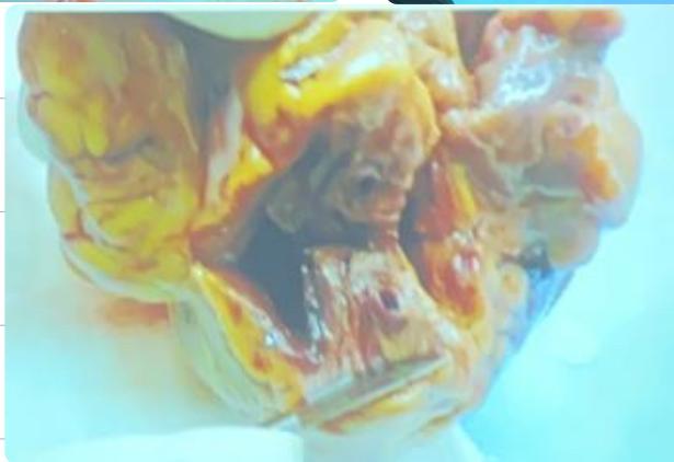
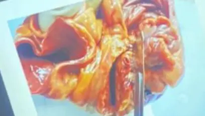
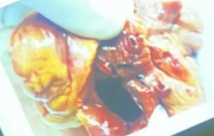
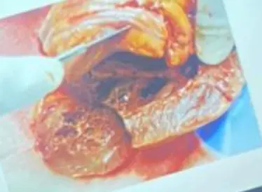
Station 17: Coronary Artery
- Finding: Coronary artery occlusion >75% causes death
- Images:
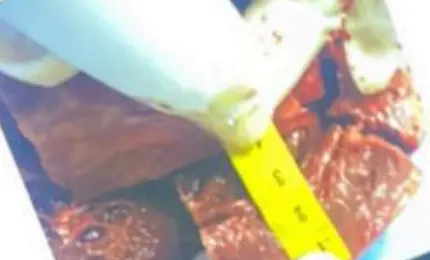
Station 18: Heart Measurements
- Questions:
- Normal Left Ventricular Thickness?
- Maximum Left Ventricular Thickness?
- Proportion of Left Ventricle to Right Ventricle?
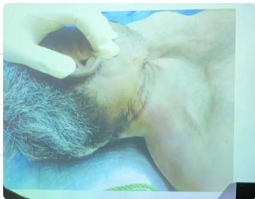
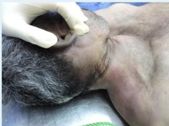
Station 19-20: Hanging
- Findings:
- Ligature mark: Pressure abrasion, oblique direction (typical hanging)
- Points to Note:
- Gender?
- What is the Person Wearing?
- Position of Knot (Occipital = Typical, Others = Atypical)
- Mark Features: Color, depth, pattern of rope
- Anatomical Relations: Above/Below Hyoid, Distance from Mandible/Ears/Hairline
- Postmortem Staining: Distribution
- Types of Strangulation:
- Strangulation: Constricting neck by external force without suspension.
- Throttling: Manual strangulation by hands.
- Mugging: Strangulation by Elbow.
- Garrotting: Strangulation by Device (Spanish Method) or by Thrown Ligature (Indian Method).
- Cherry Red Color: Indicates Carbon Monoxide Poisoning (Anemic Anoxia) if present