Fetal Growth Restriction
Overview
Fetal Growth Restriction (FGR), also known as Intrauterine Growth Restriction (IUGR), occurs when a fetus does not grow at a normal rate inside the womb.
Definition
IUGR: When the birth weight of newborn infant is below the 10th percentile for a given gestational age
Low birth weight: (<2500 grams)
Small for Gestational Age (SGA) indicates that a fetus or neonate is below a defined reference range of weight for a gestational age.
Etiology of IUGR
Maternal Factors
-
Behavioral:
- Poor nutritional intake
- Cigarette smoking
- Drug abuse
-
Medical:
- Hereditary thrombophilia
- Early cardiovascular disease
- Hypertension
- Diabetes
- Obesity (associated with leptin resistance)
- Alcoholism
- Cyanotic heart disease
- Pulmonary insufficiency
- Antiphospholipid syndrome
Other Factors
-
Teratogen Exposure: Exposure to harmful substances during pregnancy.
-
Extreme Maternal Age: Being at the extremes of reproductive age.
-
Fetal Factors:
- Intrauterine infection (listeriosis and TORCH)
- Congenital anomalies
-
Placental Factors:
- Poor implantation
- Placenta previa
- Umbilical cord abnormality (Mcikeymouse, 2 artery, 1 vein normally, single down usually)
Types of IUGR
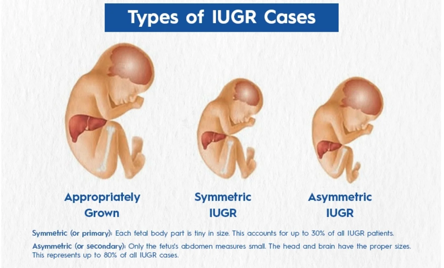
Symmetric vs. Asymmetric IUGR
- Symmetric (or Primary) IUGR: Each fetal body part is tiny in size, accounting for up to 30% of all IUGR patients.
- Asymmetric (or Secondary) IUGR: Only the fetus’s abdomen measures small, with the head and brain having proper sizes, representing up to 80% of all IUGR cases.
Classification Based on Gestational Age
- Early FGR: Less than 32 weeks.
- Late-Onset FGR: Diagnosed at or after 32 weeks.
Diagnosis
- Serial uterine fundal height measurements: Fundal height lags more than 3 cm behind expectations.
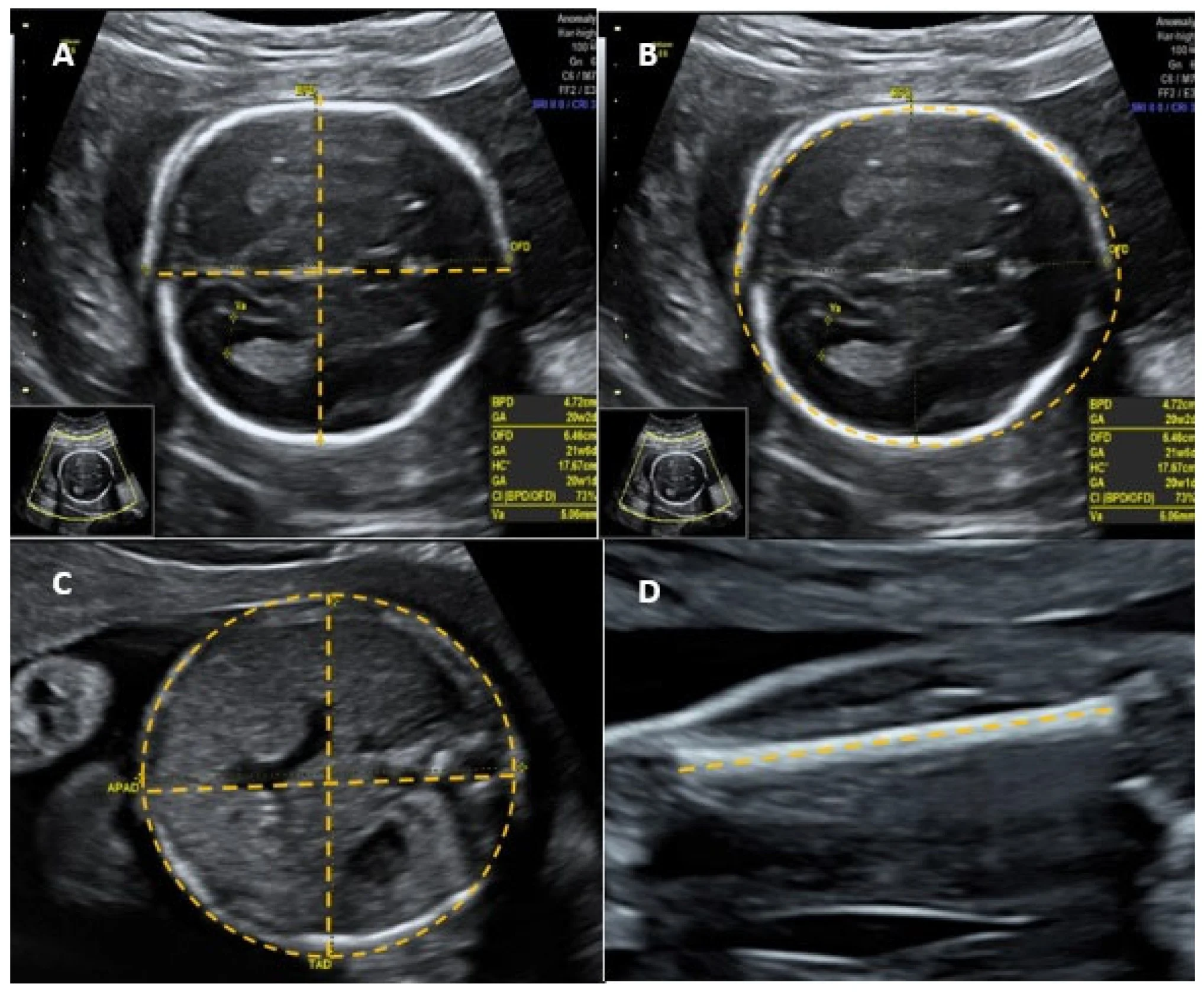
- Ultrasound:
- Fetal biometry: biparietal diameter (BPD), head circumference, abdominal circumference, femoral length, calculated fetal weight.
- Amniotic fluid volume. (index 5-25; divide abdomen in four, no fetal or cord part, measure. vertical from four areas, take ratio of total of >25 poly <5 oligio //// - >8 poly <2 olig pockets of air)
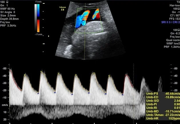
- Umbilical artery Doppler: low, absent, or reversal of diastolic flow.
Ultrasound Images of a Fetal Head
Umbilical Artery Doppler Ultrasound
 FGr at term deliver, before 37wk, assess doppler if abnormal deliver
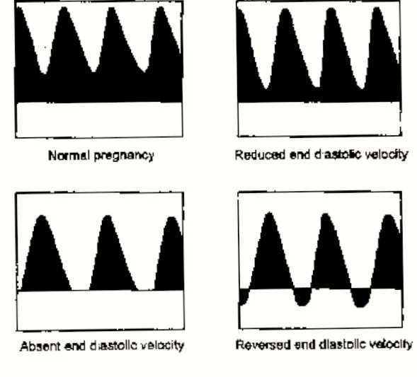
Progression of Umbilical Artery Doppler Changes
- Normal pregnancy
- Reduced end diastolic velocity
- Absent end diastolic velocity
- Reversed end diastolic velocity (Dexamethasone + Deliver now)
Key Diagnostic Parameters
- Abdominal circumference is the single most effective parameter for predicting fetal weight. Z
- Maternal lab tests: serology, amniocentesis.
Management
Pre-Pregnancy
- Stop smoking and alcohol consumption.
- Improve nutrition.
- Aspirin 81mg/day. z
Antepartum
- Stop smoking.
- Improve nutrition.
- Work leave. z
- Teach the patient about fetal kick count. z
- Hospitalization. (if absent fetal kick or reduced, if doppler is absent must be done twice week in such patient, medically unstsable, severe IUGR, if patient lives far away-;, biophysical profile is done)
Monitoring
- Twice-weekly nonstress test (NST) and biophysical profile (U/S, fetal movement, breathing, amniotic fluid, gross movement, ). - modified (Amniotic fluid index + CTG) - normal 8-10 - 4-6 suspicious, <4 abnormal CC?
- Umbilical artery Doppler weekly.
Labor and Delivery
Treatment of a fetus with suspected IUGR hinges on the clinical context, especially gestational age.
-
Gestational age < 34 weeks – administer a course of antenatal corticosteroids to promote lung maturation.
-
IUGR itself is not a contraindication to induction of labor; however, maintain a low threshold for proceeding to caesarean section if maternal or fetal status warrants.
Complications
- Increased chance of C-section and early birth.
- Breathing and feeding issues during birth.
- Hypoxia.
- Hypoglycemia.
- Having trouble regulating body temperature.
- Meconium aspiration.
- Polycythemia.
- Infection.
- NICU admission.
Clinical Scenarios Z
Case 1
 -D 2-3 weeks difference in size estimatino, doppler twice weekly or once
Case 2
 -A basic = fetal biomentry
biometry initially, biophysical detailed.