Examination of the Knee
Prof. Mamoun Kremli
Orthopedic Examination
The system to use:
- Look
- Feel
- Move
- Special tests
Summary
Knee Examination Systematic Approach
-
Look
- General → on patient
- General → local (knee, thigh, leg): Position, Major deformity, swelling, Extra
- Anatomic local
-
Feel
- Tenderness, Temperature, Anatomical
-
Move
- Active, Passive
-
Special Tests
- Patella-related
- Effusion
- Ligaments
- Collaterals
- Cruciate
- Menisci
- Gait – walking
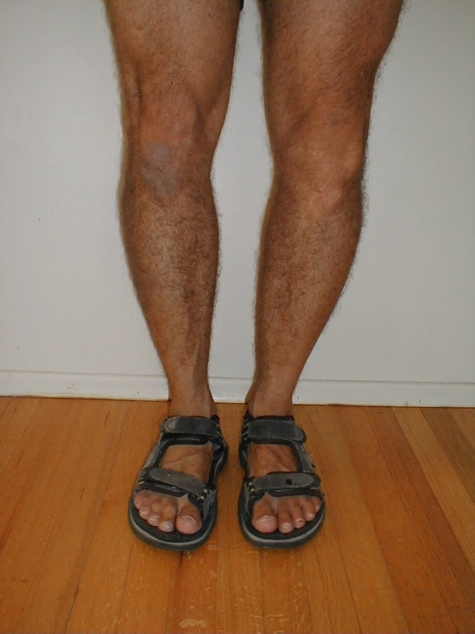
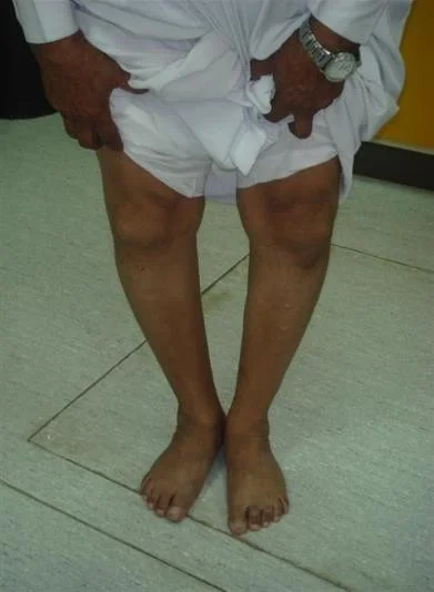
Look
General → on patient
- Proper exposure
- The patient should be in either a gown or shorts
- Rolled up pant legs do not provide good exposure!
-
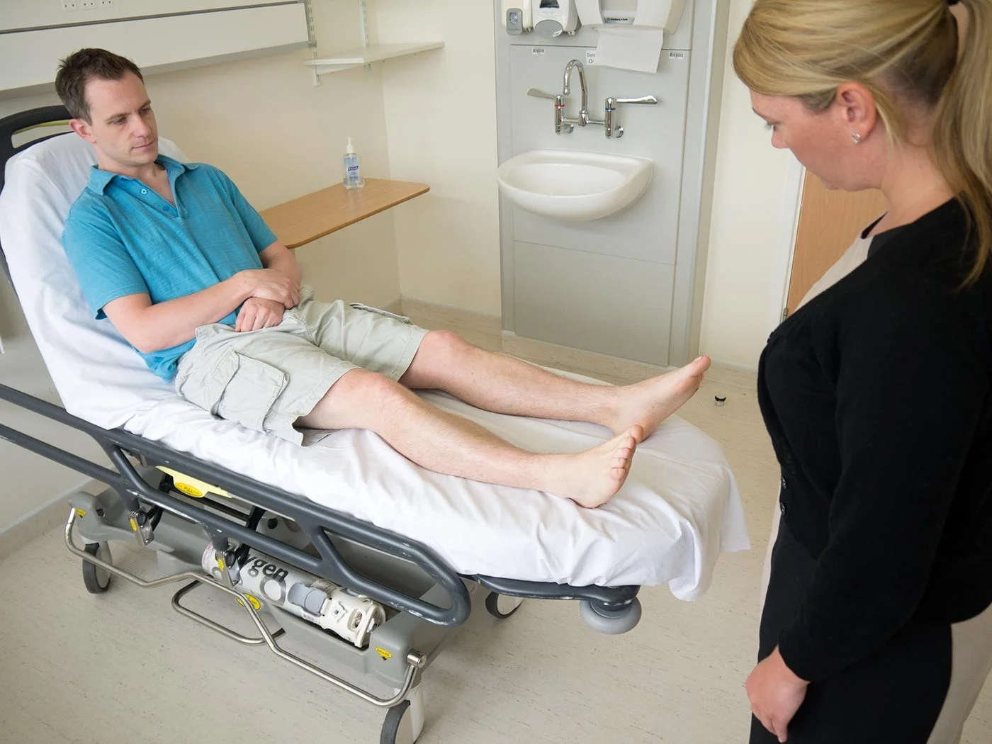
Position examples:
- Sitting comfortably on edge of couch, with knees flexed 90°

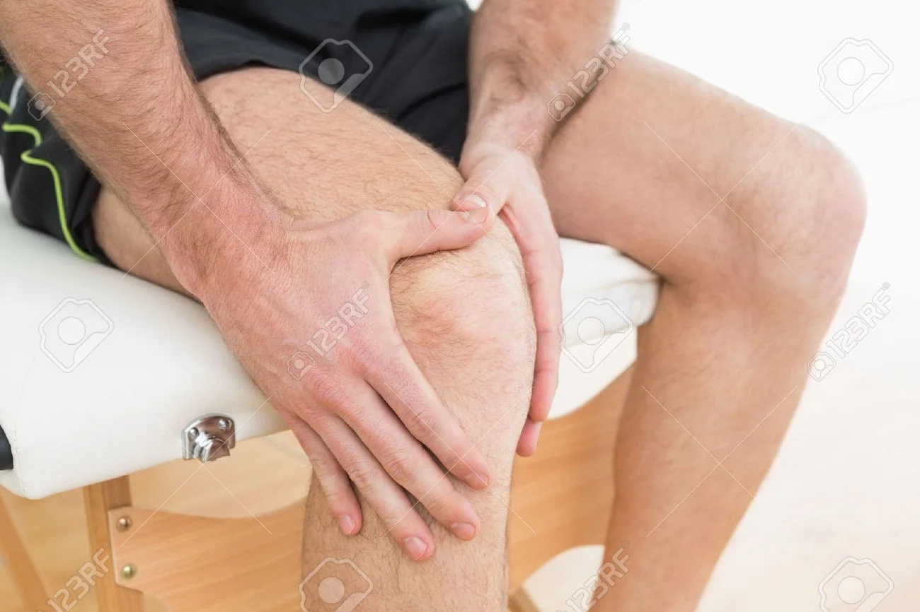
- Sitting, in pain, with both hands around flexed knee
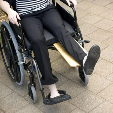
- Sitting on a wheelchair, with knee extended on a board
General → local (knee, thigh, leg)
- Position and alignment
-
Flexion / Extension
-
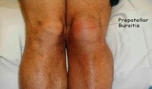
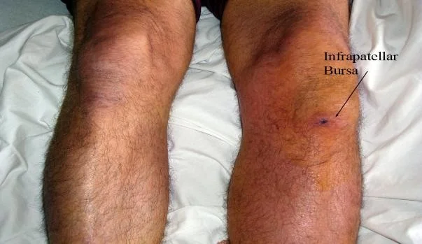
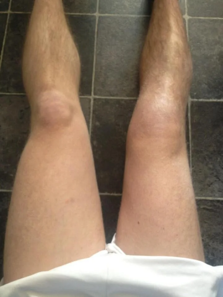
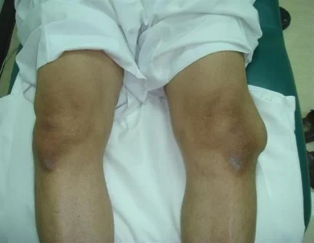
- Major deformity and swelling
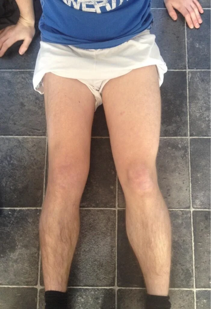
- Varus / Valgus - Best assessed in standing
- Swelling / Masses

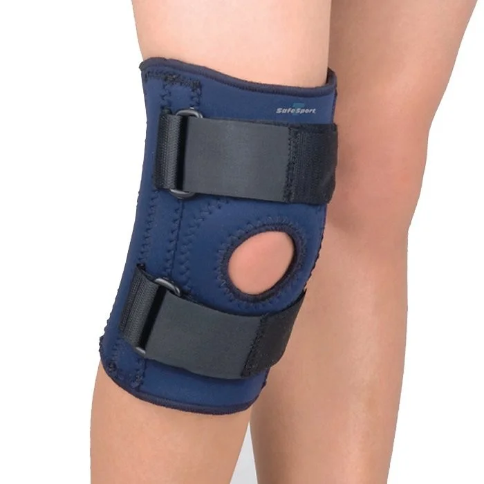
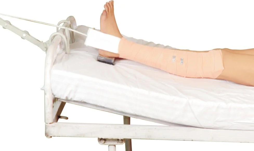
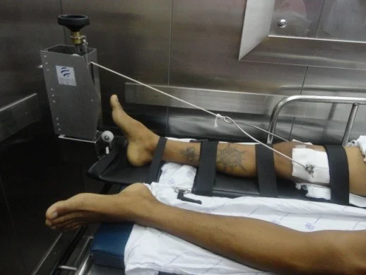
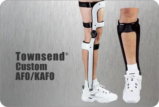
- Extra equipment and devices
- Cast, splint, dressing
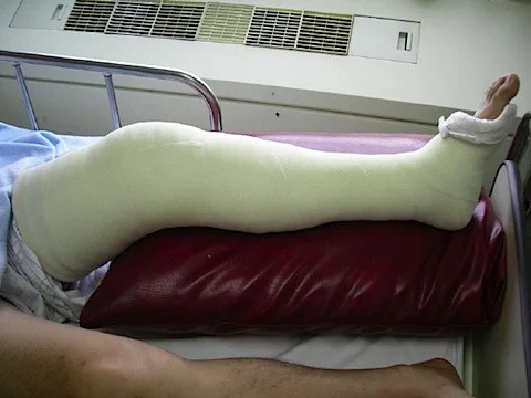
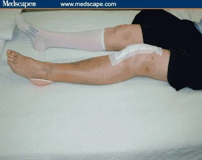
- Skin traction, skeletal traction
- Orthotics
- AFO (Ankle-Foot Orthosis)
- KAFO (Knee-Ankle-Foot Orthosis)
- HKAFO (Hip-Knee-Ankle-Foot Orthosis)
Anatomic local
- Systematic examination:
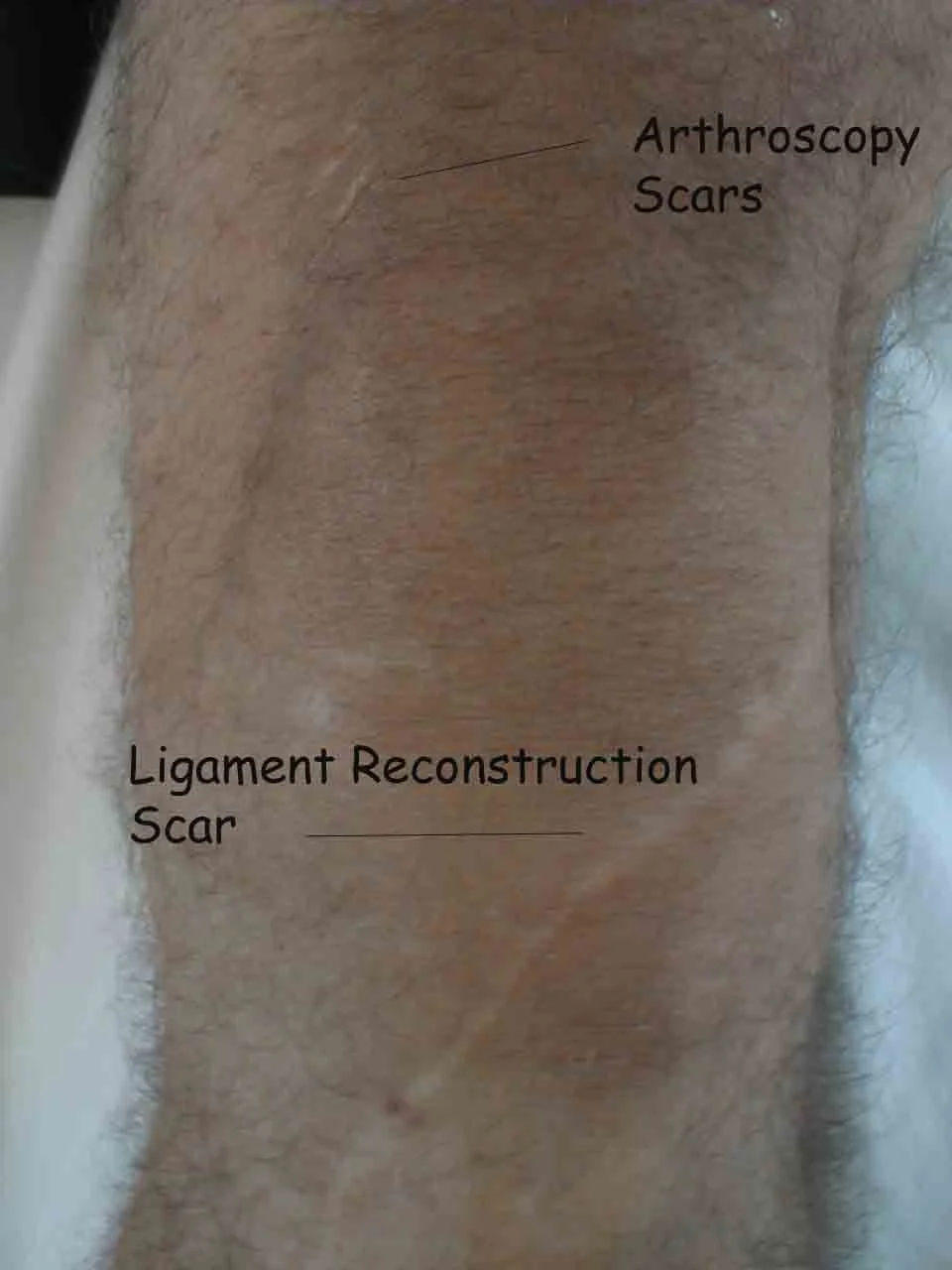
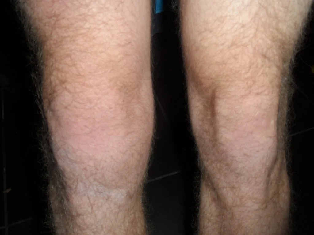
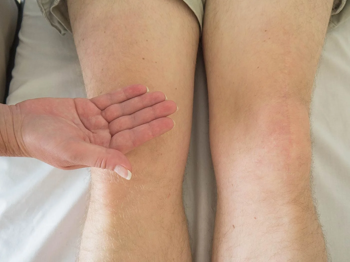
- Skin: swelling, scars, color, hair, dryness
- Subcutaneous: lymph nodes, veins, nerves, tendons
- Muscles: bulk, wasting, twitches
- Bones: landmarks, swelling, angulation, deformity
- Joints: position, swelling, redness
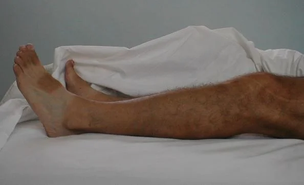
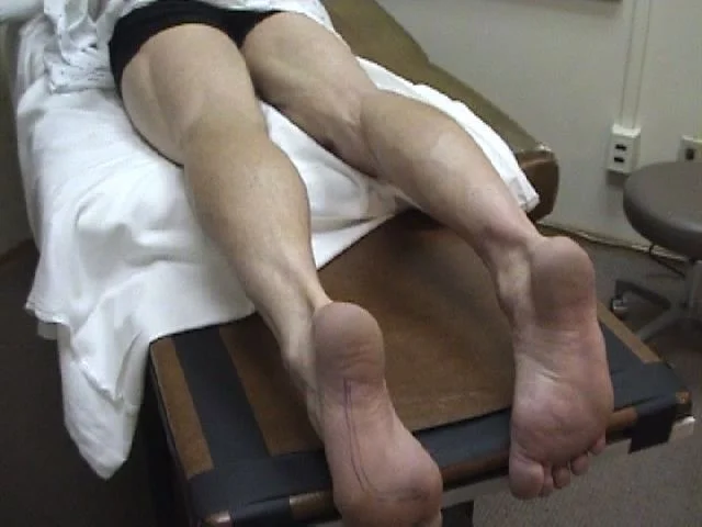
(Do not forget the posterior aspect!) (All patients have a posterior aspect!)
-
Visual examples:
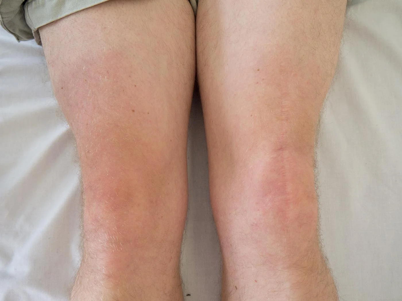


- Posterior aspect
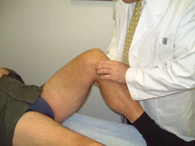
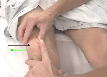
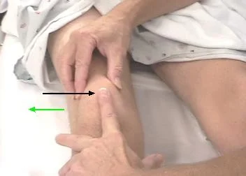
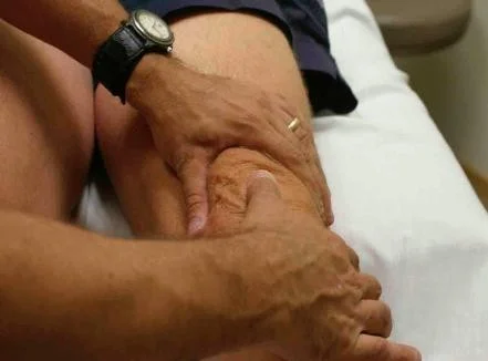
Feel
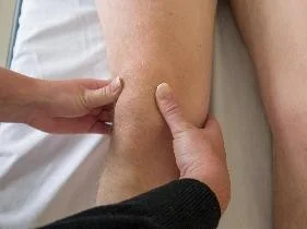
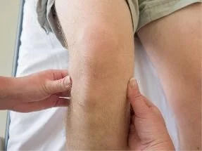
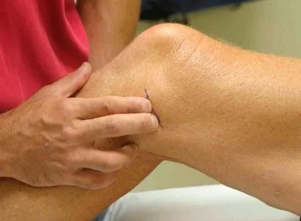
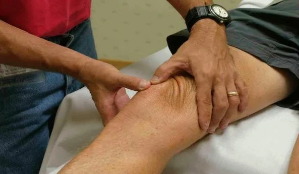
Tenderness
- Generalized: to start with
- Specific points:
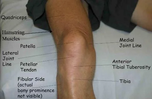
- Quadriceps muscle
- Patella
- Patellar tendon
- Tibial tuberosity
- Joint lines: medial/lateral
- Collateral ligaments
- Head of Fibula
- Popliteal fossa
- Hamstrings
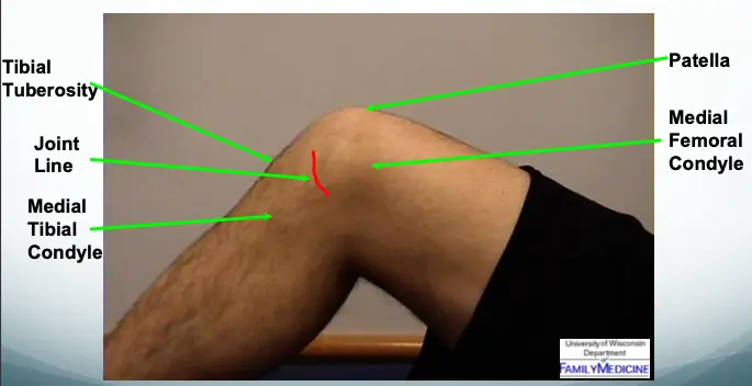
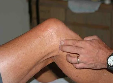
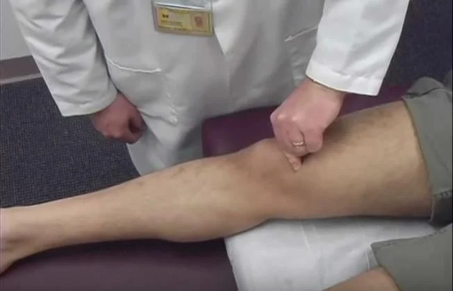
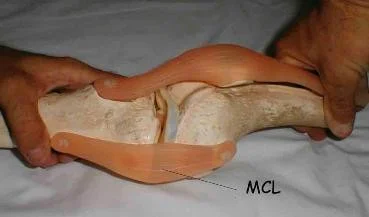
Knee - Medial Structures
- Tibial Tuberosity
- Joint Line
- Medial Tibial Condyle
- Patella
- Medial Femoral Condyle
- Joint Space - The space between the Medial Femoral Condyle and Medial Tibial Condyle
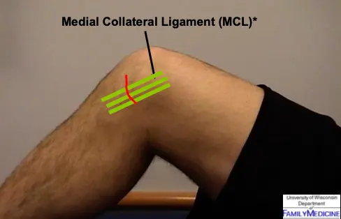
- Medial Collateral Ligament (MCL)
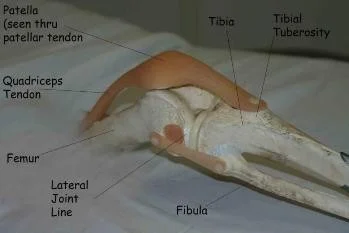
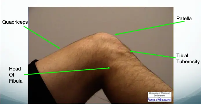
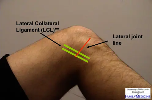
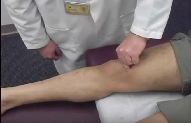
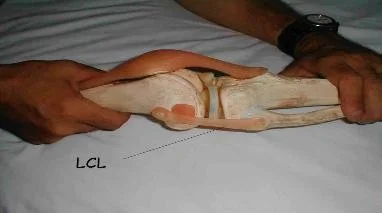
Knee - Lateral Structures
- Quadriceps
- Patella
- Tibial Tuberosity
- Head of Fibula
- Lateral Collateral Ligament (LCL)
- Lateral joint line
Temperature
- Compare distal/proximal and right/left sides
Anatomic Examination
- Skin: dryness, hyper/hyposthesia, scars
- Subcutaneous: lymph nodes, nerves, vessels, tendons, nodules
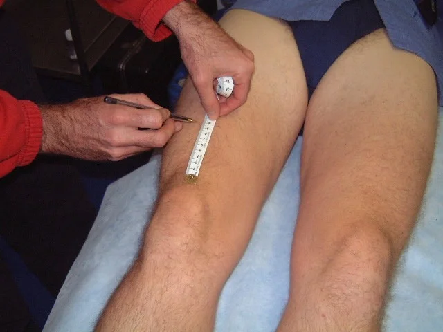
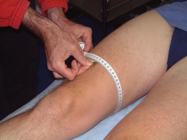
- Muscle (Quadriceps): tone, bulk, twitches, gaps, tenderness (measure girth if needed)
- Bone: landmarks (Patella, Tibial Tuberosity, head of Fibula) tenderness, mass, crepitus

- Joint lines: anterior, medial, lateral
- Joint assessment: swelling, effusion, crepitation, synovial thickening, joint line tenderness
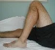
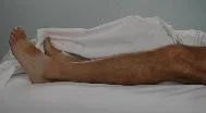
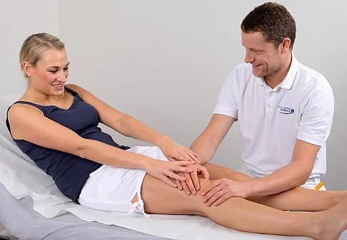
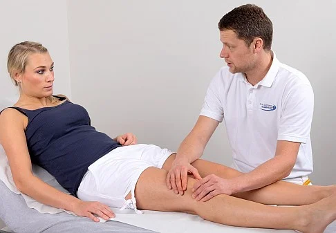
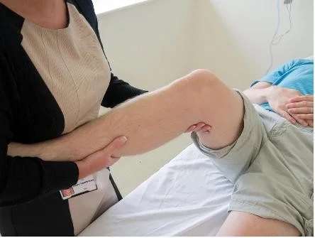
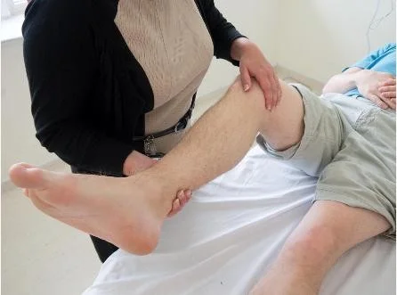
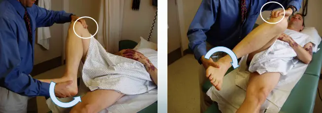
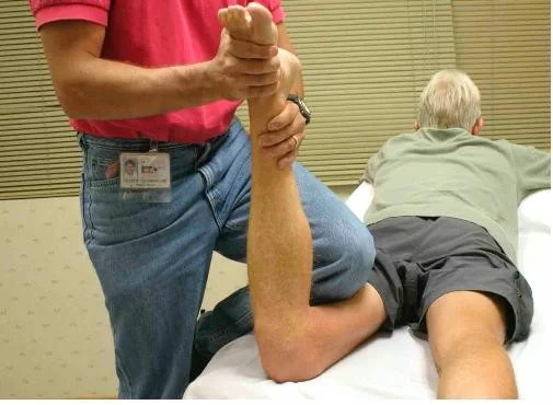
Move
- Active Vs. Passive
- Better to start with active
- Need to assess the range of motion
- painless / painful
- Extension: 0° → Flexion: 140°
 →
→ 
- Describe loss of degrees of extension
- Example: “lacks 5 degrees of extension”
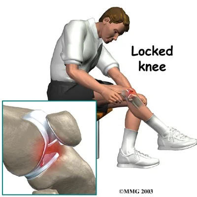
- Locking:
- Patient unable to fully extend or flex knee due to a mechanical blockage in the knee (i.e., loose body, bucket-handle meniscus tear)
- Normal range:
- Flexion / Extension
- Abnormal range:
- Anything less
- Increased
- Within normal direction:
- hyper-extension
- Abnormal direction:
- unstable

Special Tests
Patella-related Tests
- Patellar tracking: while knee flexes

- Patellofemoral grind test (chondromalacia, osteoarthritis)
- Compress patella down while quadriceps tightens
- Positive when painful
- Patellofemoral resistance test (chondromalacia, osteoarthritis)
- Resist patellar motion while quadriceps tightens
- Patellar apprehension test
Summary of patellar tests:
- Patellar tracking
- Patellar grinding
- Apprehension test
Effusion Tests
-
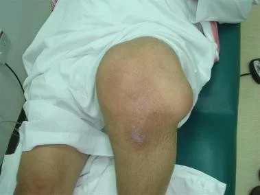
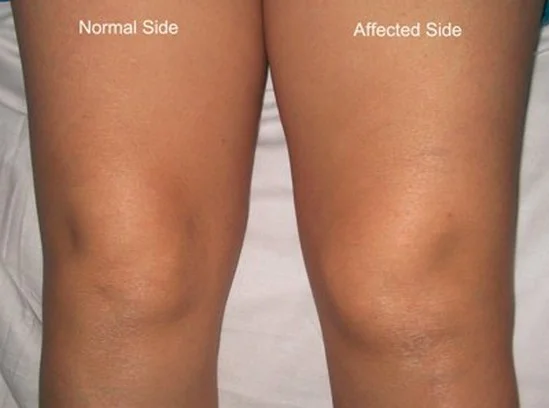
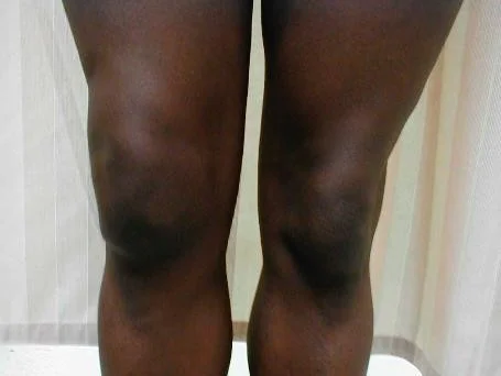
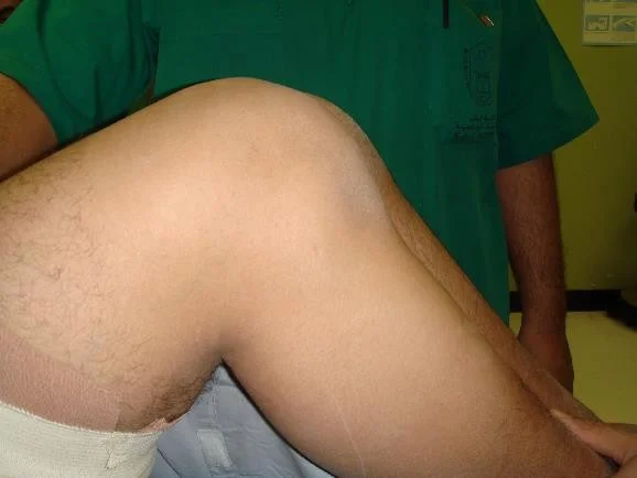
Large effusion is easy to see
-
Assessment of effusion severity:
-
Look at normal depression medial to patella
-
Minimal effusion: If depression present, “milking” test may demonstrate fluid
-
Mild effusion: If obliterated, fluid can be pushed away but reappears as hand is removed
-
Moderate effusion: If obliterated and fluid cannot be pushed away by hand
-
Moderate/severe effusion: Positive patellar tap (Balloting)
-
CC VID
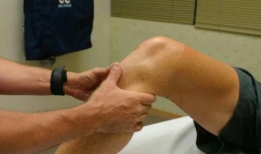
Ligament Tests
- Collateral ligaments: Medial / Lateral
- Cruciate ligaments: Anterior / Posterior
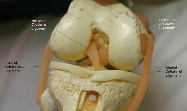
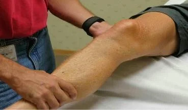
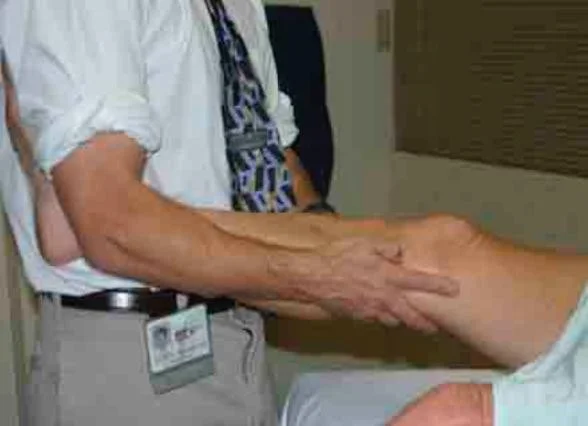
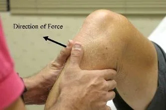
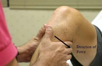
Collateral Ligament Testing
- Stress test with knee slightly flexed
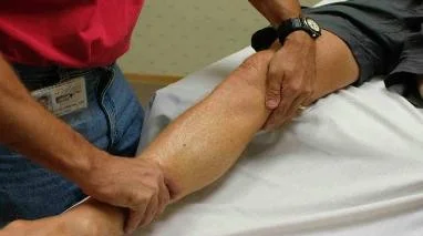
- Alternative technique for collateral ligament testing
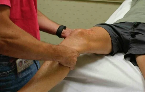
Cruciate Ligament Testing
- Anterior/Posterior Drawer Test
- Lachman’s test
- Sagging sign: indicates Posterior cruciate ligament tear
Meniscus Tests
-
McMurray’s test for medial & lateral meniscus
-
Apley grinding test
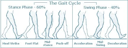
Gait Assessment
- Walking gait analysis
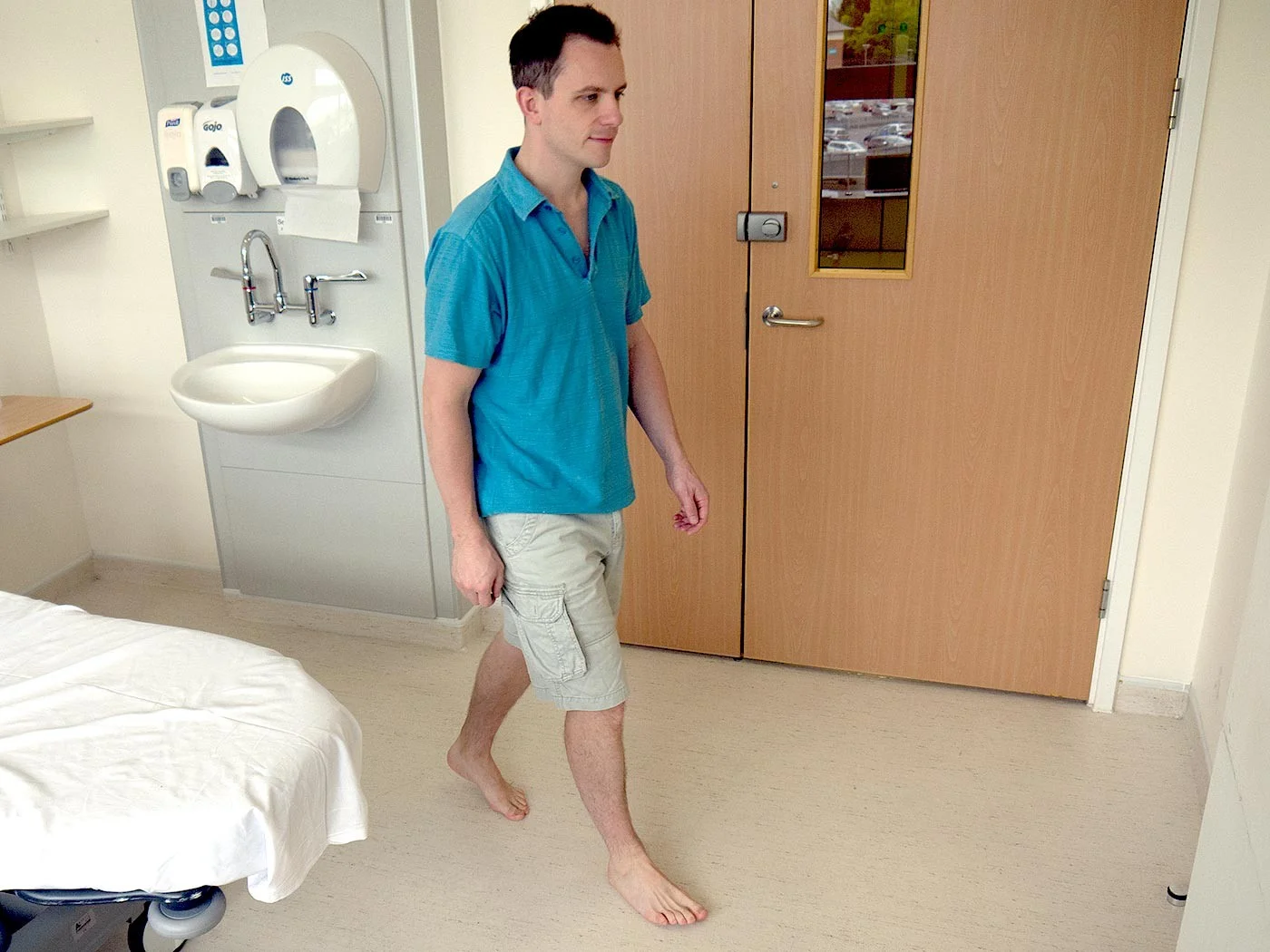
Special Tests: Gait Analysis
The Normal Gait Cycle
Stance phase (60%)
- Heel strike
- Foot flat - mid-stance
- Push off
Swing phase (40%)
- Acceleration
- Mid-swing
- Deceleration
Gait Patterns and Interpretation
| Gait Type | Explanation |
|---|---|
| Normal | Normal stance and swing phases |
| Antalgic | Painful to weight-bear – short stance phase |
| Lurch | Shortening – painless limping – normal stance period |
| Circumduction | Stiff hip – motion of pelvis compensates |
| High Step | Foot drop – more hip & knee flexion needed to free toes from ground |
| Tip-toe | Heel off the ground |