اللهم يا معلّم موسى علّمني، ويا مفهم سليمان فهّمني، ويا مؤتي لقمان الحكمة وفصل الخطاب آتني الحكمة وفصل الخطاب اللهم اجعل ألستنا عامرة بذكرك، وقلوبنا بخشيتك، وأسرارنا بطاعتك، إنك على كل شيء قدير، حسبنا الله ونعم الوكيل
Updates through https://medatlax.com/Clinical/Level-11/Orthopedics/OSPE/OSPE-Ortho/OSPE-Ortho - will be highlighted yellow - If there is any adjustment or edits please let me know Dr.alkharji@proton.me
collected from previous batches, Dania abuobaid, Alzahraa Ismaeel, Deema alsarawi, ~FA, Mujahid Almuhaydib, and Ola Al-Rayes
Principles of Fracture
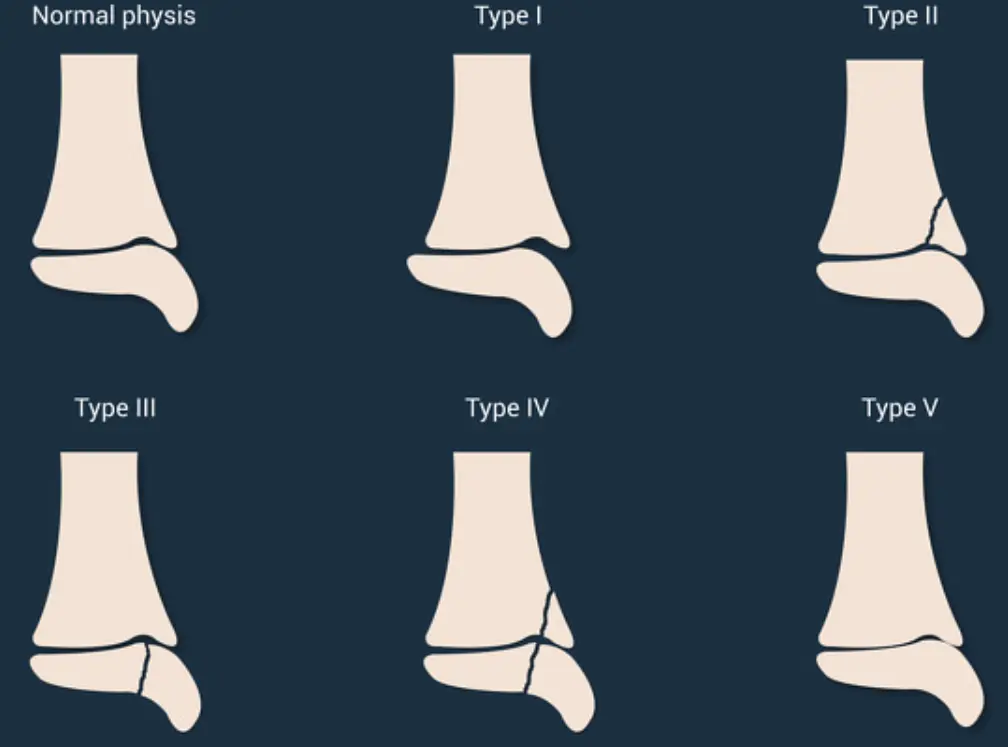
Salter-Harris Fractures
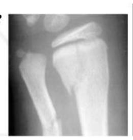
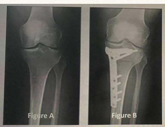
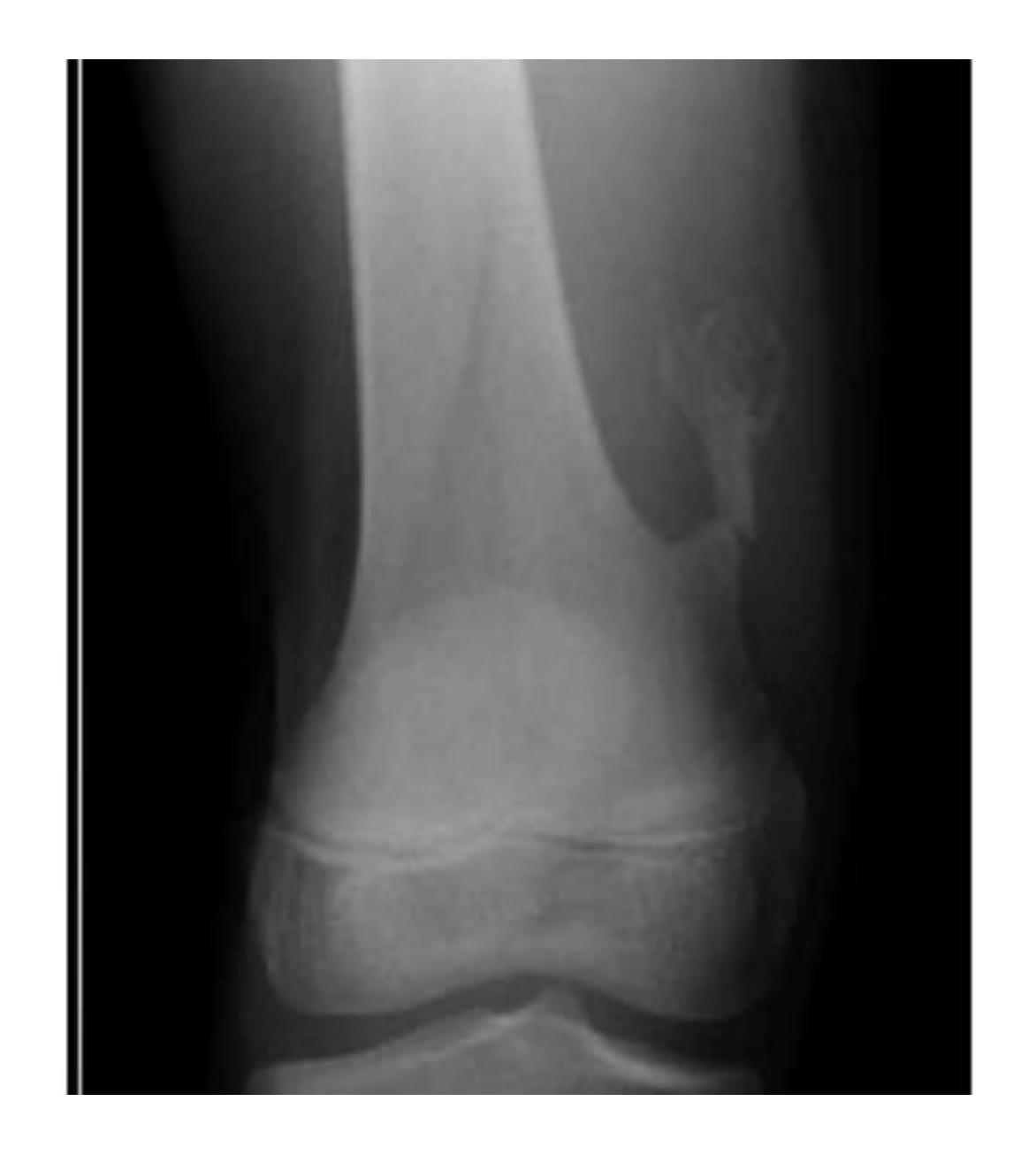
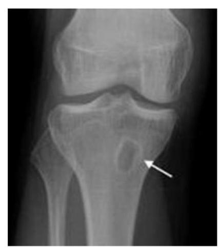
Q8/ What is the type of fracture shown in these pictures?
- A- Salter harris fracture II
Associated Injuries with Fractures
Q6/ For patients with this following fracture, you have to rule out the most associated injury which is:
- A. Spine fracture
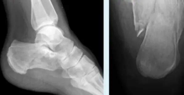
QUESTION 9: A case about a patient who fell from a high building. “X-ray showed a calcaneus fracture”, what is the most likely fracture to be associated with this injury?
Fracture Identification and Pain Causes
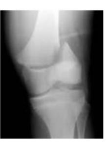
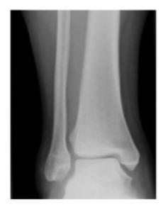
Q5/ A patient came with pain and the physician ordered X-ray to figure out the cause. According to his X-Ray, what could be the cause of his pain?
- C- Fracture association
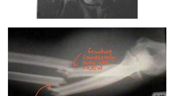
QUESTION 10: Patient come with pain and had this x-ray, what could be the cause of pain?
- a. Fracture
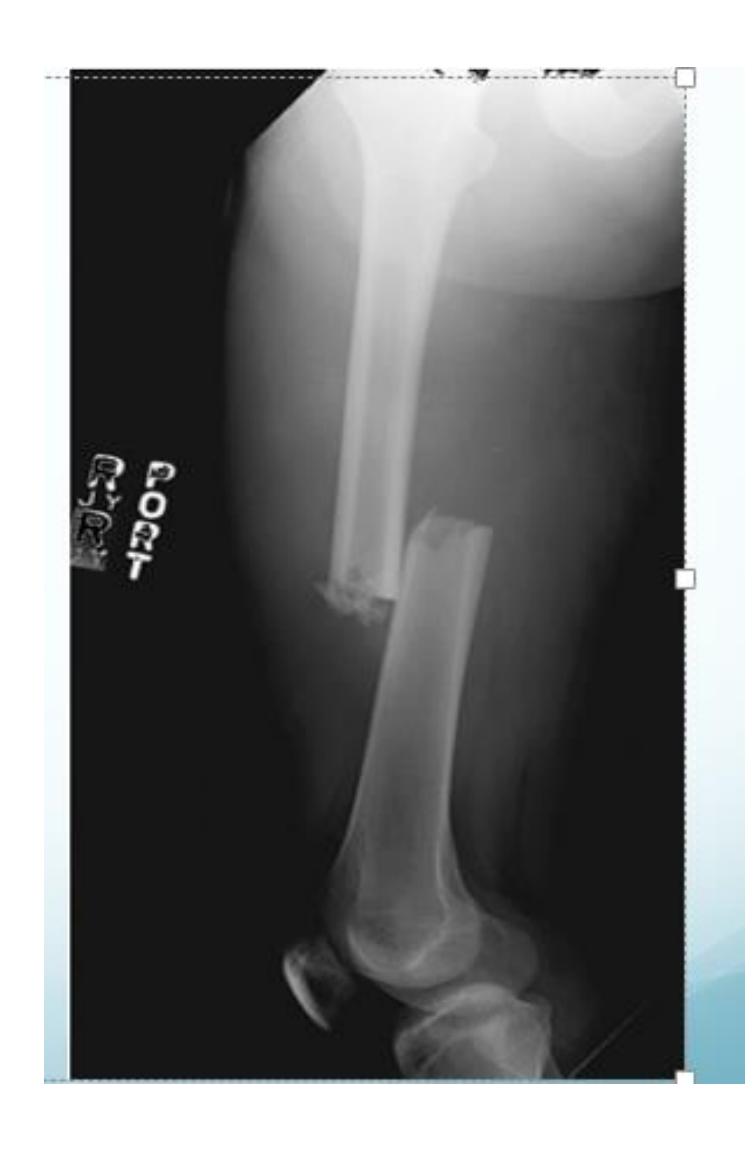
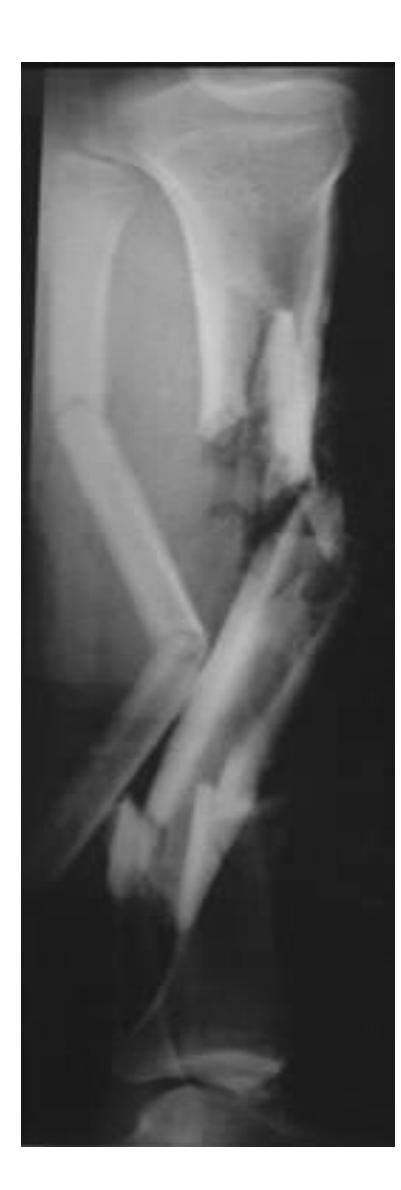
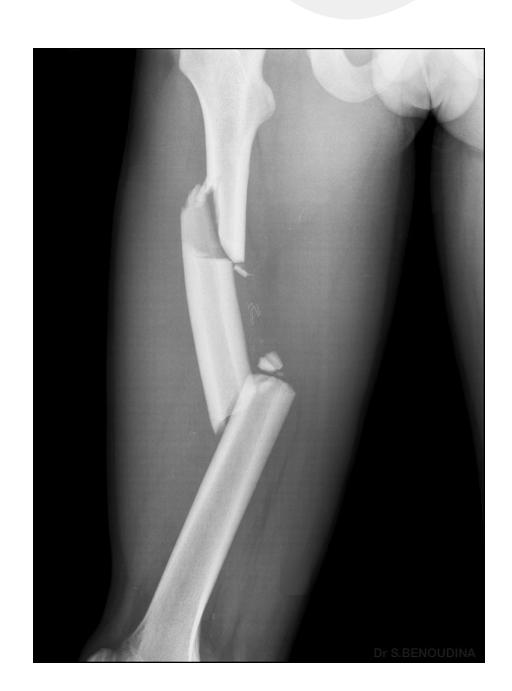
Fracture Patterns and Types
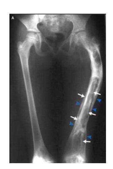
Which pattern of fractures is demonstrated in the attached X-ray?
- B. Segmental
Q19- what would be the bleeding source in the injury observed in this x ray?
- The answer: Venous plexus
Fracture Management Considerations
Management Factor
- Neurovascular deficit would affect management
Complications of Fractures
Fat Embolism Case
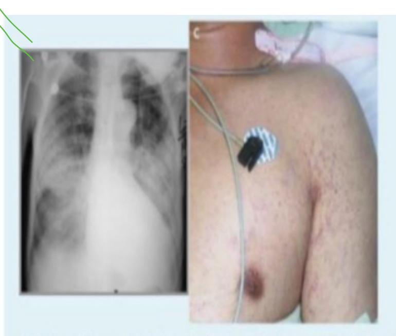
A 32-year-old man who had sustained a traumatic fracture of the left femoral shaft 20 hours earlier was referred for a sudden deterioration of consciousness. He was unresponsive to verbal stimuli but no focal neurologic abnormalities were found.
Q1. What is the most probable diagnosis?
- Fat embolism (petechial hemorrhage)
Q2: What is the treatment of choice?
- Respiratory support, early recognition, call the anesthesia and transfer to ICU
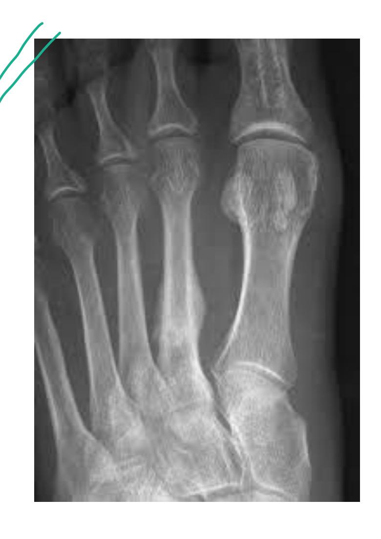
Stress Fractures
Stress Fracture Characteristics
- Stress fracture
- In Soldiers, athletes,…
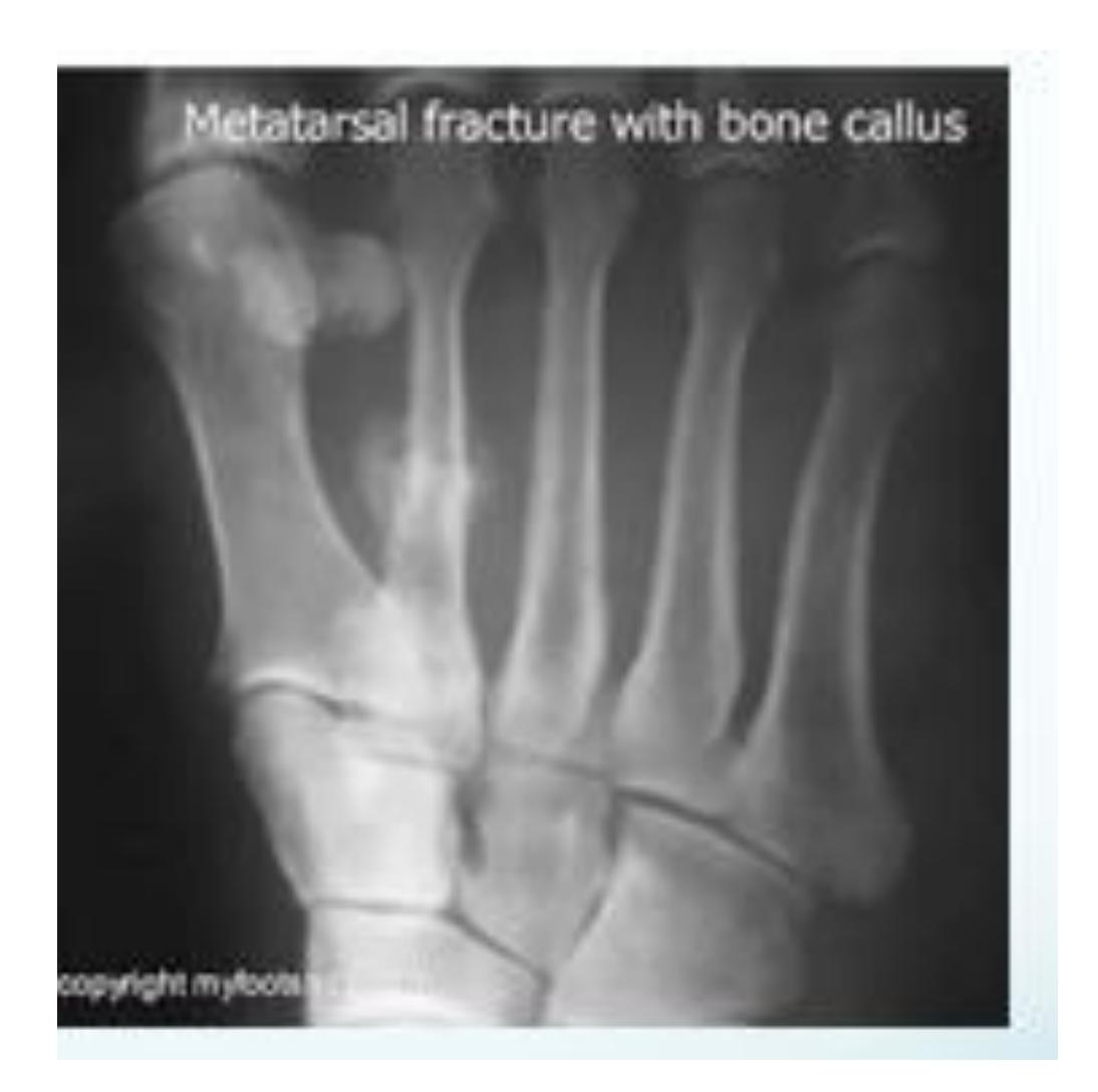
Stress Fracture Example
- Callous formation is 2nd metatarsal
- Looks like stress fracture
Principles of Treatment, Traction, Casting, Implants, Splints
Clinical Questions
Q5: 35-year-old male with fracture - Treatment
A 35-year-old male with this fracture, how would you treat it?
- Open reduction and internal fixation
Q8: 70-year-old female with hip injury
A 70-year-old inactive female presents to the emergency department after falling down in the bathroom. She’s been complaining from hip pain and inability to bear weight. Which of the following is the most appropriate choice of treatment?
- Bipolar hemiarthroplasty
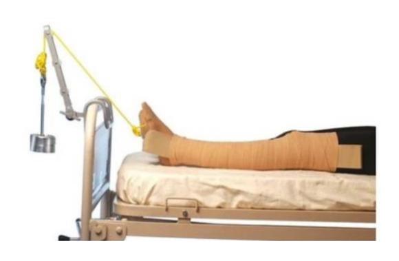
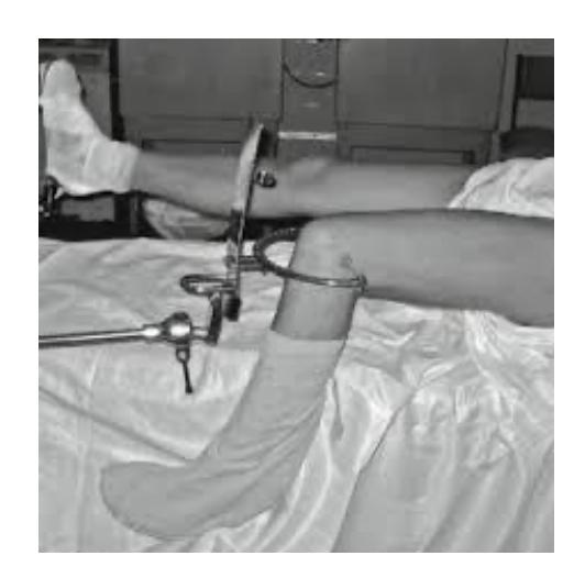
Q10: 30-year-old with femur fracture - Traction weight
A 30-year-old was involved in a road traffic accident. He sustained a closed femur fracture. The apparatus seen was applied to him. What is the maximum weight that can be used if his weight is 80 kg?
- 8 kg
Q12: Skeletal traction weight calculation
Assume that a patient weighs 80 kilograms, what is the maximum weight that can be applied in such form of traction? 4.5 kg or less
Q15: 35-year-old male with fracture - Treatment
35 years old male with this fracture, how would you treat it:
- Open reduction and internal fixation of ulna - Closed reduction of head of radius +/- fixation
Q17: 5-year-old boy with closed injury - Treatment
A 5-year-old boy sustains an isolated, closed injury. He weighs 55 kg and is otherwise healthy. What is the best treatment option for this patient?
- Fixation and intramedullary nailing
Q19: Traction weight calculation
Assume that a patient weighs 60 kilograms, what is the maximum weight that can be applied in such form of traction? 4.5
Compartment Syndrome
Diagnostic Criteria
Which of the following pressures is diagnostic of compartment syndrome?
- Delta pressure of 20 mmHg
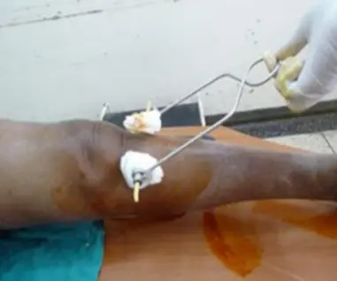
Fasciotomy Indications
- High risk patients
- Prophylactic with major corrective osteotomy of the leg & forearm
- S&S not resolved within 30-60 min. of appropriate precautions
- Significant tissue injury
- Suspicion: Equivocal clinical findings
- Based on measurements:
- Delta pressure (DBP - compartment P.) 25 mm Hg.
- Compartment pressure > 30mm Hg.
Case: A man involved in a road traffic accident (RTA) where a car ran over his forearm. X-ray showed fracture of the forearm.
Diagnosis?
- Compartment syndrome
What are the clinical features?
- The 5 P’s:
- Pallor
- Paresthesia
- Pain (out of proportion to injury)
- Paralysis
- Pulselessness
Management?
- ABC’s (Airway, Breathing, Circulation)
- Correct hypotension
- Remove circumferential bandages & cast
- Position limb at level of the heart
- Supplemental oxygen
- Fasciotomy (definitive treatment)
Q22: 30-year-old active patient with femoral neck fracture
30-year-old active patient presents to ER after history of falling down from second floor. Diagnosed with displaced femoral neck - he has only mild controlled hypertension - what is the next step for his management?
- Anatomical reduction and cannulated screws
Q23: Traction weight calculation
Assume this patient weighs 80 kg. What is the maximum weight that can be applied in such form of traction?
- 8 kg
Additional Questions
35-year-old male with fracture
A 35-year-old male fell and had this fracture - what is appropriate treatment?
- ORIF
Management of imaging finding
What is the management of the finding in the image?
- Screw fixation

Skeletal traction weight guidelines
A male patient who weighs 80 kg has a fracture, skeletal traction is applied. What is the maximum weight for traction?
- 10% of body weight
- 8 kg
Orthopedic Devices and Principles
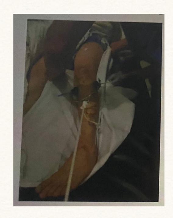
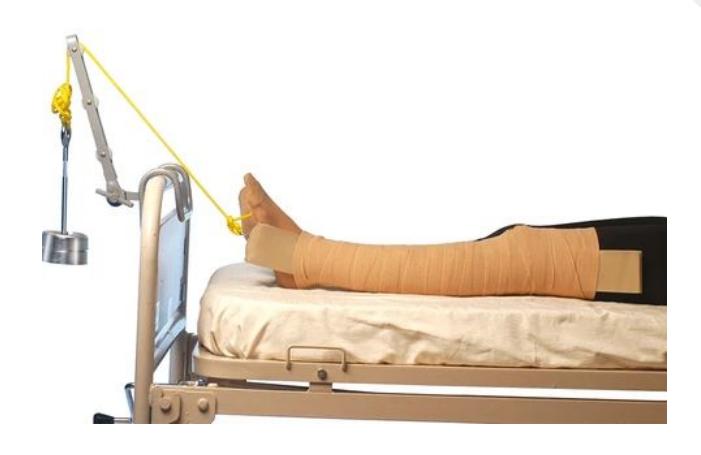
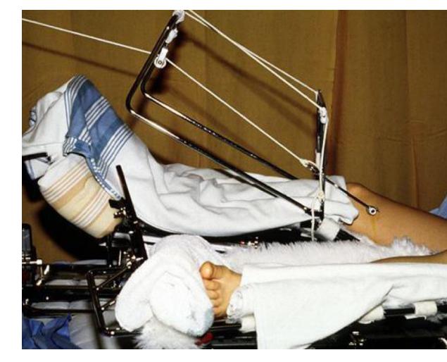
Skeletal Traction
1. Skeletal Traction
- Indication: When a strong force is needed (weight more than 4-5 kg) in adults
- Complications: Infection, over distraction of bone fragments
Synthetic Fiberglass Cast
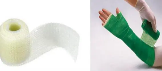
1. Synthetic Fiberglass
- Disadvantages: Sharp edges, not moldable
- Advantages: Strong, quickly sets
Thomas Splint
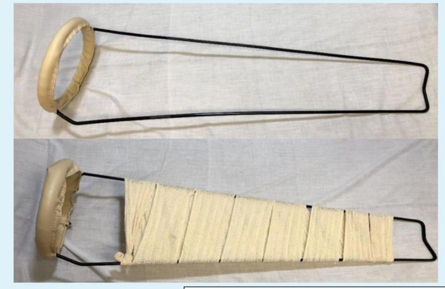
Name of device: Thomas splint
Indication of use: Transport of patient with femoral shaft fracture
Locked Head Screws and Locked Plate

Name of device: Locked head screws and locked plate
Used in: Osteoporotic bone
Synthetic Fiberglass Cast
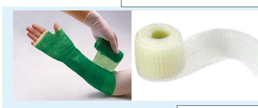
Name of cast: Synthetic Fiberglass Cast
Advantages:
- Strong
- Water resistant
Disadvantages:
- Costly
- Sharp edges
Hip Spica
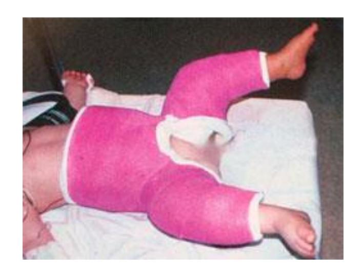
Q1) Hip spica
Q2) Indications:
- DDH
- Femoral fracture
Thomas Splint Applications
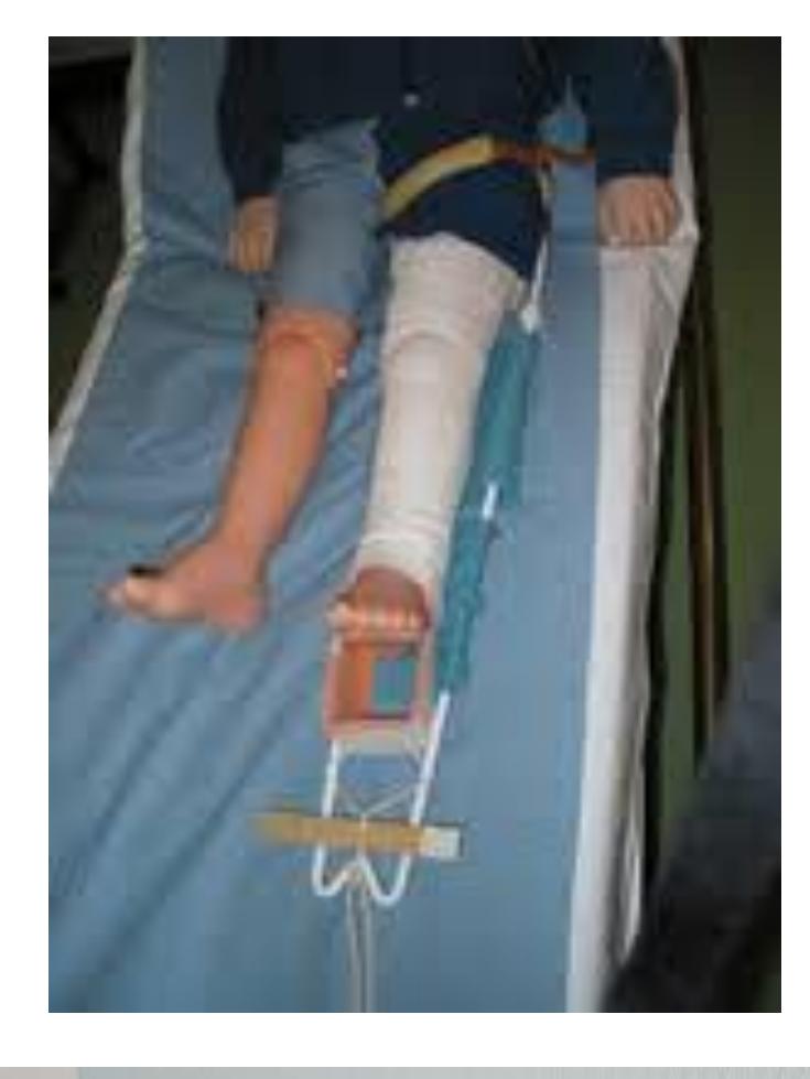
Name of splint: Thomas splint
Used for:
- Fixed traction for transport
- Can also be used as balanced traction for treatment
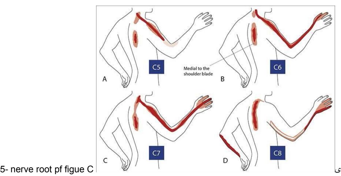
Orthopedic Clinical Examination
Neurological Examination
Dermatomes and Myotomes
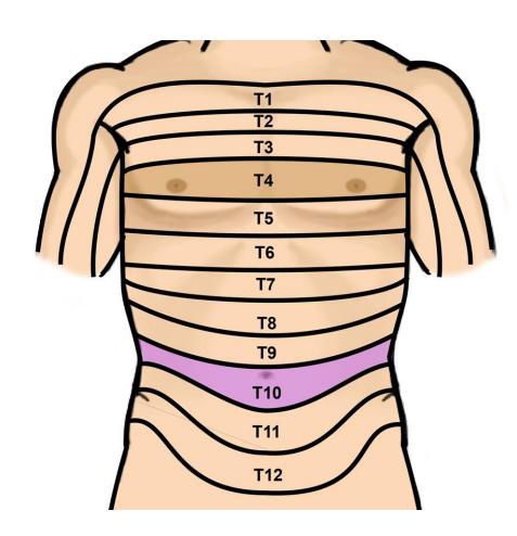
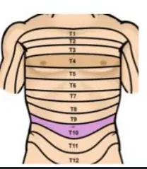
What’s the dermatome at the level of the umbilicus?
- D. T10
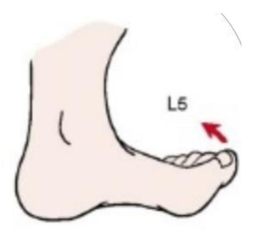
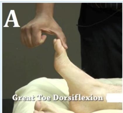
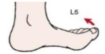
What’s the myotome of the shown structure (Big toe)?
- C. L5
Which nerve root is responsible for the reflex of the shown structure (Achilles tendon)?
- D. S1

Nerve root examination in reflex testing:
- Picture A: L5, S1
- Picture B: S1, S2
Additional nerve testing:
- C7 testing
- Nerve root of umbilicus: T10
Hand and Peripheral Nerve Testing
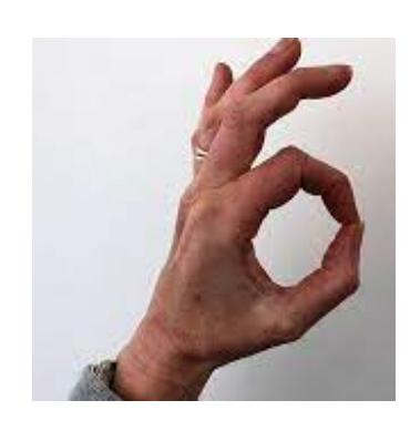
OK Sign Test
- What is being tested with the OK sign?
- AIN (Anterior Interosseous Nerve)
- Loss of OK sign in right hand indicates:
- Inability to flex DIP of thumb and index
- AIN (branch of median nerve) injury
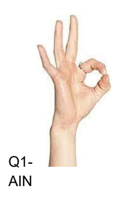
During clinical examination with OK sign:
- Interpretation: Loss of OK sign
- Peripheral nerve affected: Anterior interosseous nerve (branch of median nerve)
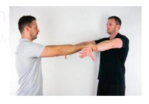
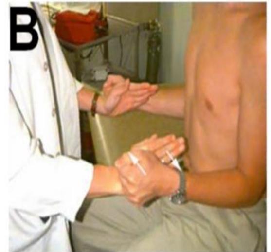
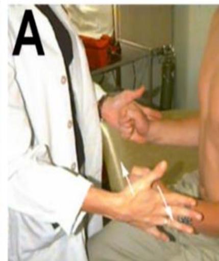
Froment’s Sign
- What does the positive sign signify?
- Positive Froment’s sign
- Ulnar nerve dysfunction
Carpal Tunnel Syndrome (Phalen’s Test)
- Diagnosis when patient holds hands in position:
- CTS (Carpal Tunnel Syndrome)
- Positive sign criteria:
- Reproducing symptoms within 1 minute (numbness, tingling, and burning sensation)

- Reproducing symptoms within 1 minute (numbness, tingling, and burning sensation)
Shoulder Examination
Rotator Cuff Testing
Supraspinatus Muscle Testing
- What muscle is being examined?
- A. Supraspinatus
- A. Supraspinatus
Jobe Test (Empty Can Test)
- Test being performed:
- Jobe test
- Tests: Supraspinatus muscle
Rotator Cuff Strength Testing
- External rotators strength (Image A)
- Internal rotators strength (Image B)
Shoulder Instability Tests
Shoulder Apprehension Test
- Test name: Shoulder apprehension test
- Positive findings: Shoulder instability (previous shoulder dislocation)
- What the test shows: Patient feels pain (apprehension) when pushing the shoulder from behind in this position
- Associated condition: Shoulder dislocation (recurrent most probably)
Biceps Tendon Testing
Biceps Tendinitis Test
- Pain over anterior shoulder during this test indicates:
- A. Biceps tendinitis
- A. Biceps tendinitis
Shoulder Range of Motion
Apley’s Scratch Test
- Range of movement tested in images:
- Adduction and internal rotation
- Which photograph is abnormal?
- A
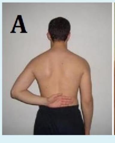
- A
Scapular Testing
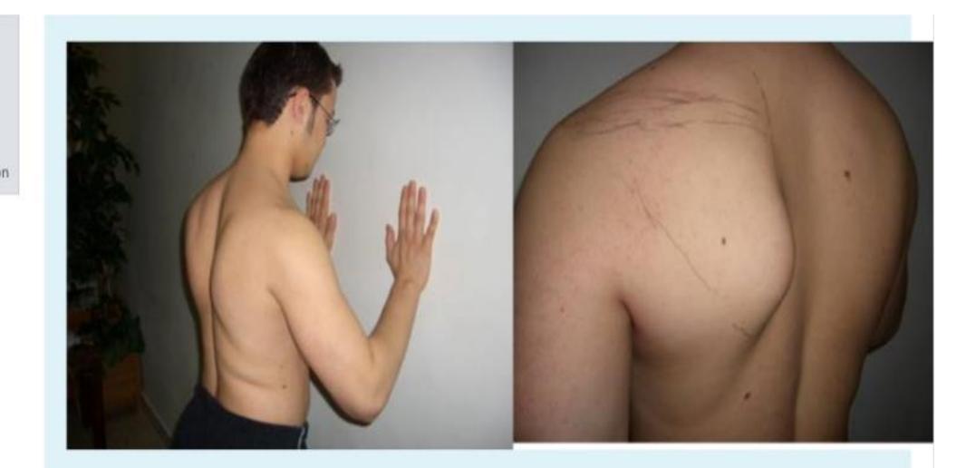
Winged Scapula Test
- During wall pushing test:
- Sign name: Winged scapula
- Affected nerve: Long thoracic nerve
Hip and Pelvic Examination
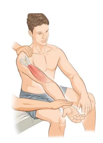
Hip Flexor Testing
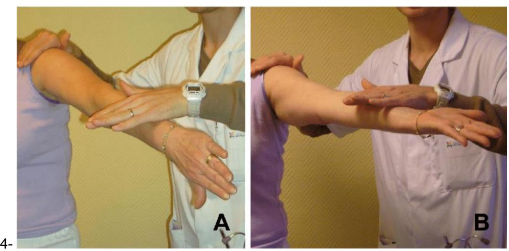
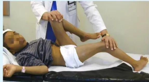
Thomas Test
- Test name: Thomas test
- Positive findings: Fixed flexion deformity of the hip
Iliopsoas Testing
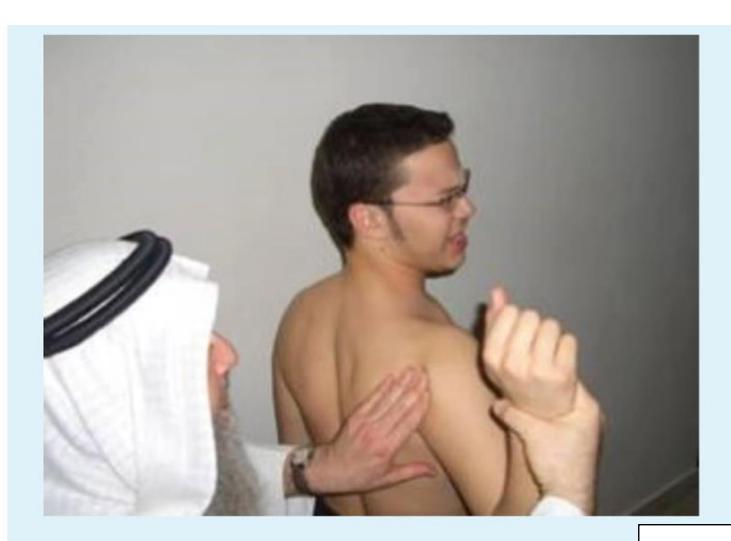
- Which structure is being examined?
- Left psoas
- Which muscle is attached to the pointed structure?
- Iliopsoas
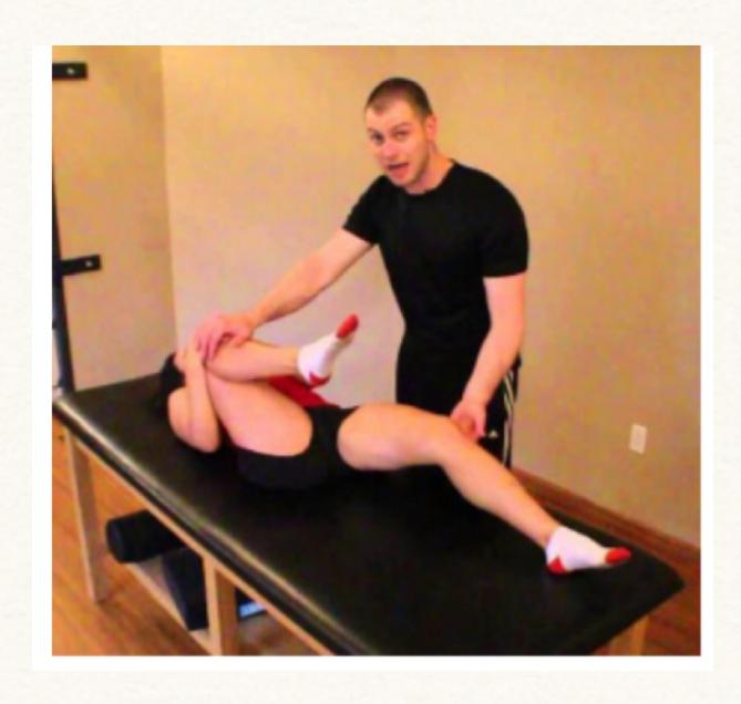
- Iliopsoas
- Which muscle is tested during this test?
- Left psoas
Iliopsoas Tightness Test
- What does this picture examine?
- A. Iliopsoas tightness
Hip Abductor Testing
Trendelenburg Test
- Test name: Trendelenburg test
- Positive if: Weak hip abductors, hip dislocation, OA
- True statement regarding Trendelenburg sign:
- A. Contralateral pelvic tilt, and ipsilateral shoulder tilt towards the affected side

- A. Contralateral pelvic tilt, and ipsilateral shoulder tilt towards the affected side
Sacroiliac Joint Testing
Sacroiliac Joint Stress Test
- Test name: Sacroiliac joint stress test
- Positive finding: Sacroiliac joint instability

Lower Limb Examination
Hamstring Testing
Hamstring Muscle Attachment
- Which muscle is attached to the fractured structure?
- d. Hamstring
- d. Hamstring
Achilles Tendon Testing
Achilles Tendon Examination
- Which tendon is being tested?
- A. Achilles tendon
- A. Achilles tendon
Achilles Tendon Rupture Sign
- This sign is seen in:
- a. Achilles tendon rupture
- a. Achilles tendon rupture
Radiological Views
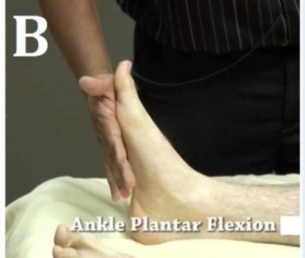
Ankle Views
- Mortise view:
- c. anterior posterior with 15 degree internal rotation
Normal vs Abnormal Findings
Normal vs Abnormal Comparison
- What is shown, indicate what?
- b. Normal
- b. Normal
Upper Limb
Clavicle Fractures
Z-shaped Clavicle Fracture
- Treatment: Surgery
Clavicle Fracture Treatment

- Figure A: Figure-of-eight brace (bandage) and shoulder sling
- Figure B: Shoulder sling
Humerus Fractures
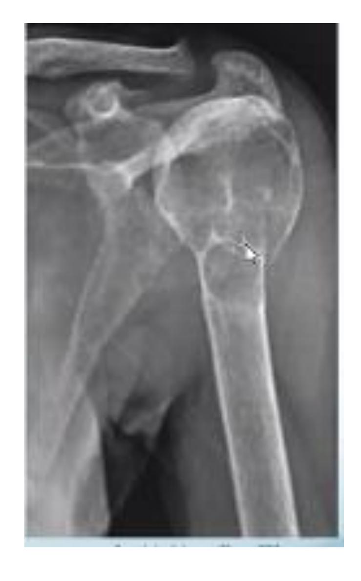
GT (Greater Tuberosity) Fracture
Surgical Neck of Humerus Fracture

Humeral Shaft Fracture
- Clinical Examination: Check for radial nerve injury by asking the patient to extend the wrist and fingers
Distal Humerus Fracture
Forearm Fractures
Galeazzi Fracture

-
Treatment: Surgery -
Monteggia Fracture
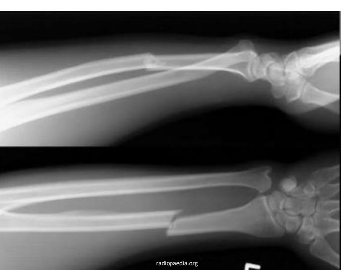
- Location: Proximal ulna fracture w/ radial head dislocation fracture of the proximal (or mid‑shaft) ulna with dislocation of the radial head at the elbow.
Wrist and Hand Fractures
Colles’ Fracture
- Deformity: Dinner-fork deformity
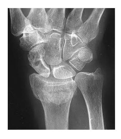
Scaphoid Fracture
Scaphoid Fracture Treatment
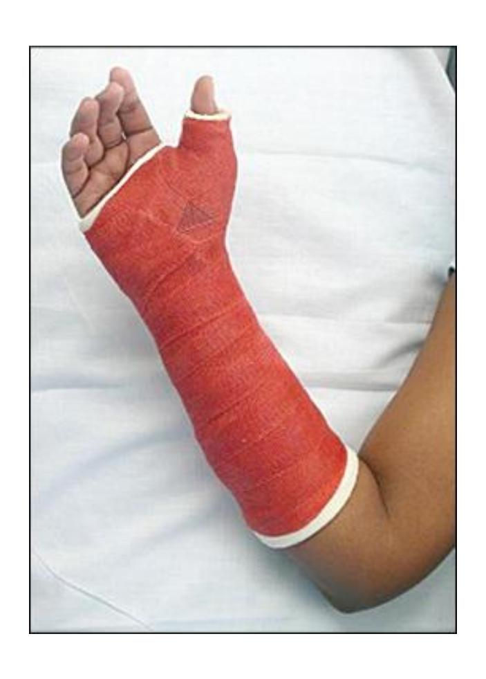
- Treatment: Thumb spica cast
Mallet Finger
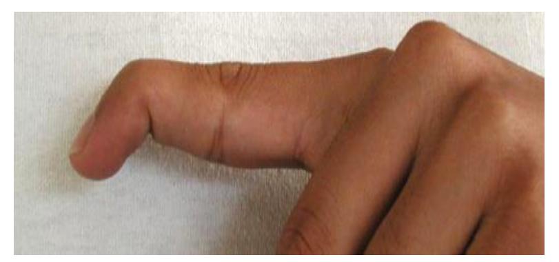
- Treatment: Splint (hyperextender) for 6 continuous weeks OR surgical fixation with nail removal after 6 weeks
Lower Limb
Hip and Pelvis Injuries
Hip Fractures
A 70-year-old inactive female presents to the emergency department after falling down in the bathroom. She’s been complaining from hip pain and inability to bear weight. Which of the following is the most appropriate choice of treatment?
- C. Bipolar hemiarthroplasty
Management Question 8
- B) Total hip arthroplasty
Muscle Attachments and Hip Pain
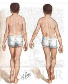
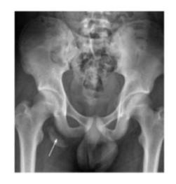
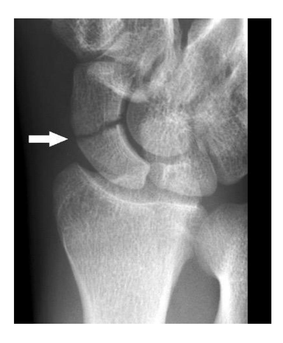
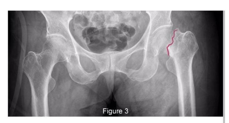
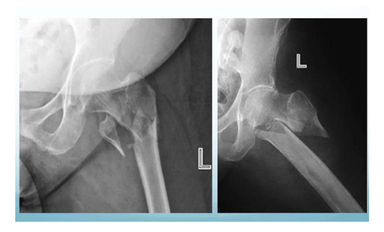
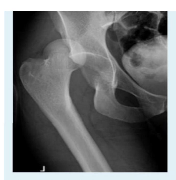
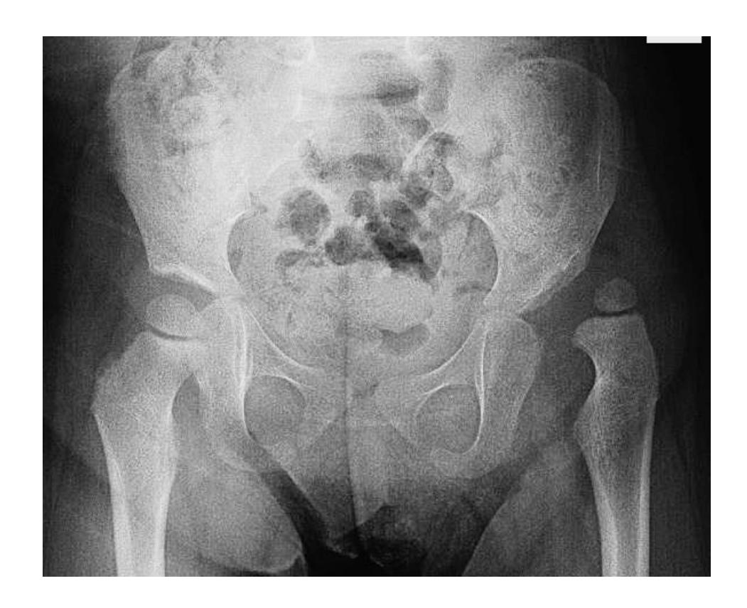
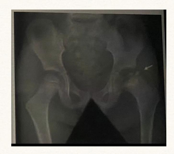
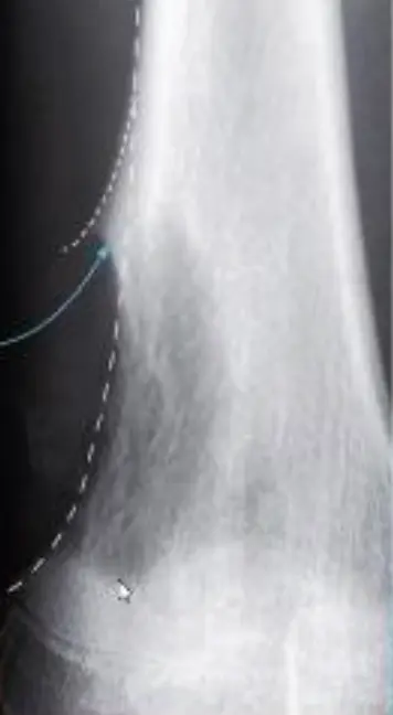
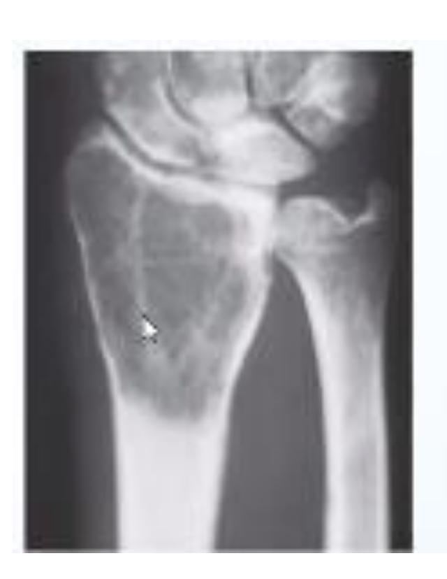
A teenager with acute onset of pain in the right hip during a run. He sustained the injury seen in the x-rays with a white arrow. This injury usually occurs due to a forceful eccentric contraction of which of the following muscles?
- C. Rectus femoris
- Sartorius
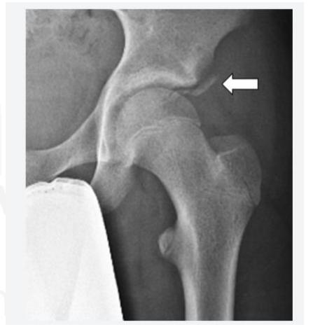
Pelvic Anatomy
Which of the following muscles is attached to the structure pointed by the arrow?
- C. Sartorius
Note: Arrow was pointing at ASIS (Anterior Superior Iliac Spine)
- A. Hamstring

Femoral Fractures
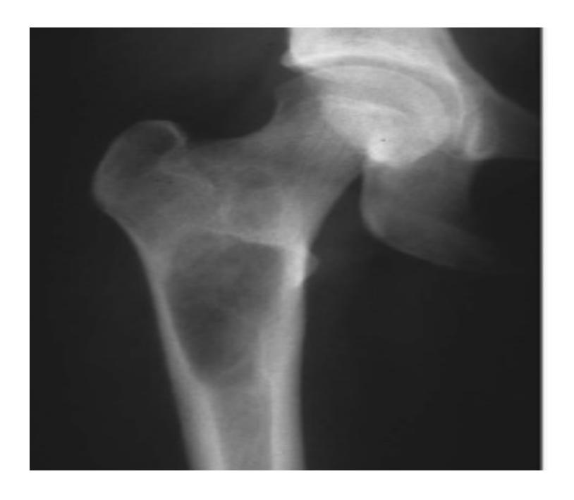
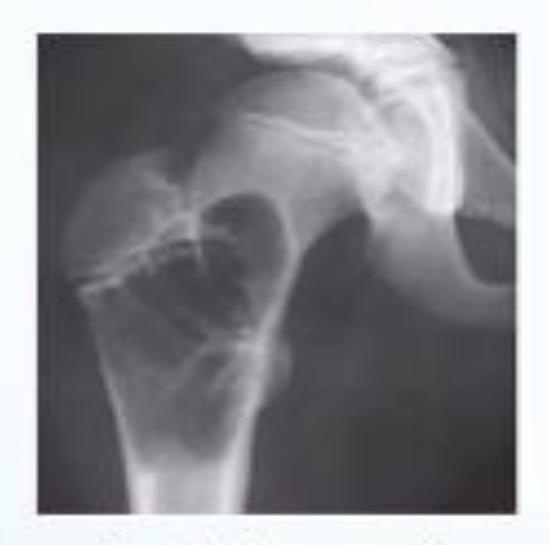
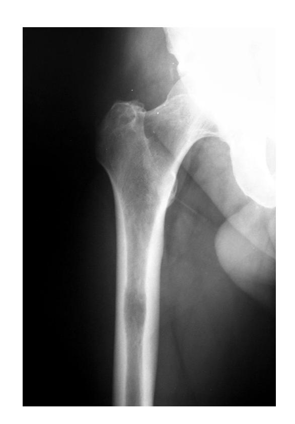
Intertrochanteric Fracture
Diagnosis: Intertrochanteric fracture of neck of femur Treatment: Dynamic hip screw or proximal femoral nail
Femoral Shaft Fracture
Diagnosis: Femoral shaft fracture Management:
- IV line, fluids, blood transfusion
- 2 views and 2 joints x-rays (must see hip joint)
- Treatment of choice: Locked intramedullary nail
Knee and Thigh Injuries
Patellar Fractures
Fracture of Patella
Treatment of choice: Open Reduction Internal Fixation (ORIF) because it’s intra-articular with muscle pulling, using wires/screws and tension bands
Ankle and Distal Lower Leg
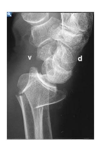
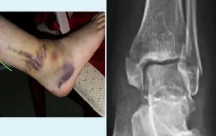
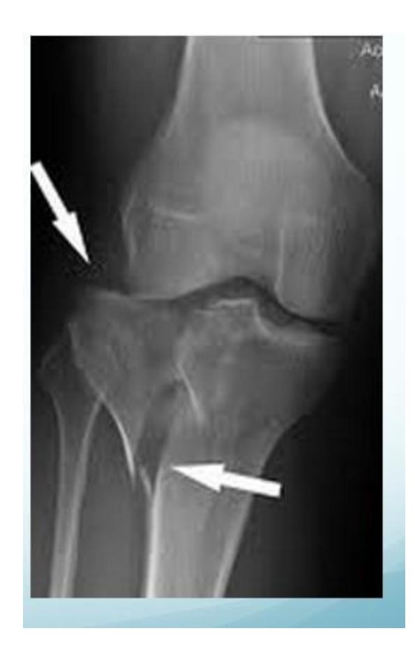
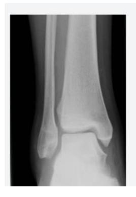
Syndesmotic Injuries
A 17-year-old male had a high ankle sprain followed by pain in his leg. X-ray films were obtained. According to the attached X-ray, which structure has been injured?
- B. Syndesmosis ligament
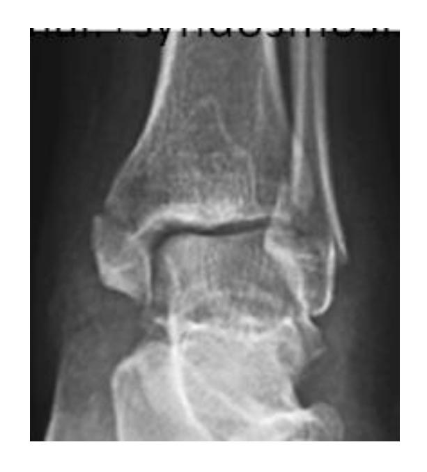
A 23-year-old male injured his ankle after a twisting ankle injury 2 days back. What is the most likely pathology based on shown x-ray?
Management Question: What is the management of the finding in the image?
- A. Screw fixation for syndesmosis injury

Ankle Fractures
Ankle Fracture Cases
Answer 1: Ankle fracture (Suprasyndesmotic fracture in distal fibula type C with medial malleolus fracture)
Answer 2: ORIF by plate in fibula and syndesmotic screw, and screw for medial malleolus fixation
- 1: Ankle fracture (transyndesmotic fracture in distal fibula type B and medial malleolus fracture)
- 2: Open reduction internal fixation by plate in fibula and syndesmotic screw and screw for medial malleolus fixation
Twisting Injury Cases

Q1: What is the mechanism of this injury?
- Spiral fracture caused by indirect twisting injury
Q2: What is the treatment of choice?
- Surgical treatment is best
- Conservative options if minimally displaced: Long leg cast for 4-6 weeks, Patella-bearing cast (Sarmiento) or fracture brace
- Average union time is 16±4 weeks
- Operative treatment: Intramedullary (IM) Nailing is the treatment of choice
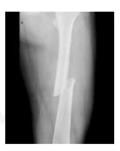
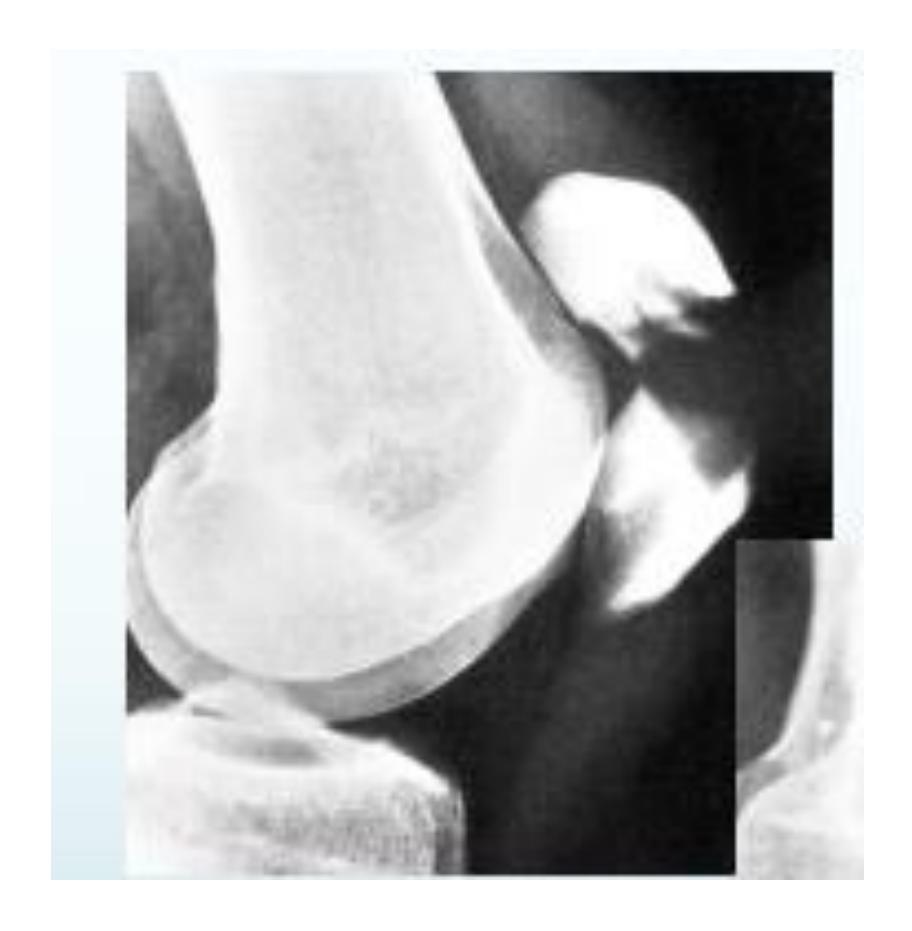
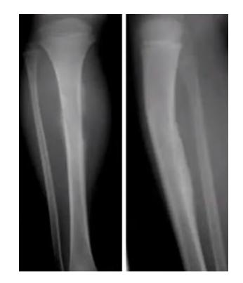
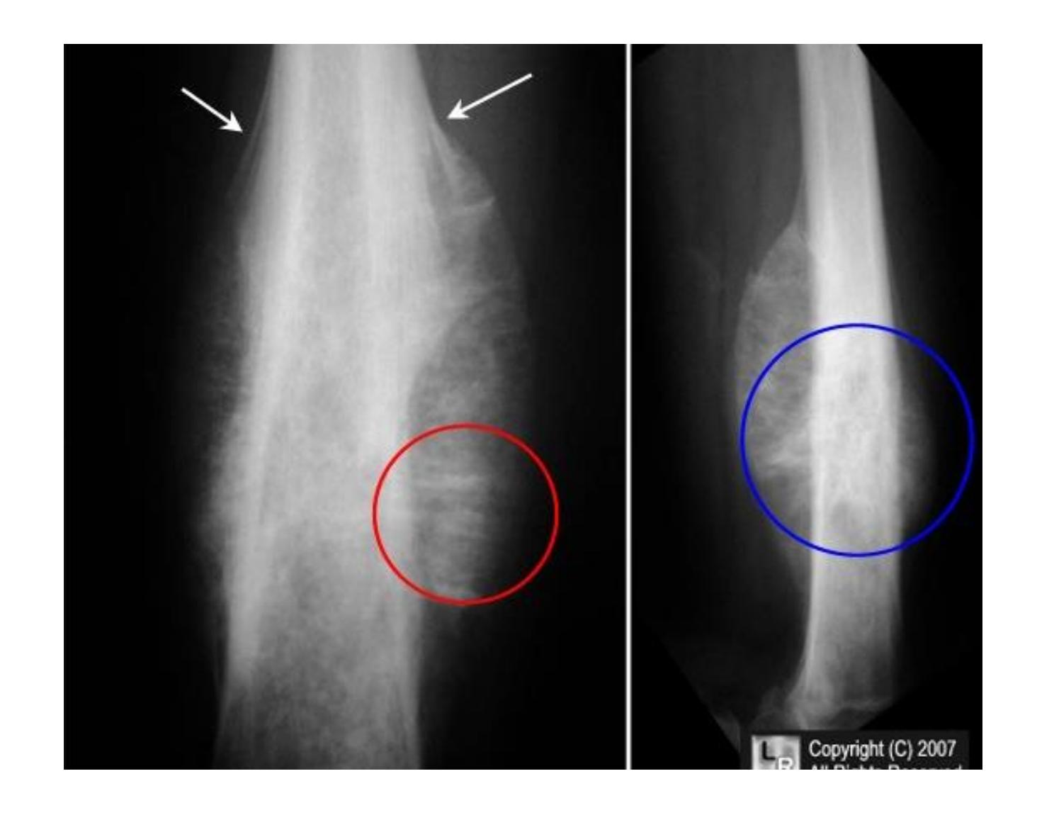
Tibial Fractures
Tibia and Fibula Fracture
Diagnosis: Segmental and comminuted fracture of tibia and fibula
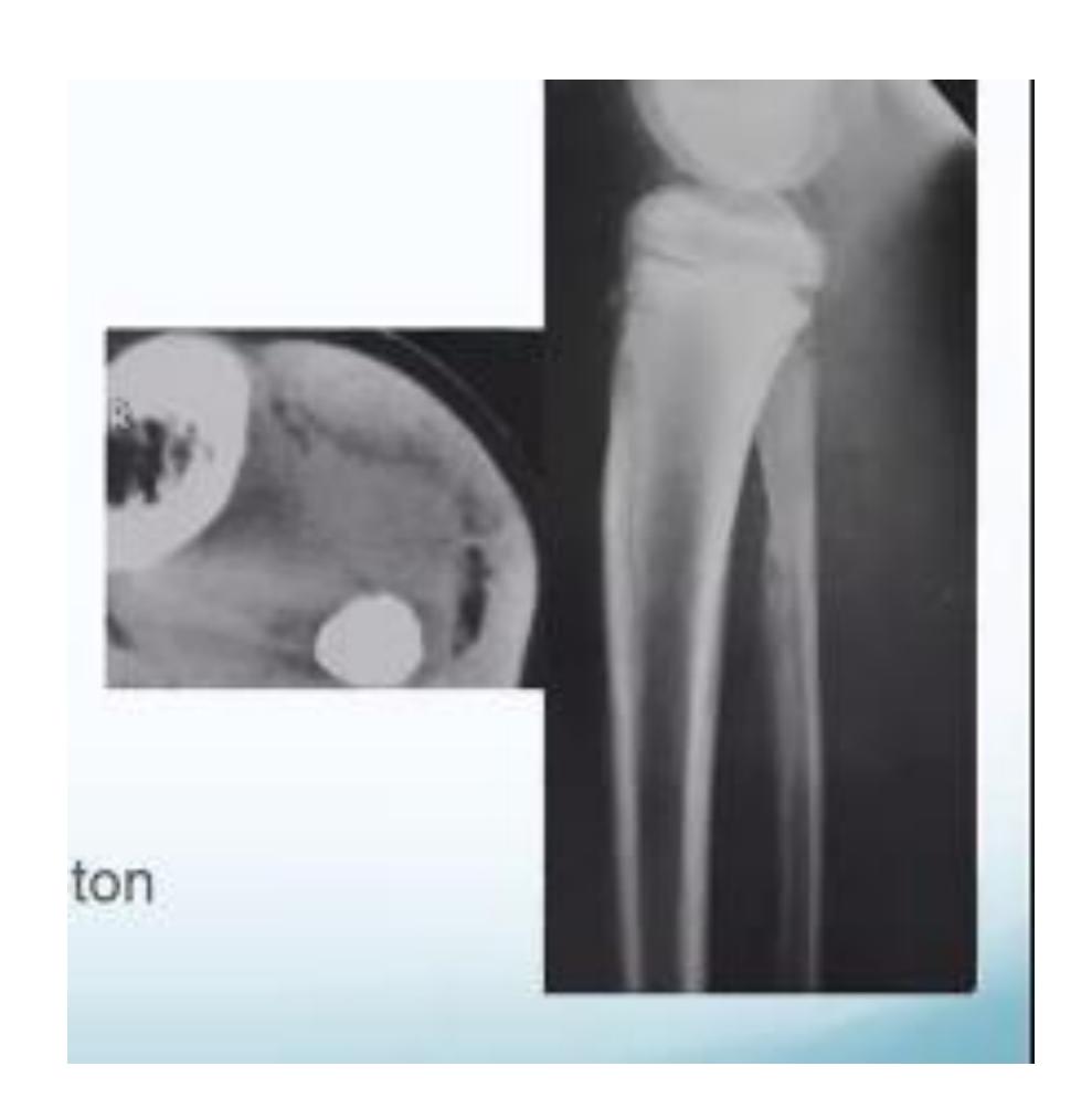
Tibial Plateau Fracture
Diagnosis: Tibial Plateau fracture Characteristics: Intra-articular Treatment: ORIF CT scan is necessary for evaluation
Sarmiento Brace
Description: Sarmiento brace (patella-bearing cast) used for tibial fractures
Ankle Specific Fractures
General Ankle Fracture
Diagnosis: Ankle fracture Mechanism: Twisting injury Common in: Old people
Mortise View of the Ankle

Foot Injuries
Talus Fractures
Fractured Talus
Complications: If displaced, can lead to avascular necrosis Treatment for displaced: ORIF or percutaneous fixation
Avascular Necrosis in Talus
Q1. What is the diagnosis?
- Fracture talus
Q2. Write complications specific to this fracture?
- Avascular necrosis
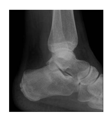
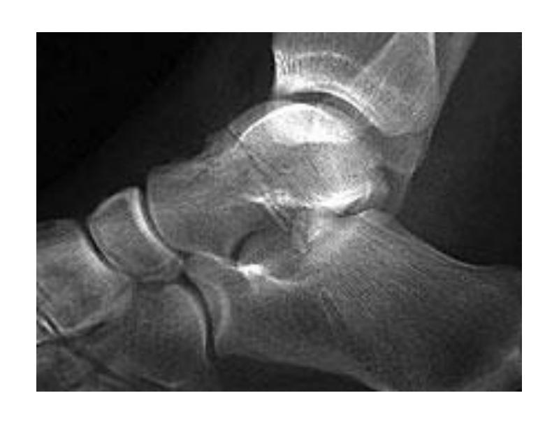
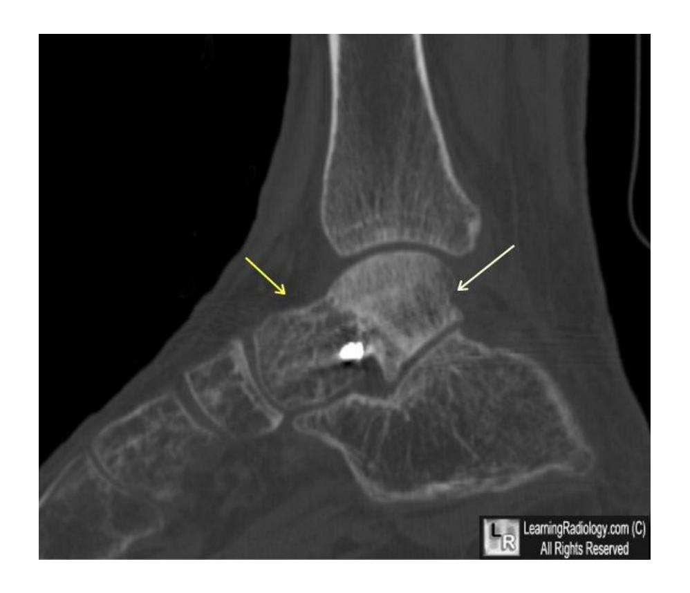
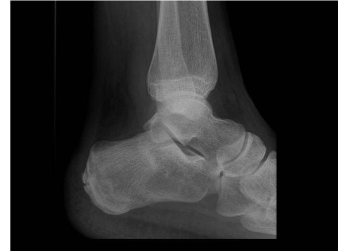
Calcaneal Fractures
Calcaneal Fracture
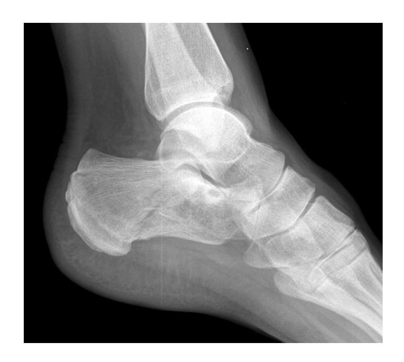
Complications: If displaced, can lead to osteoarthritis of subtalar joint which is painful Evaluation: CT scan to assess displacement Treatment: Conservative management
Calcaneal Fracture Evaluation
Q2: What other image you need for better evaluation of fracture?
- Calcaneal fracture
- CT scan to show the displacement
Pelvis
Pelvic Anatomy
What does this view demonstrate?
- Anterior column
Pelvic Fractures and Radiological Findings
Give 2 abnormalities in this X-ray?
- Fracture of pubic bones
- Disrupted sacroiliac joint on the same side
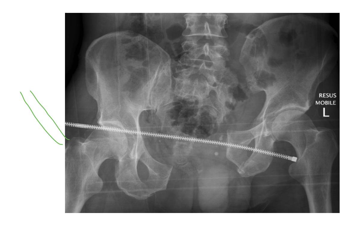
Emergency Management of Pelvic Trauma
Give first 2 measurements you need for saving life?
- ATLAS protocol (ABC)
- IV fluid administration
- IV analgesia
- Pelvic sheet application
Spine & Back
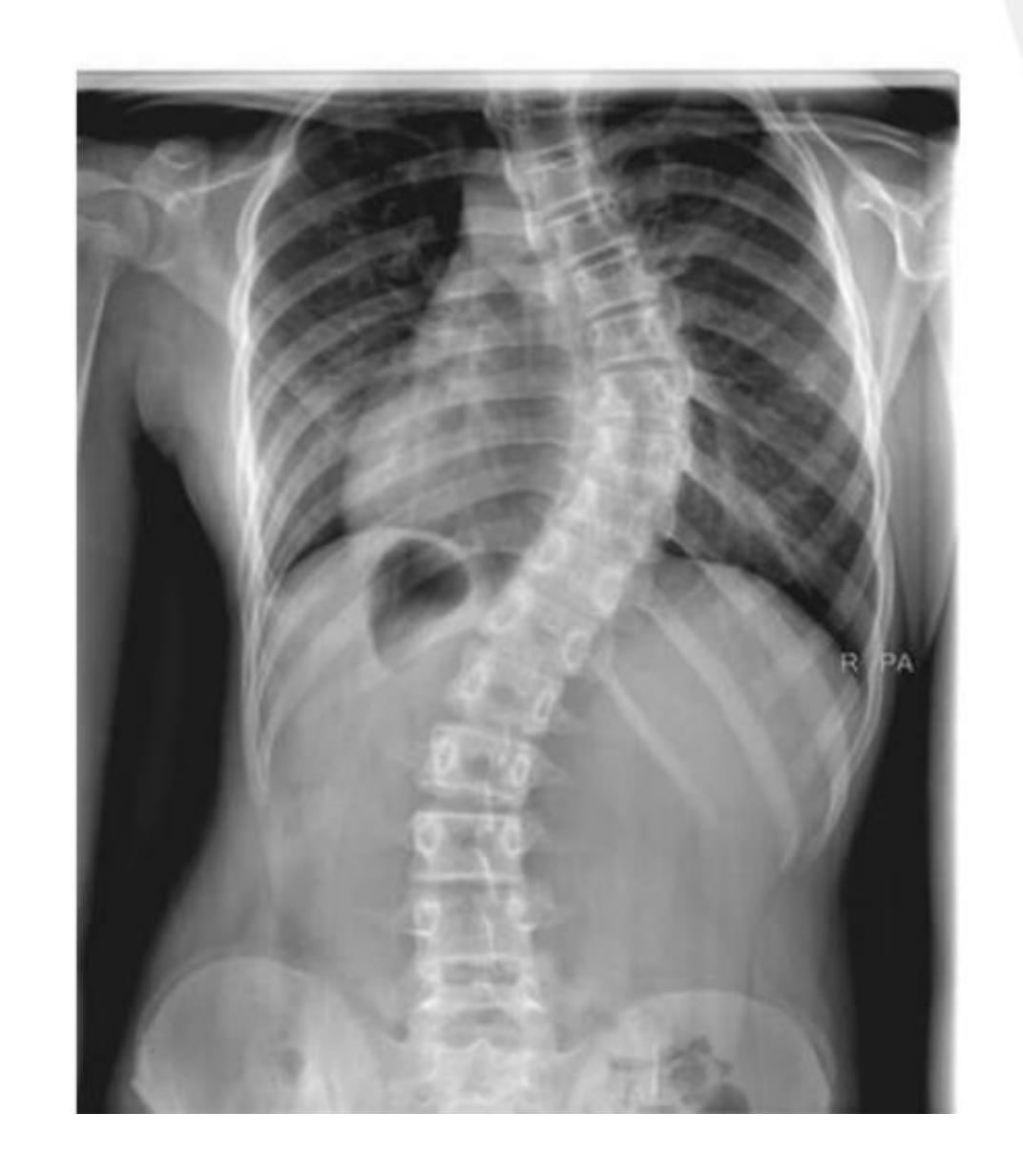
Scoliosis Cases
Which of the following is associated with this finding?
- Adolescent Idiopathic Scoliosis
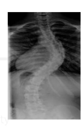
A 14-year-old girl presented to the clinic after her parents noticed a deformity in her back. Adam’s test was positive. Which of the following pathologies represents this case?
- Idiopathic scoliosis
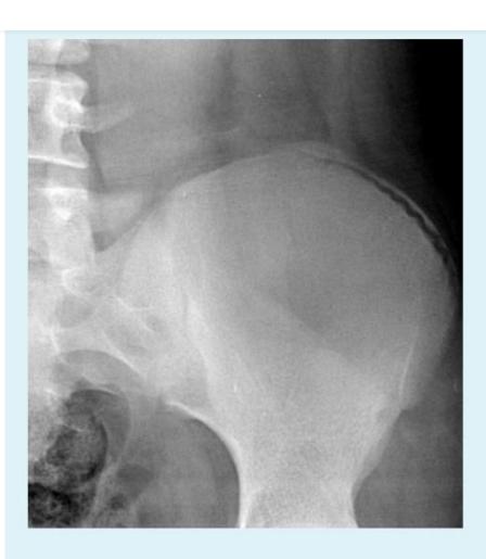
A 13 years old patient, she is known to have idiopathic scoliosis. X-ray pelvis was requested for her.
- 1. Mention the name of the sign seen the X-ray.
- Risser sign
- 2. How it can help in taking decision the management?
- By assessing potential of growth to evaluate potential deformity progression (e.g. scoliosis)
Growth Assessment Signs
Rosser’s sign
1-Describe? Rosser’s sign
2-What is clinical importance of this sign? Measure progress of bony fusion of iliac apophysis. The lower the grade, higher the potential for progression.

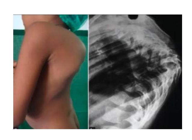
Spinal Deformities
What is this deformity?
- Gibbus deformity
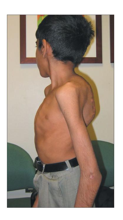
What is the deformity in this picture?
- Gibbus deformity
What is the most common cause?
- TB, congenital POTTS

Spinal Trauma & Mechanisms
Which of the following is the mechanism of this injury; MOI?
- Flexion distraction injury
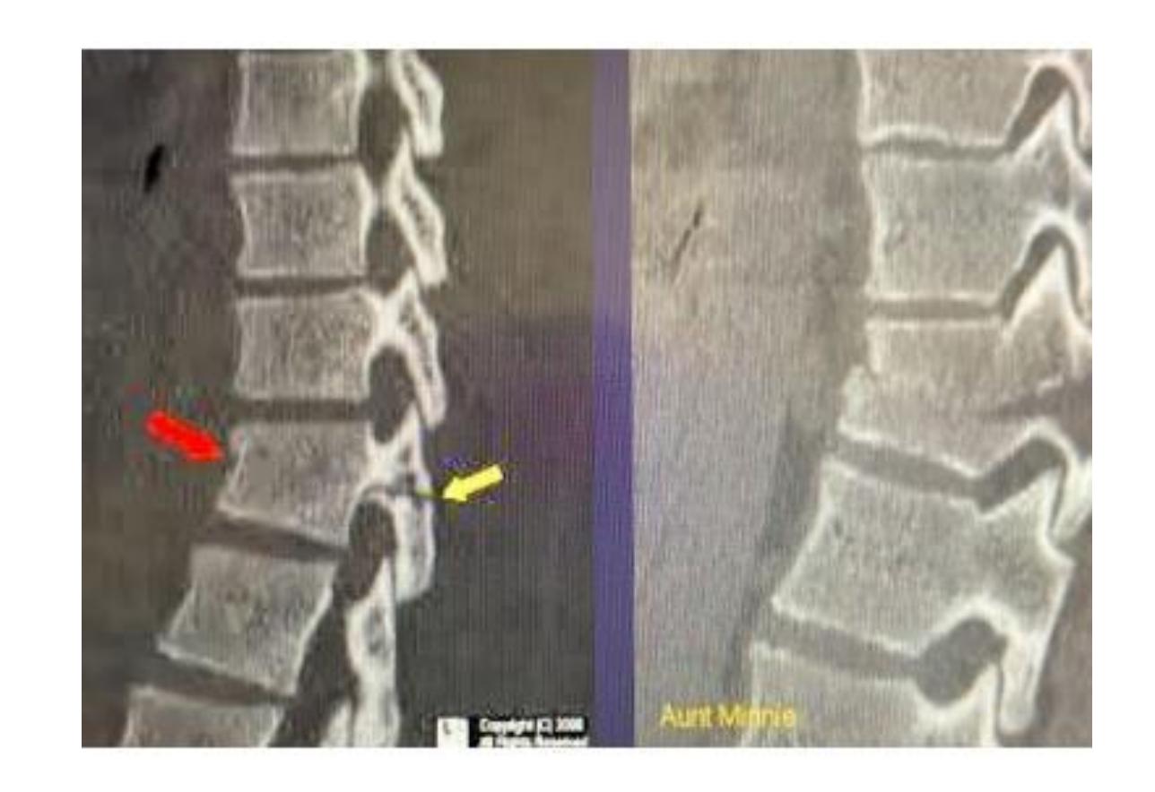
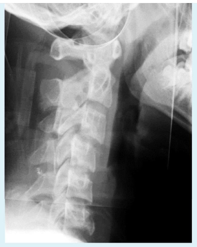
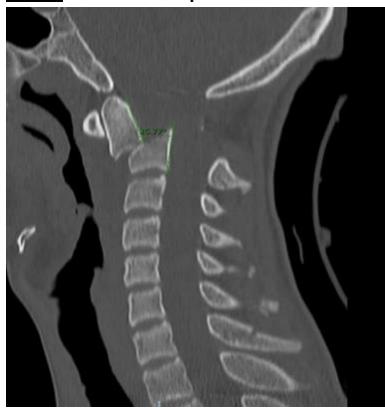
You are evaluating a 17 years old adolescent girl who was in an unrestrained motor vehicle accident. Imaging is demonstrated. And she has significant posterior midline neck pain. What is the diagnosis and MOI?
- Jumped facet Hyperflexion
A case about a patient who fell from a high building. “X-ray showed a calcaneus fracture”, what is the most likely fracture to be associated with this injury?
- Spinal fracture
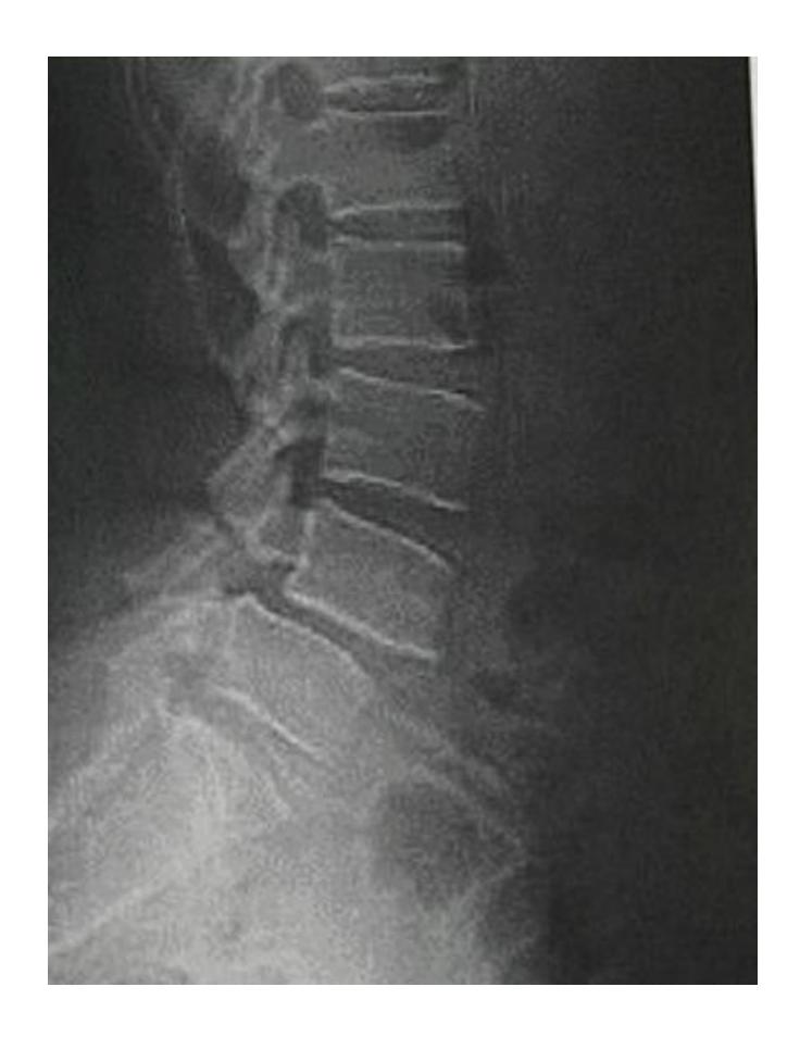
Spondylolisthesis
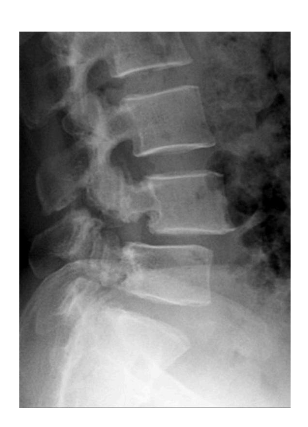
Lady came to the clinic complaining of long standing low back pain
Description Lateral x-ray of lumbar spine showing a fracture in the pedicle & slipped L4 over L5
Diagnosis: Spondylolisthesis
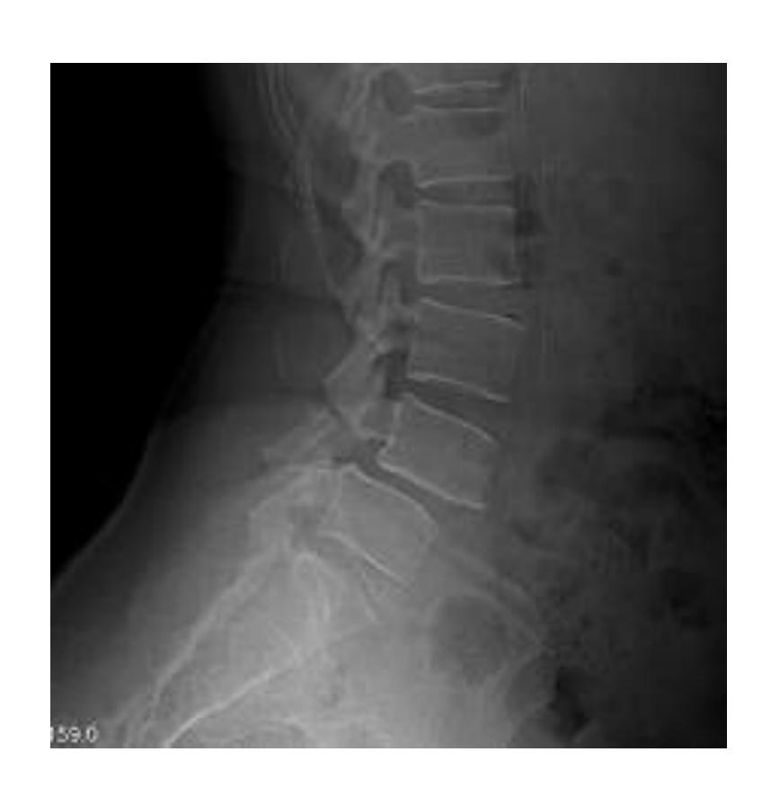
Diagnosis: Spondylolisthesis Next step? Oblique view - to see Scotty dog
Findings? X-ray of cervical spine should include all C vertebrae and T1. Anterior slippage of C4 over C5 and pars-interarticularis fracture (decapitated scotty dog)
Diagnosis? Pars interarticularis fracture (decapitated scotty dog)
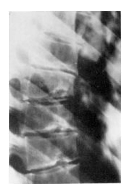
Anterior displacement of L4 over L5, pars interarticularis disruption, osteopenia
Nerve compression may cause inability to control bowel and bladder sphincter, chronic pain and numbness
Scheuermann’s Disease
Describe: Schmorl’s Node etc.
Diagnosis: Scheuermann’s Disease
Chronic Back Pain Cases
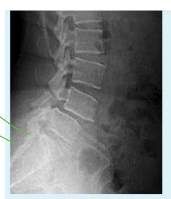
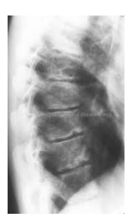
A 36-year old patient presented with low back pain for two years that has recently increased.
Q15: Write TWO abnormal findings you can see on the x-ray
- Slipped lumbar spine – over the other lumbar spine “Not sure what is the vertebrae number”
Q16: What is the most probable complication?
- Nerve root injury
Other Cases
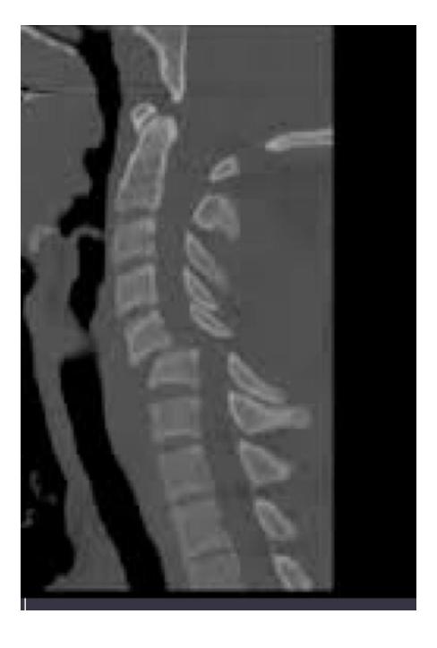
MRI of spine fracture at top of spine
 Cervical Facet Dislocations
Same as pic A but the choices didn’t have C2-C3 dislocation.
Cervical Facet Dislocations
Same as pic A but the choices didn’t have C2-C3 dislocation.
Young patient presents with limbing leg length discrepancy x-ray is shown. What is the diagnosis?
- Fibrous dysplasia
Segmental
Knee Examination and Pathology
Meniscal Injuries
Meniscal Tear Identification
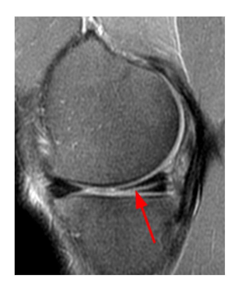
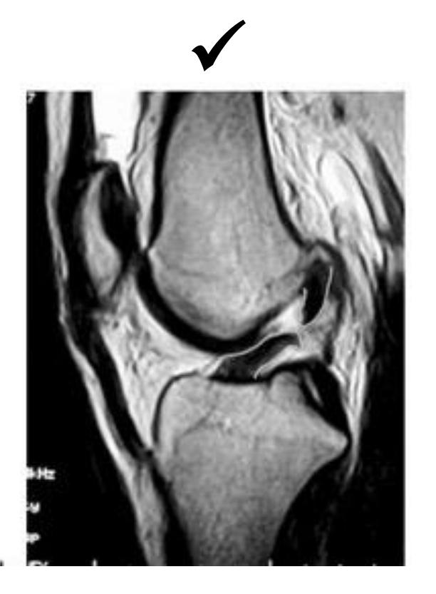
What pathology is seen in this MRI of an 18-year-old male who twisted his knee?
- Posterior horn meniscal tear
- Bucket-handle meniscal tear
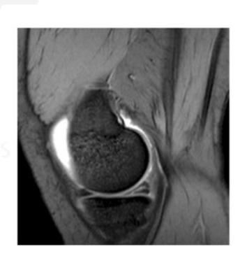
What is the significance of the double PCL sign?
- Indicates bucket-handle meniscal tear
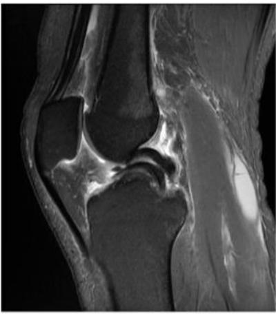
What does the white arrow indicate in this image?
- Posterior meniscus horn
What is the diagnosis shown in these images?
- Posterior meniscus horn tear
Clinical Correlation
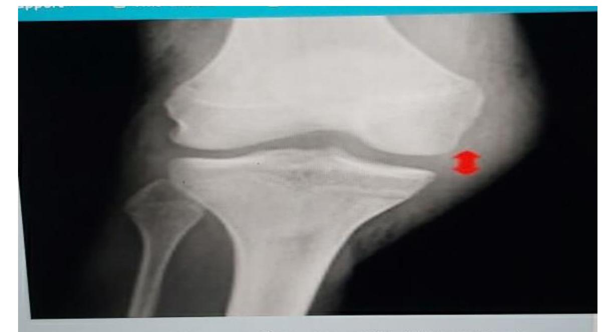
In an 18-year-old male who twisted his knee during sports, which physical finding is most likely to be found on MRI?
- Joint line tenderness
Cruciate Ligament Injuries
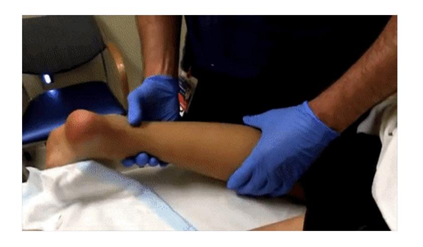
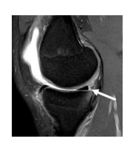
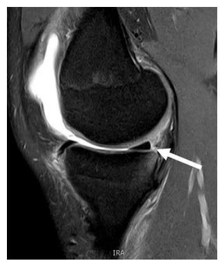
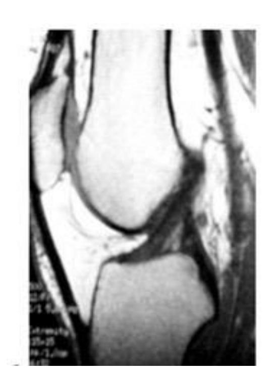
Posterior Cruciate Ligament (PCL)
What does this sign indicate?
- Posterior cruciate ligament injury

What is the clinical significance of the posterior sag sign?
- Seen in PCL injury
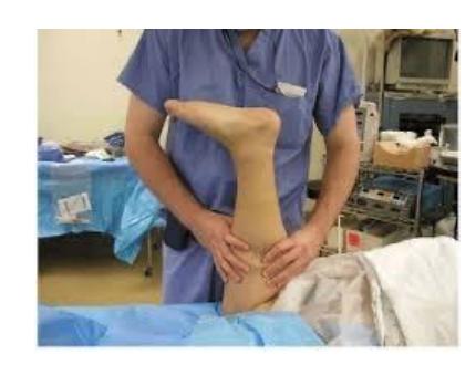
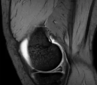
Anterior Cruciate Ligament (ACL)
What special test is being performed in this image?
- Anterior drawer test
What is the name of this test and what does it indicate when positive?
- Name: Lachman test
- Positive meaning: Tear (instability) of the ACL
What is the imaging modality and diagnosis?
- Modality: MRI
- Diagnosis: ACL tear
What finding is shown in this MRI?
-
Torn ACL
What special test would you perform during clinical examination to confirm ACL instability?
- Valgus stress test
- Most probable diagnosis: Tear (instability) of the ACL
Knee Dislocations and Traumatic Injuries
Knee Dislocation Cases
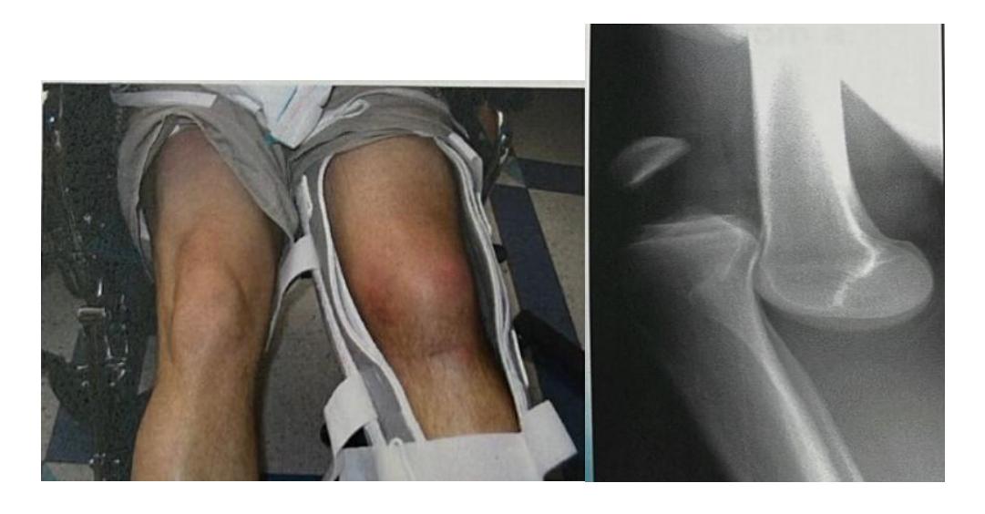
Case 1: 28-year-old parachute jumper
- Injury mechanism: Severe injury to left knee while landing in wrong position with force
What is the diagnosis?
- Anterior knee dislocation
What are TWO expected serious associated injuries?
- Popliteal artery injury
- Common peroneal nerve injury
Case 2: 27-year-old male who fell from first floor balcony
- Presentation: Pain and inability to move left knee
Special Clinical Tests
Patellofemoral Examination
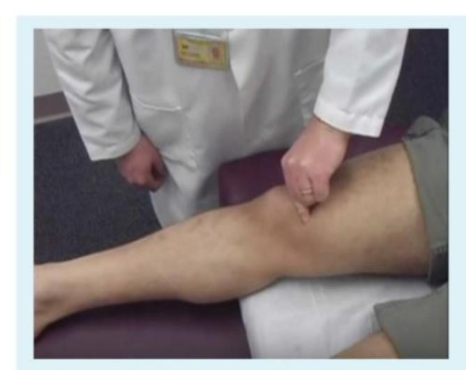
What test is being performed and what conditions does it diagnose?
- Test: Patellofemoral grind test (examiner resists patellar motion while quadriceps tightens)
- Diagnoses: Chondromalacia, osteoarthritis
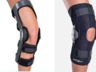
Collateral Ligament Testing
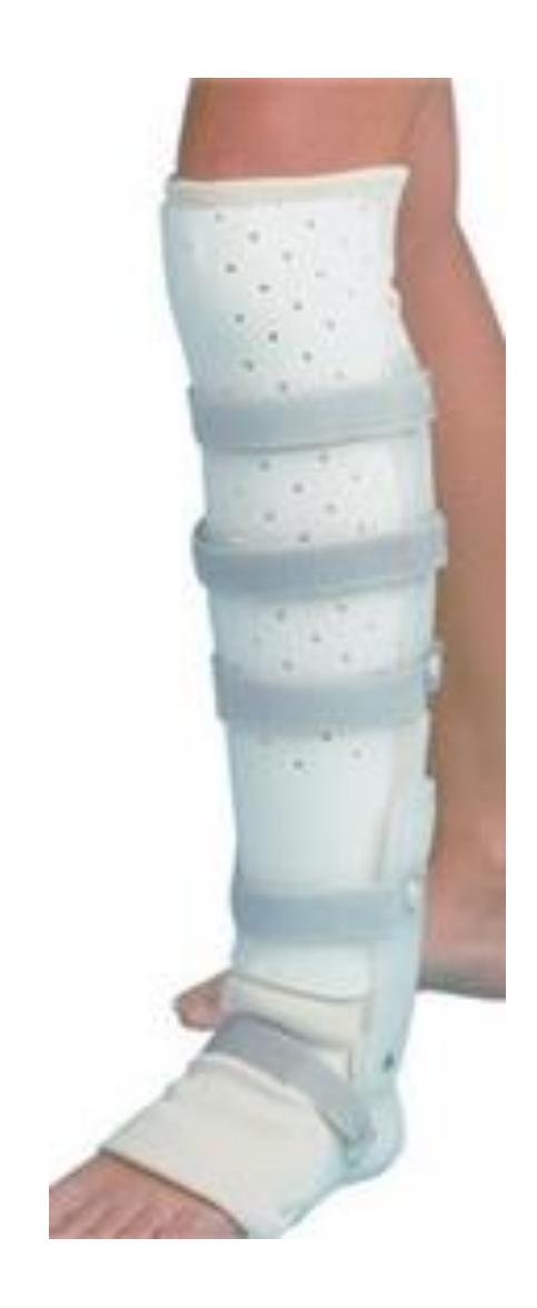
What would be indicated for a patient with potential collateral ligament injuries?
- Hinged knee splint
- Collateral ligament injuries
Pediatric and Growth-Related Conditions
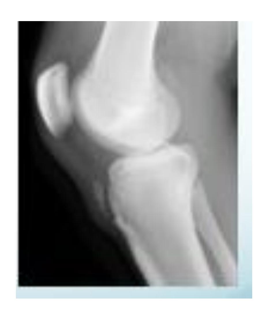
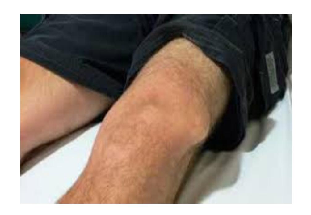
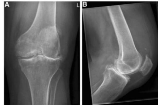
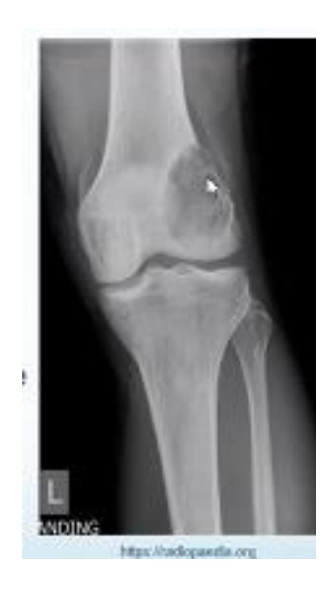
Osgood-Schlatter Disease
What is shown in this image and how should it be managed?
- Diagnosis: Osgood-Schlatter disease (tendon avulsion injury - partial avulsion)
- Characteristics: Not painful, might be tender
- Nature: Benign lesion
- Management: Ignore unless very tender and painful, reassure patient
- Treatment: Conservative
Video References
Lachman Test Demonstration
- Lachman Test Video
- Test name: Lachman test
- Structure tested: ACL
Acute Joint Dislocations
Shoulder Dislocations
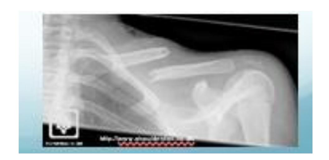
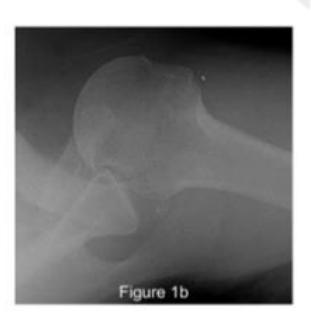
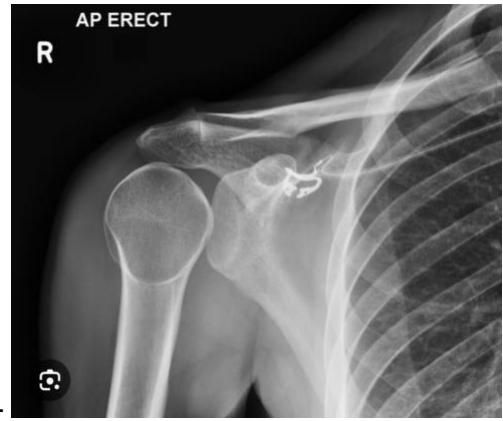
Anterior Shoulder Dislocation
What is the diagnosis shown in the image?
- Anterior Shoulder Dislocation
What is the most common complication of anterior shoulder dislocation?
- Axillary nerve injury
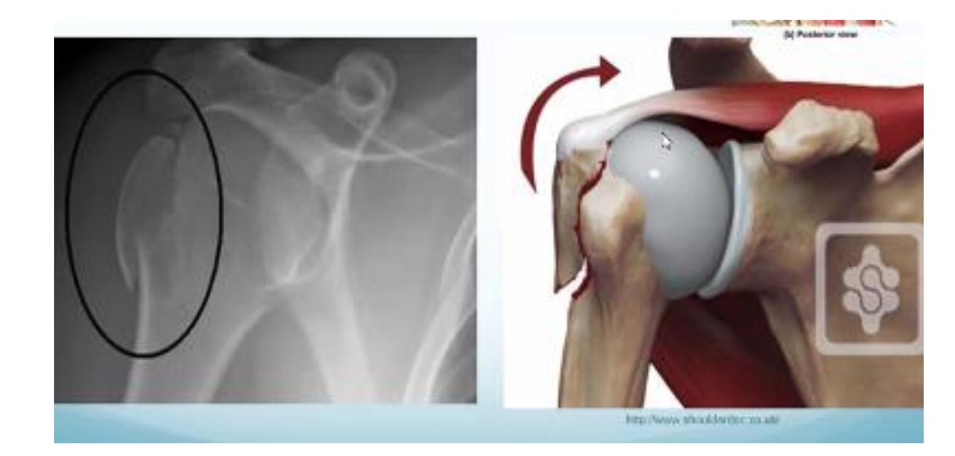
Hill-Sachs Lesion
What does this photo show?
- Hill-Sachs lesion
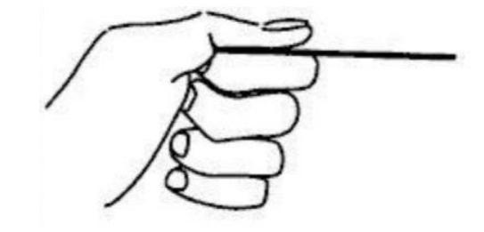
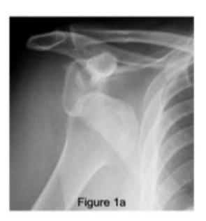
 18-year-old male presented after sport injury and unable to do internal rotation of right shoulder. What is the name of the injury seen in the figure?
- Hill-Sach’s defect
18-year-old male presented after sport injury and unable to internally rotate his left shoulder. What is the diagnosis?
- Hill-Sachs lesion
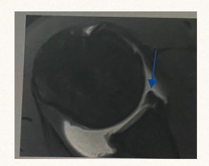
A case scenario about a male with anterior shoulder dislocation. What does the figure 1b show?
- Hill-Sachs lesion
reverse - Hill sachs lesion
What do you see in this image?
- Hill sachs lesion
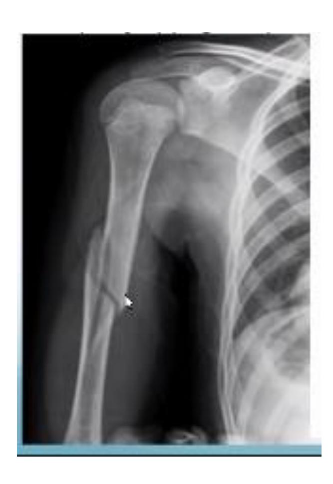
Associated Injuries
What is the associated injury in anterior shoulder dislocation?
- Lesser tuberosity fracture
18-year-old male presented after sport injury and unable to internally rotate his left shoulder. What is the likely finding?
- Lesser tuberosity fracture
Hip Dislocations
Posterior Hip Dislocation
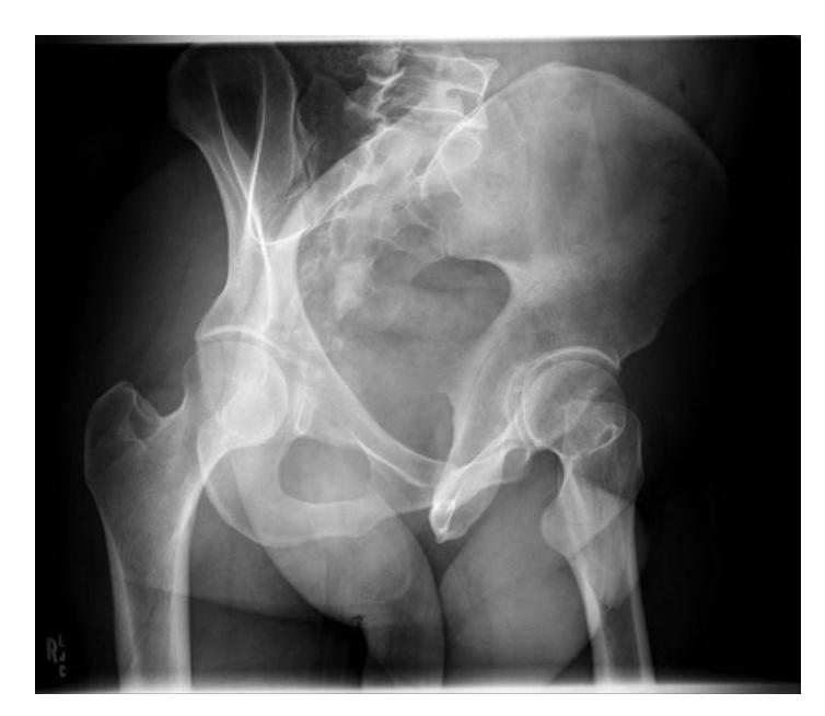
A 22-year-old man involved in RTA presented to emergency room with pain at left hip, the lower limb was adducted and internally rotated. What is the diagnosis?
- Posterior hip dislocation
What are TWO common complications of posterior hip dislocation?
- Sciatic nerve injury
- Avascular necrosis of femoral head
Posterior dislocation of the right hip position shows?
- Internal rotation of the lower limb
Knee Dislocations
Patellar Dislocation
What is the diagnosis?
- Lateral patellar dislocation
What is the treatment?
- Reduction & immobilization in above knee cast
Ankle Injuries
Syndesmotic Injury
A 23-year-old male injured his ankle after a twisting ankle injury 2 days back. What is the most likely pathology based on the shown x-ray?
- Syndesmotic ligament injury
Chronic Shoulder, Hand & Foot Disorders
Shoulder Disorders
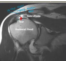
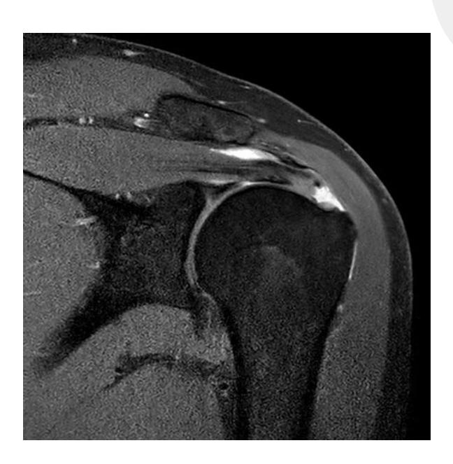
Q21 - What causes pain during overhead activities when MRI shows rotator cuff muscle tear?
- Subscapularis and infraspinatus muscle tears
- Note: Supraspinatus was not included in the answer choices
Q34 - What impingement syndrome is most likely associated with abduction and external rotation?
- Impingement during overhead activities
Q35 - What condition is associated with a prominent coracoid process?
- Prominent coracoid process
Question 56 - A 45-year-old male with anterior shoulder dislocation. Which injury is demonstrated in the attached MRI?
- d. Supraspinatus tear
Foot Disorders
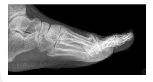
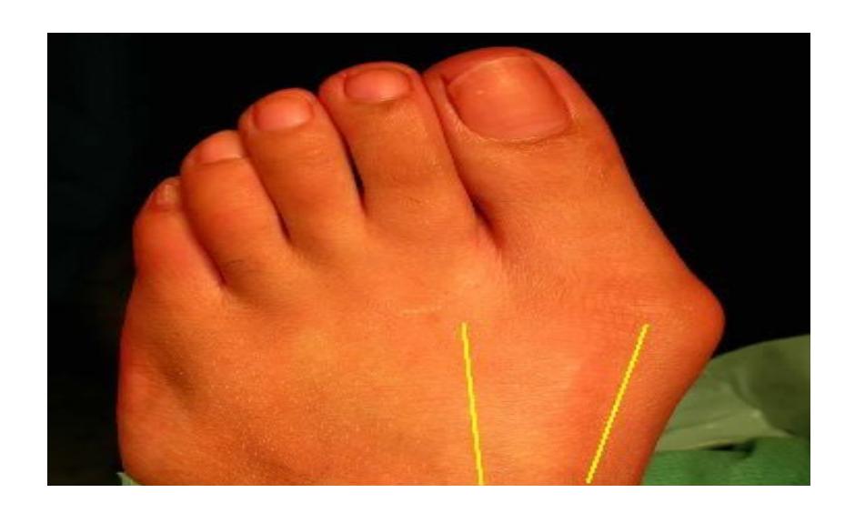
Q31 - In a foot X-ray showing possible hallux valgus, which measurement is abnormal?
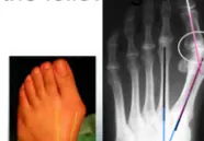
- Lateral deviation of intermetatarsal angle by 14°
Q32 - In a case of painful bunion, what deformity is observed in the big toe?
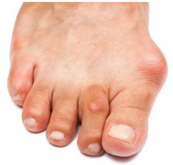
- Pronation of the great toe + lateral deviation (valgus)
Q5 - A 55-year-old patient has significant foot pain. What pathology is shown on the X-ray?
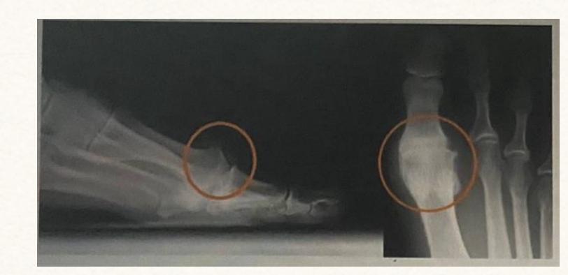
- Hallux rigidus
Elderly patient foot pain diagnosis
- Diagnosis: Hallux rigidus
Question 20 - An elderly patient came with foot pain. What is the diagnosis?
- Hallux rigidus
Key Points Summary
Hallux Rigidus
- Arthritic condition affecting the big toe joint
- Causes stiffness and pain in the great toe
- Characterized by osteophyte formation and joint space narrowing on X-ray
Hallux Valgus (Bunion)
- Lateral deviation of the great toe
- Associated with increased intermetatarsal angle (>14°)
- Often accompanied by pronation of the great toe
Rotator Cuff Pathology
- Common cause of shoulder pain during overhead activities
- May involve tears in subscapularis, infraspinatus, or supraspinatus muscles
- Diagnosis confirmed with MRI imaging
Shoulder Disorders
Question: A case scenario that was suggestive of an anterior shoulder dislocation in a 45-year-old male. Which of the following injuries is demonstrated in the attached MRI?
- Answer: Supraspinatus tear
Hand and Finger Disorders
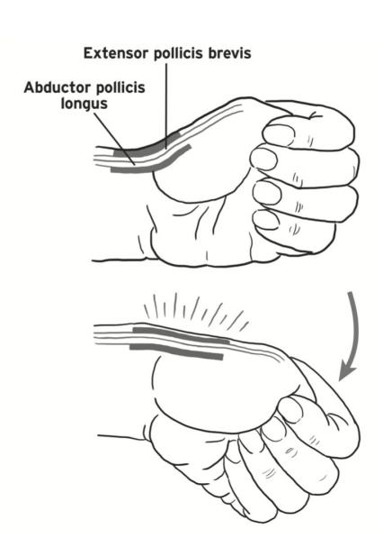
Question: De Quervain’s Tenosynovitis is known as the inflammation of which extensor compartment of the hand? Question: This test is used to assess tendonitis in which compartment?
- Answer: 1st compartment
Question: What is the name of the deformity in the distal interphalangeal joints and what is the cause?
- Answer: Heberden’s nodes
- Cause: Osteophytes in case of osteoarthritis
Elbow Disorders
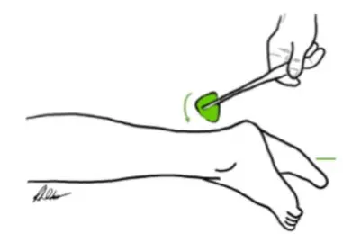
Question: A 30-year-old carpenter presented with pain at the outer aspect of his elbow since 4 weeks. Clinically he has pain on resisted wrist extension. Local steroid injection was decided.
What is the lesion pathology located?
- Lateral epicondyle (origin of wrist extensor muscles) - tennis elbow
Where do you have to inject exactly?
- Distal to the epicondyle at maximum area of tenderness

Hip Disorders
Question: A 40-year-old female reports a painful snapping in her hip in which she’s able to hear it. The snapping is associated with a clicking sensation. She’s been on conservative treatment for 5 years with no signs of improvement. Which of the following is the treatment of choice for her condition?
- Answer: Arthroscopic debridement for synovial chondromatosis
Foot Disorders
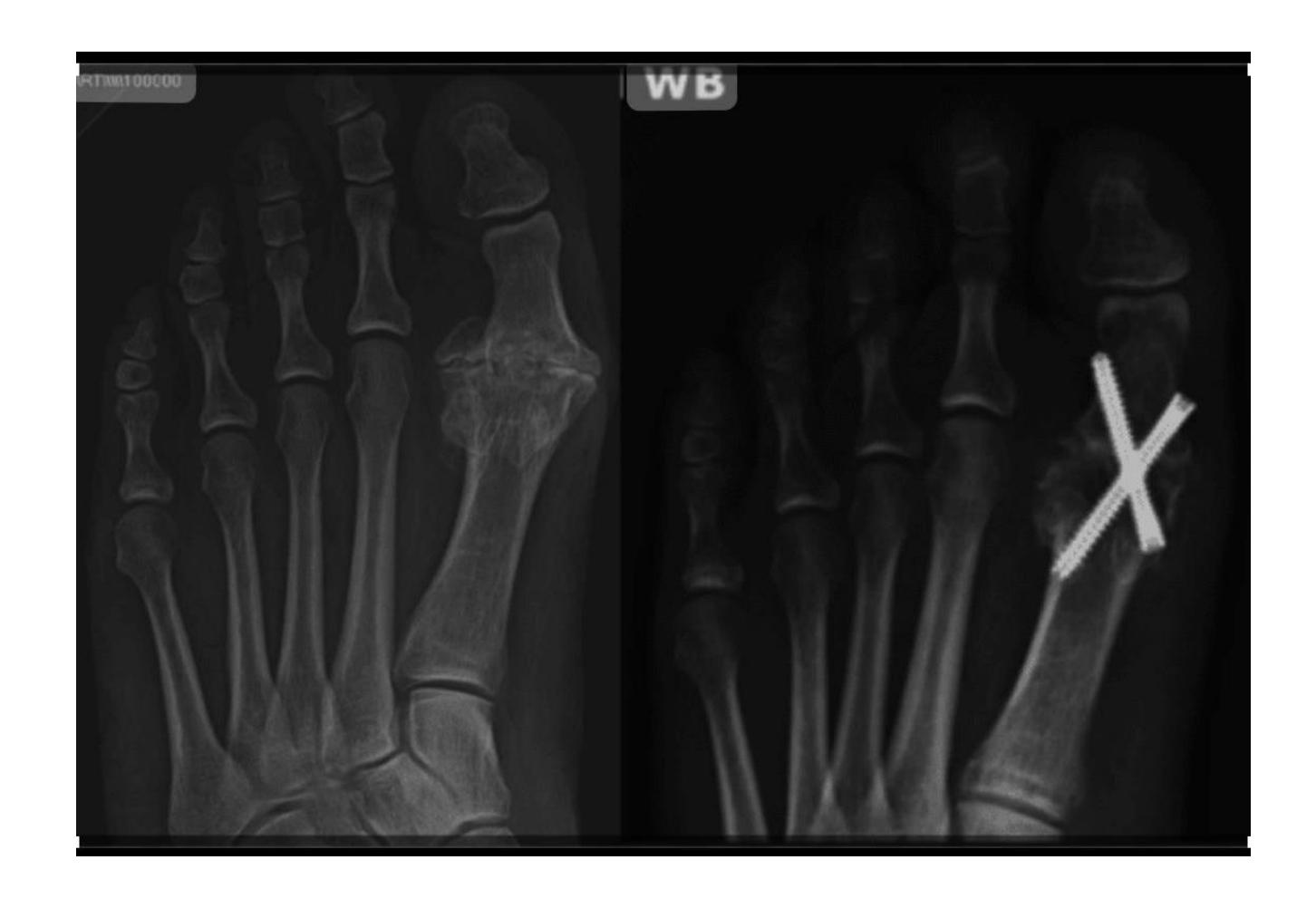
Question 1: What is the deformity called in this picture?
- Answer: Hallux Valgus
Question 2: Patient complaint of severe pain and it was inflamed, what is the possible treatment?
- Answer: Surgery (Re-align 1st metatarsal, correct valgus deformity of big toe, and soft tissue balancing)
X-rays
Introduction
Rheumatology-related Imaging
Gout
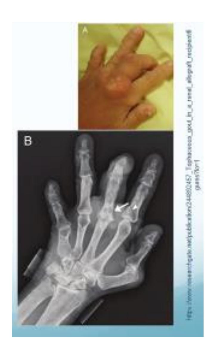
- Tophus formation visible on radiograph
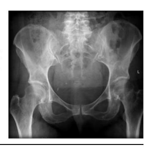
Hip and Pelvis
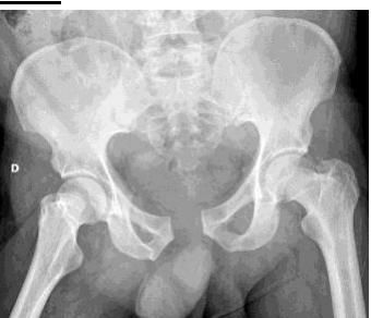
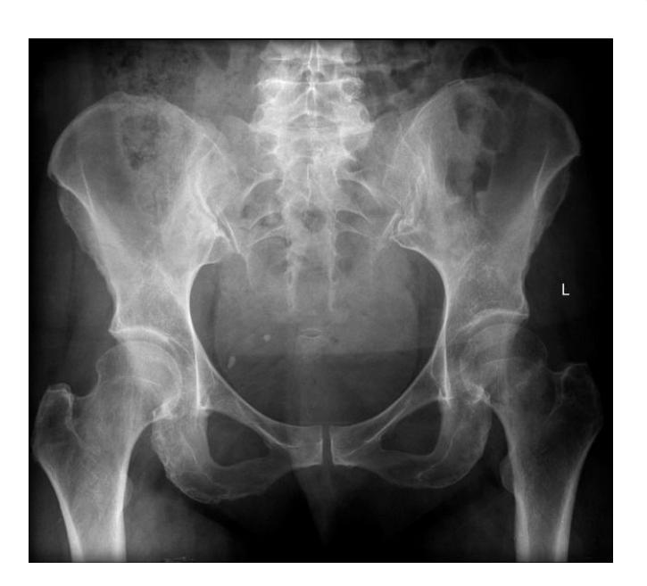
Bilateral Neck of Femur Fracture

Findings:
- Adult patient
- Osteoporotic bone changes
- Bilateral fracture neck of femur
Recommended additional imaging:
- CT scan for further evaluation
Knee and Patella
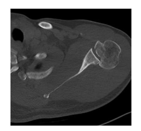
Patellar Subluxation
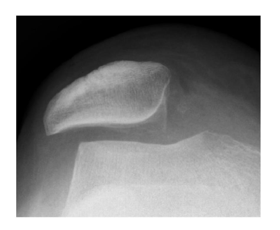
Findings:
- Skyline view of the patella
- Subluxation of patella with trochlear dysplasia
Advanced Imaging
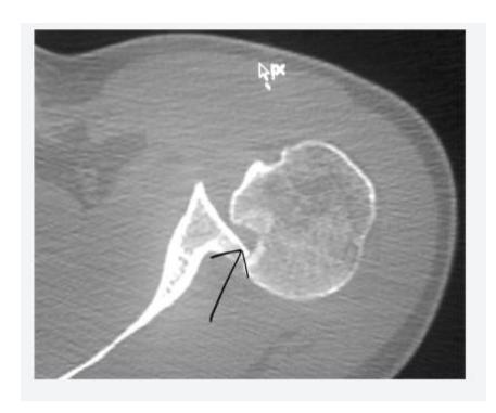
CT Scan for Joint Assessment
Clinical application:
- CT scan is essential to assess the joint line before starting the management plan
- Provides detailed anatomical information for surgical planning
Peripheral Nerve
Posterior Interosseous Nerve Questions
Which nerve is responsible for this action?
- Posterior interosseous nerve

If a patient is unable to perform this sign, which nerve is affected?
- Posterior interosseous nerve
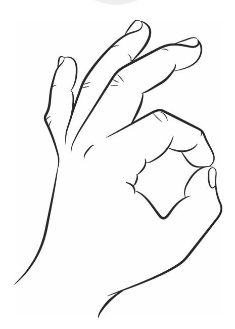
Anterior Interosseous Nerve Questions
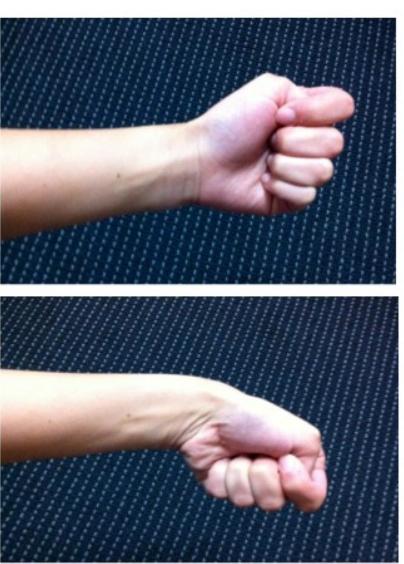
Which of the following nerves is responsible for this action? (Ok sign)
- Anterior interosseous nerve

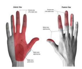
Which nerve is shown in this image?
- Median nerve
Nerve Root Injuries
What nerve root injury is present in Erb’s palsy?
- C5, C6
A weakness in elbow flexion. The herniation would be in?
- C5, C6
Dermatomes and Myotomes
What’s the dermatome at the level of the umbilicus?
- T10
What’s the myotome of the shown structure (Big toe)?
- L5
Which nerve root is responsible for the reflex of the shown structure (Achilles tendon)?
- S1
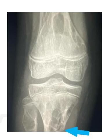
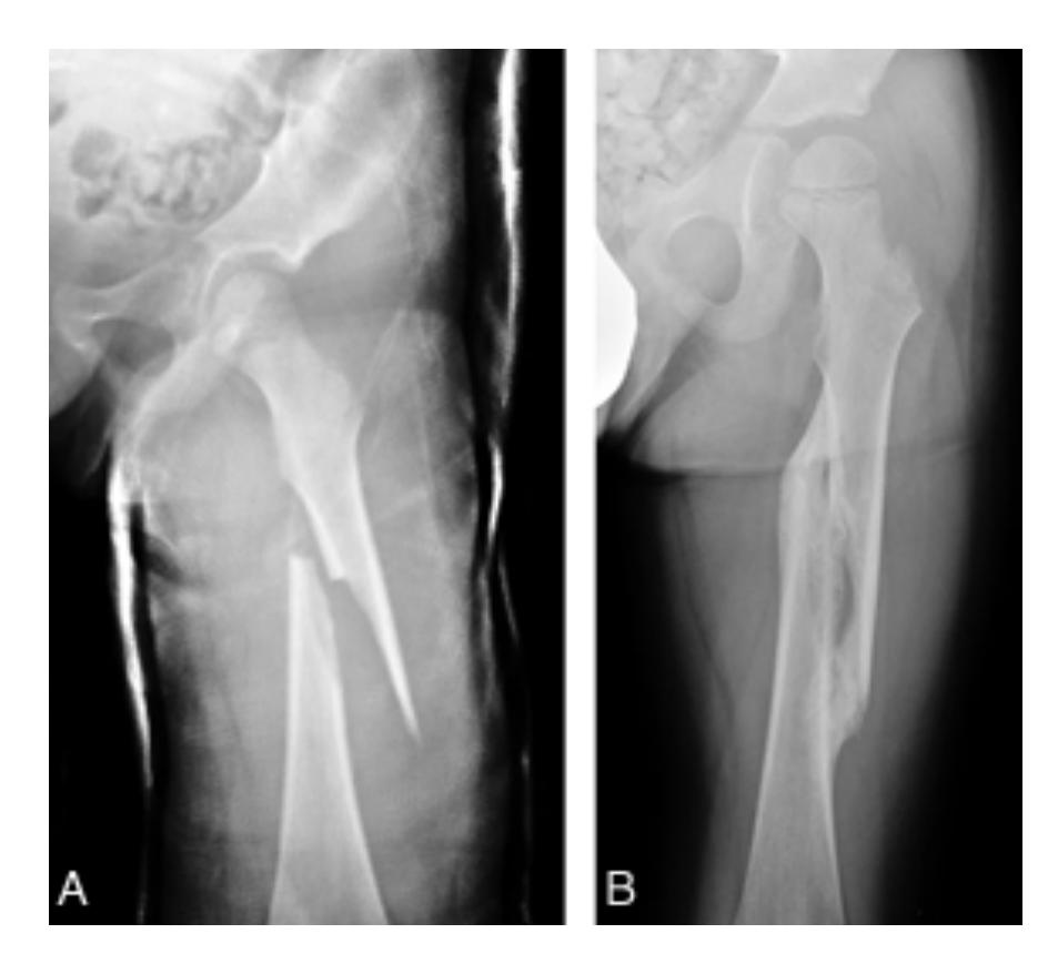
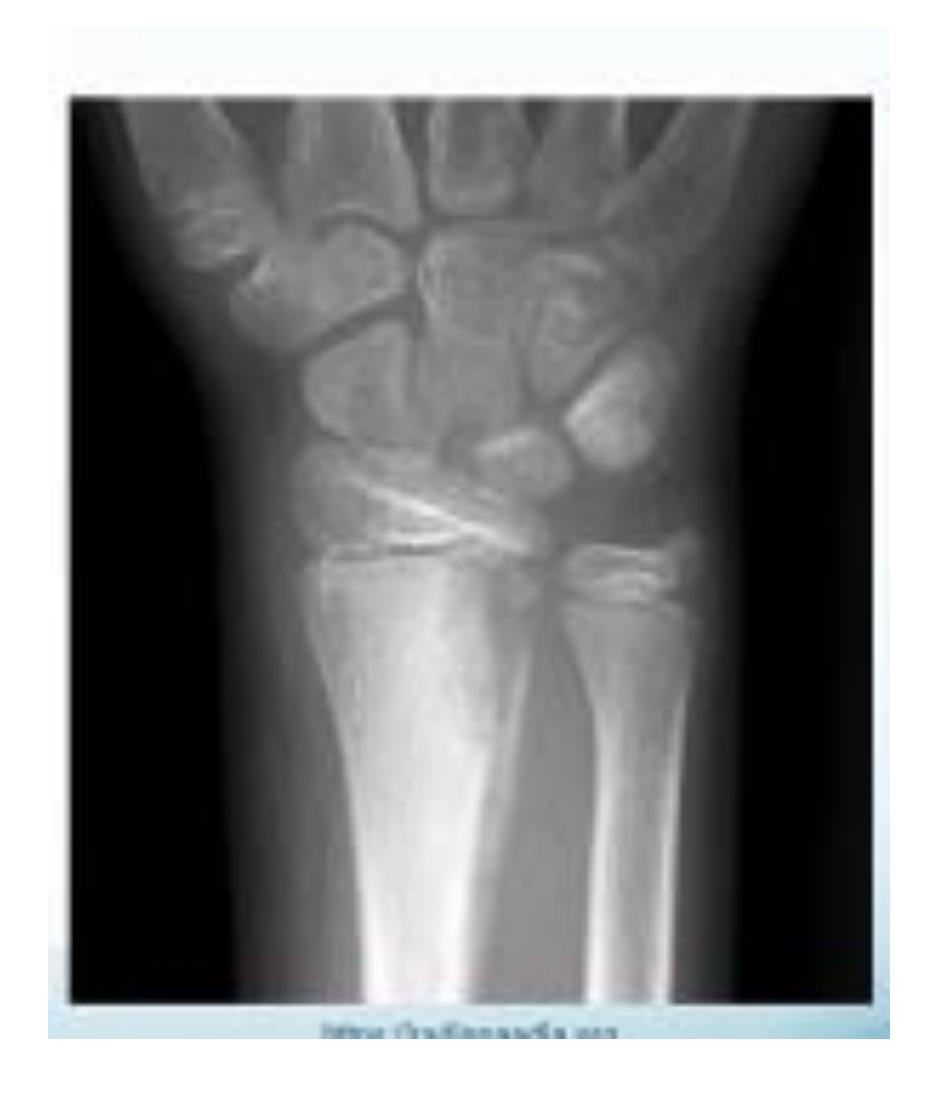
Pediatric Fractures
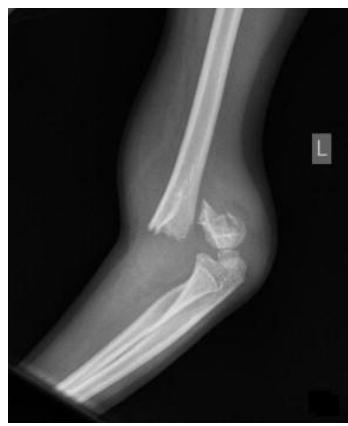
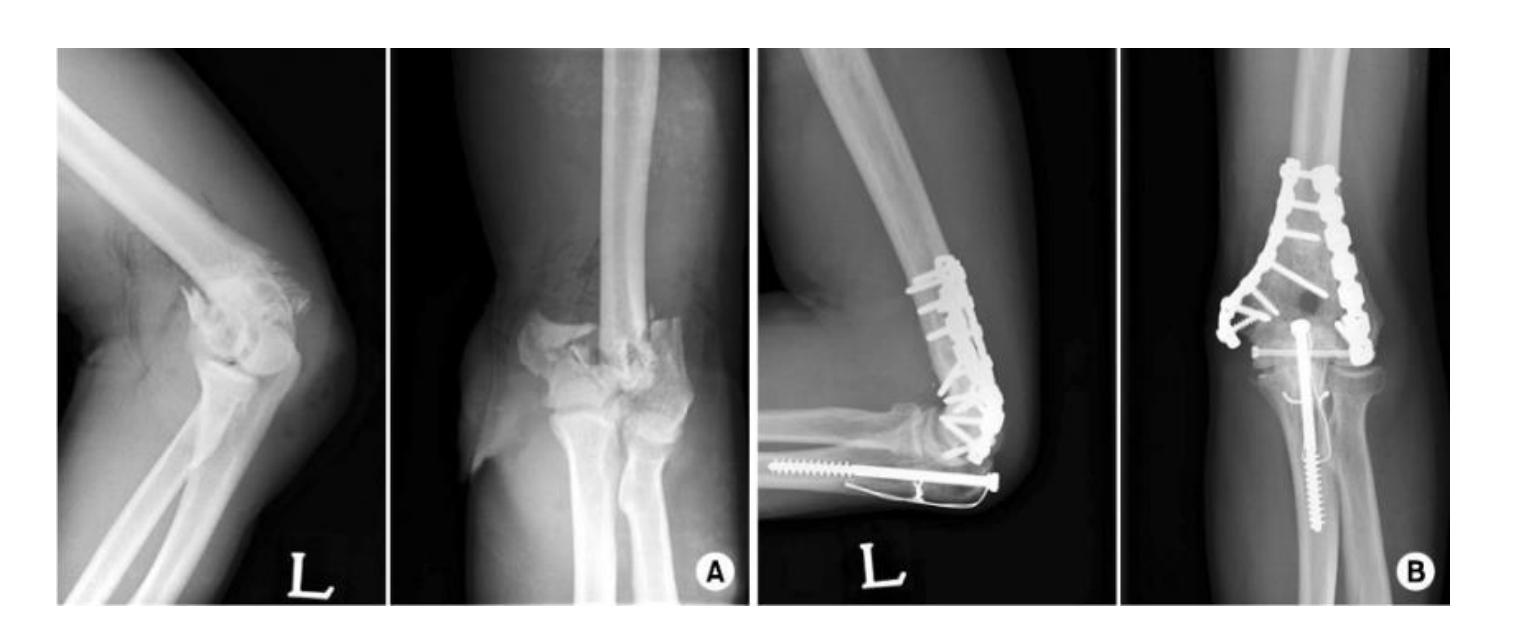
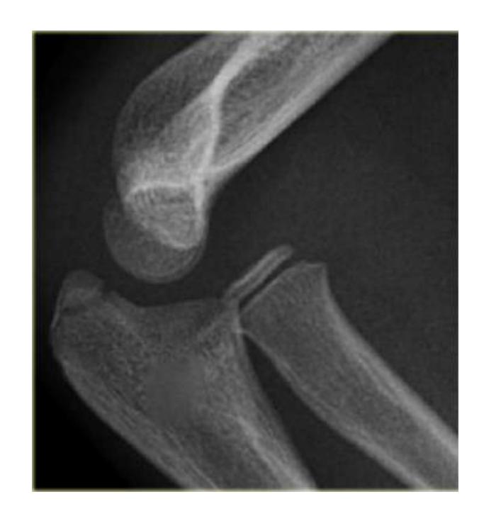
Supracondylar Fractures
Clinical Presentation
- What is the characteristic finding in supracondylar fractures? Supracondylar fracture in children with fat pad sign
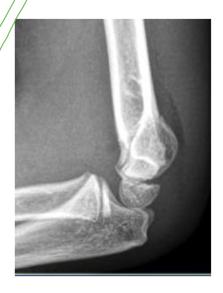
Treatment Protocol
- What are the treatment options for supracondylar fractures?
- If not displaced: Back slab above elbow (flexed elbow)
- If displaced: Closed reduction and fixation with K-wires and slab
Emergency Room Management: Back slab with flexion in 30 degrees and send for AP and L x-rays
Radial Head Dislocation
Clinical Diagnosis
- How is radial head dislocation diagnosed?
Possible dislocation of the head of radius
- Draw a line from the neck of the radius - it won’t pass through the capitellum center
Bucket-Handle Fracture
Clinical Features
- What is a bucket-handle fracture? Twisting injury
- What is the clinical significance of bucket-handle fractures? Non-accidental injury (child abuse)
Management Protocol
- What is the immediate management priority? Inform the social worker is the first thing to do
Pediatric Deformity
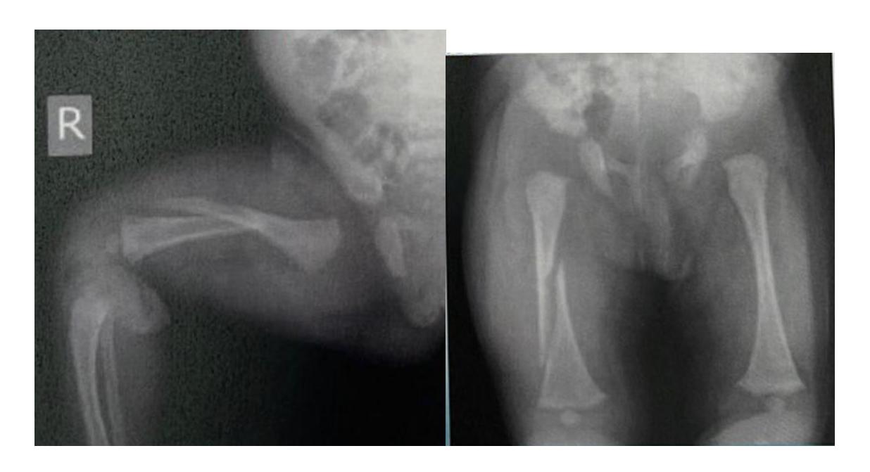
SCFE (Slipped Capital Femoral Epiphysis)
Case: A child was stated to be stuck in external rotation and abduction, with an X-ray shown.
CTEV (Clubfoot) Deformities
Varus Hindfoot Deformity
Q1: What is the deformity showing in this CTEV patient?
- C. Varus hindfoot
Q2: What is the deformity in the photo in this CTEV patient?
- C. Varus midfoot or hindfoot
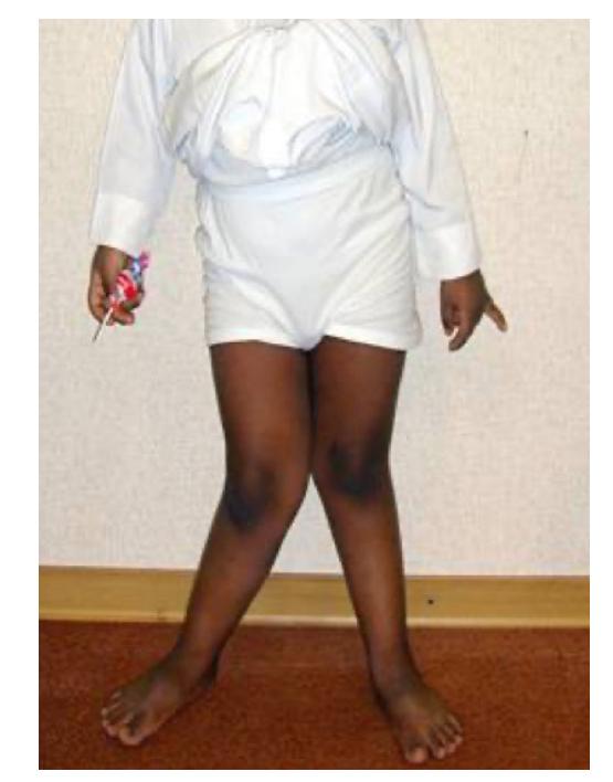
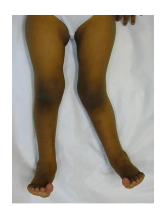
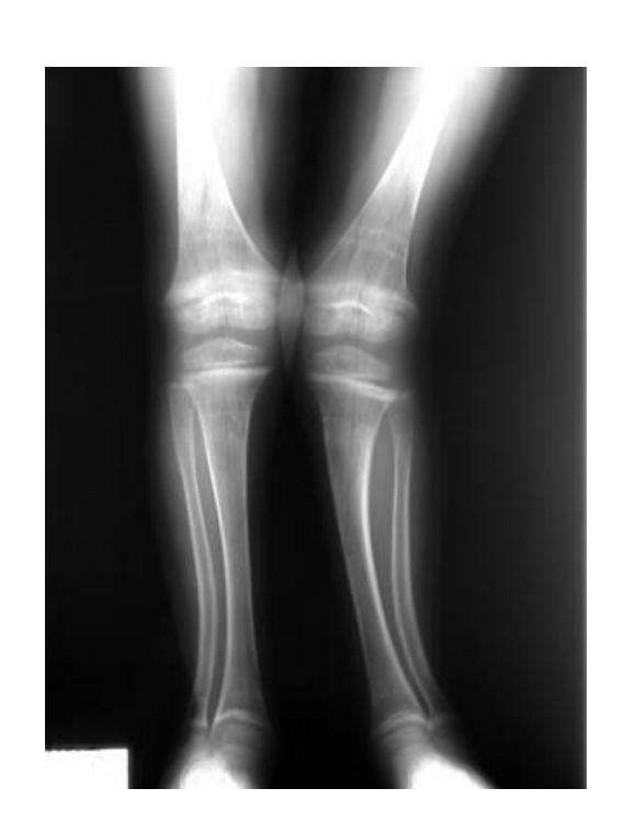
Genu Valgum
Q1: What do you see in this image?
- A. Genu valgum, Coronal view
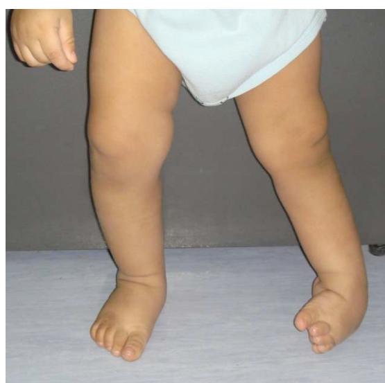
Medial Femoral Torsion (Anteversion)
Clinical Case
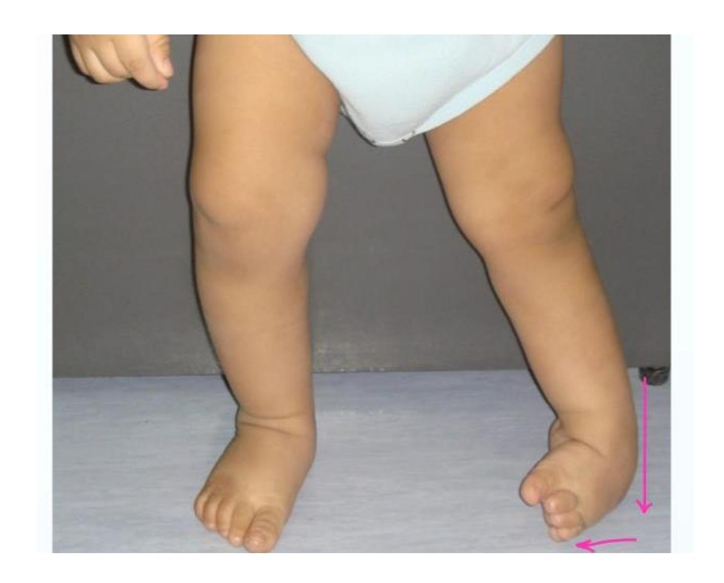
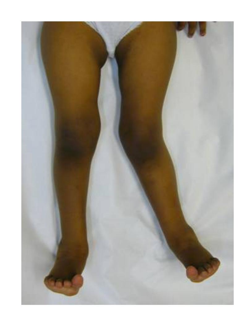
Patient: A 6-year-old girl, brought by her mother to the clinic complaining of awkward walking for 4 years.
Q1: Describe what each clinical picture shows
- Patient stands with medial rotation of the femur (“kissing patella”)
Q2: At what level is the deformity occurring?
- Medial femoral torsion (“Anteversion”)
- Starts at the age of 3 years and peaks at 4-6 years
Finding:
- Stands with knees medially rotated (kissing patellae)
- Sits in “W” position
- Runs awkwardly (egg-beater)
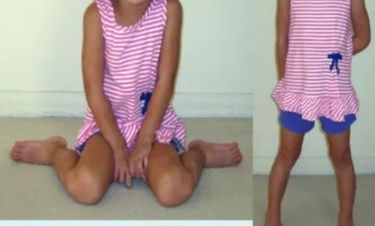
Asymmetrical Deformities
Diagnosis: Asymmetrical deformity
Pediatric Neuromuscular Disorders
Clinical Case Presentation
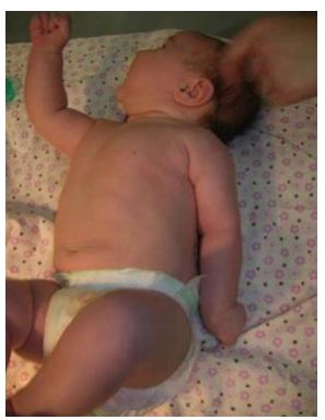
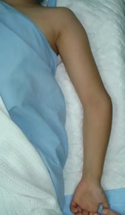
You were called to the nursery department to examine a newborn baby not moving his upper limb.
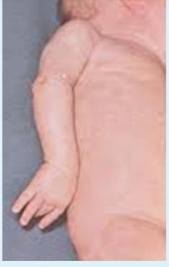
Diagnosis
Erb’s Palsy (Neonatal Brachial Plexus Injury)
Affected Muscle Groups
- Shoulder abductors
- External rotators
- Biceps
Clinical Features
Erb’s palsy is a type of neonatal brachial plexus injury that affects the upper trunk (C5-C6 nerve roots), resulting in weakness or paralysis of the shoulder and elbow muscles while preserving wrist and hand function.
Pediatric Congenital
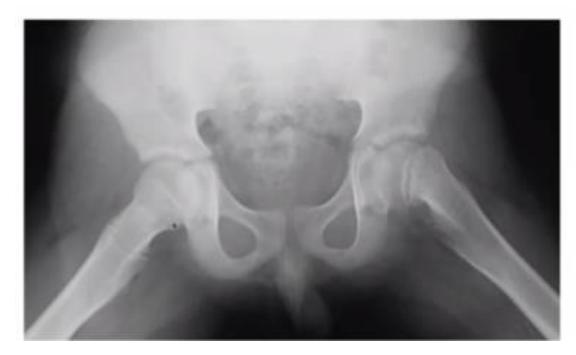
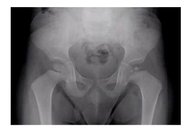
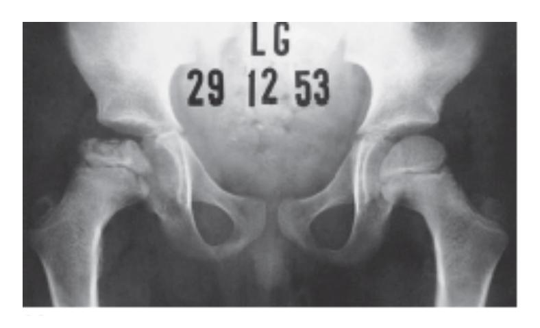
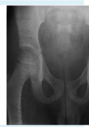
Developmental Dysplasia of the Hip (DDH)
Radiological Findings
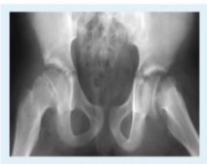
What are the four main radiological findings in DDH?
- Acetabular index more than 25°
- Broken Shenton’s line
- Smaller size of femoral head ossification center
- Lateralization of femoral head to perpendicular line
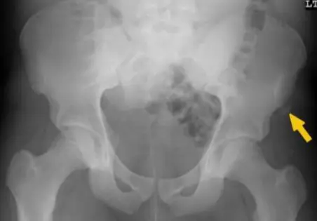
A case scenario that was suggestive of developmental dysplasia of the hip in an 18-month-old girl. Which of the following radiological signs represents this case?
- Wide acetabular index (also referred to as increased acetabular index)
Clinical Cases
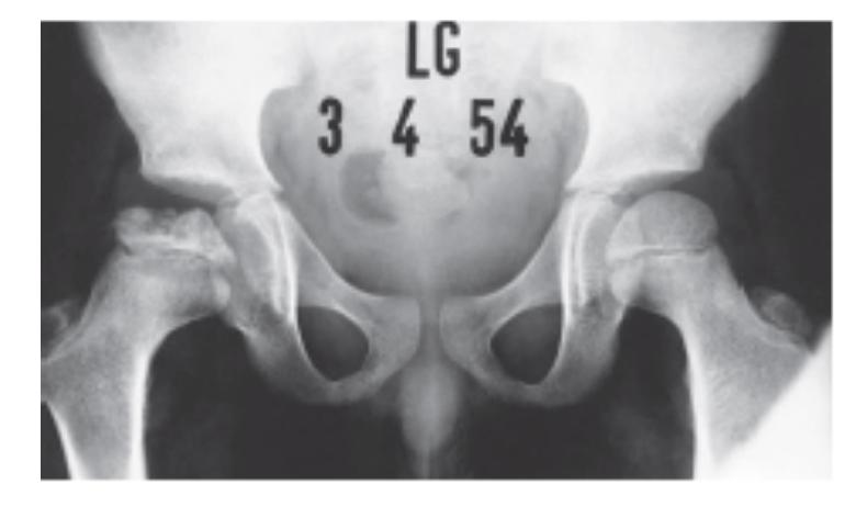
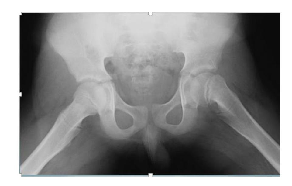
A 15-month-old child was brought to the clinic because of painless limping. There is no history of trauma. The pelvic x-ray is shown.
Q1: Write two abnormalities seen on the X-ray.
- Lateralization of the ossifying centre
- Acetabular angle more than 27°
- Broken Shenton line
- Shortening of the line from greater trochanter to the horizontal line
Q2: What is the most probable diagnosis?
- Developmental dysplasia of the hip (DDH)
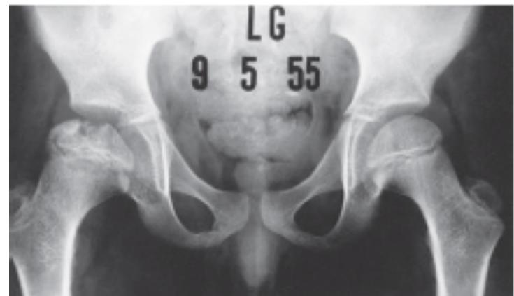
10 months old child presenting with this condition:
- Right DDH

Physical Examination
Q11/ The test that is shown in the following picture is used in which of the following conditions?
- Developmental hip dysplasia
 !
!
Perthes Disease (Legg-Calve-Perthes)
Clinical Presentation
Q4/ 7 years old boy, noticed by his parents that he had limping over last 3 months. x-ray obtained. What’s the diagnosis?
- Perthe’s disease
Diagnosis: Legg-Calve-Perthe’s Disease
- Collapse – Fragmentation - flattening (coxa plana)
A 7 year old boy presented to emergency room with painful limping right lower limb. Clinically he has limitation of abduction and internal rotation movement at his right hip.
Q1: Mention two abnormal findings:
- Collapse and fragmentation of epiphysis
- Sclerosis
Q2: What is the most probable diagnosis?
- Legg-Calve-Perthe’s disease
X-ray findings in Perthes disease:
- Fragmentation and collapsed epiphysis
- Sclerosis
- May initially look normal in x-ray
Slipped Capital Femoral Epiphysis (SCFE)
Clinical Presentation
A 13-year-old overweight boy presented to the clinic with pain in his left thigh and knee for one month. He was limping. There was no swelling and no tenderness in the thigh and knee. The hip was held in external rotation and showed limitation of internal rotation.
Q1. What is the diagnosis?
- Slipped capital femoral epiphysis (SCFE)
Q2. What is the treatment of choice?
- Fixation in situ without manipulation, and fix the other limb because there is 1/3 of cases have recurrence in the other joint
14 years old boy, limping, obese, hypogonadism
X-ray: AP and Frog lateral
Presentation: Acute: painful, Chronic: painless
Diagnosis: Slipped Capital Femoral Epiphysis (SCFE)
Signs:
- Hip externally rotates when flexed
- Limited internal rotation
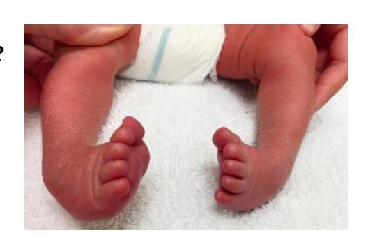
Clubfoot (Congenital Talipes Equinovarus - CTEV)
Clinical Presentation
What is the diagnosis?
- Congenital Talipes Equino Varus (CTEV)
Which of the following is the initial management of this case?
- Serial casting for clubfoot
Treatment:
- Ponseti method of serial casting for 4-6 weeks followed by tenotomy of Achilles tendon
- If failed >> surgical release


Other Pediatric Conditions
 Cubitus varus
Talipes Calcaneovalgus
Q.1: What is the deformity called in this picture?
- Talipes Calcaneovalgus
Q.2: This deformity is associated with?
- DDH
Diagnosis: Talipes Calcaneovalgus
Pediatric Fractures
An 8-month-old girl was brought by her mother with history of falling down the stairs while carrying her baby 2 days before. She noticed that her baby is not moving her right lower limb since then, with pain on changing diapers.
Q1: How would you treat this fracture?
- Conservative treatment with hip spica
Q2: What is a major concern in such a case?
- Non-accidental injury
Q.3: What should you consider in this case?
- Child abuse

An 8-month-old boy was irritable. During assessment, he was afebrile with restricted motion due to pain. X-ray flms were obtained. What is the next step of management?
B. Prepare for admission
Pediatric Deformities
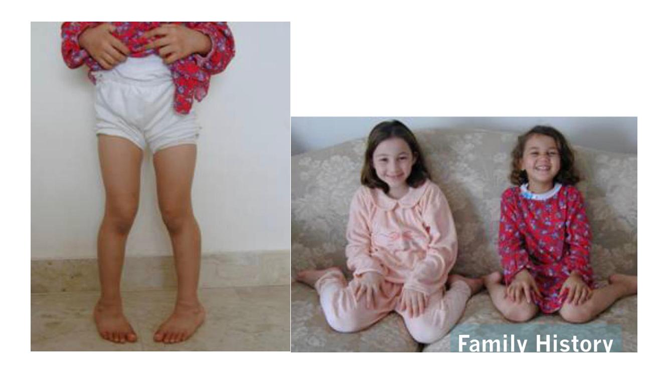
Unilateral genu valgum of the left knee in a child
Most common cause?
- A previous fracture to the distal femur with malunion
Additional Resources
Video Reference: https://youtu.be/kZm5VEHKMPQ
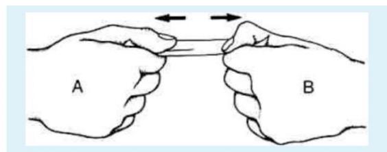
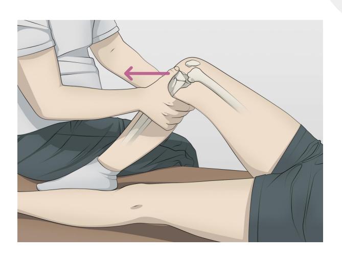
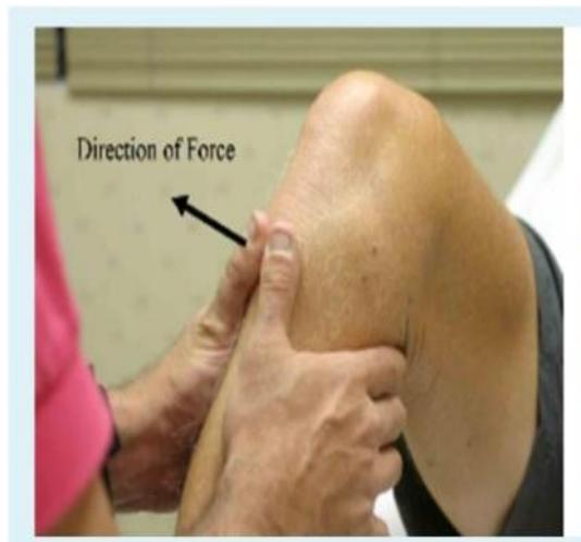
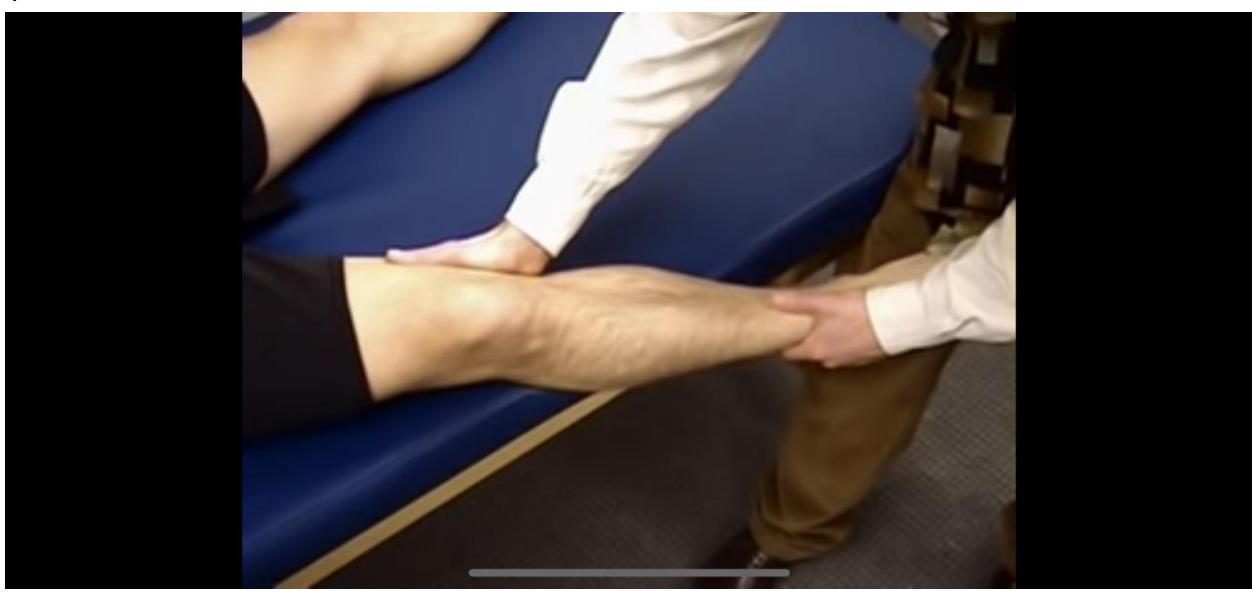
A- name of the test: varus stress test B- structure: LCL
Limping and Gait Analysis
Trendelenburg Gait Pattern
Visual Assessment
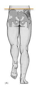
What is the Trendelenburg sign?
The Trendelenburg sign is characterized by:
- Contralateral pelvis drop (the pelvis on the opposite side drops downward)
- Ipsilateral shoulder lean (the shoulder on the affected side leans toward the involved hip)
Clinical Significance
The Trendelenburg sign indicates:
- Weakness of the hip abductor muscles, primarily the gluteus medius and gluteus minimus
- Inability to maintain pelvic level during single-leg stance
- Commonly associated with conditions affecting the hip joint or surrounding musculature
Underlying Conditions
- Hip osteoarthritis
- Hip dysplasia
- Gluteal muscle weakness or denervation
- Greater trochanteric pain syndrome
- Post-hip surgery or trauma
Assessment Notes
During gait assessment, observe for:
- Pelvic drop during stance phase
- Lateral trunk lean toward the affected side
- Compensatory mechanisms to maintain balance
- Severity often correlates with degree of muscle weakness
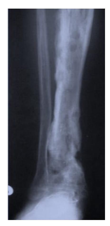
Osteomyelitis
Clinical Cases and Questions
What anatomical structure is indicated by the blue arrow in a patient with chronic osteomyelitis?

- a. Involucrum
- b. Sequestrum
16- The blue arrow show?
A. Involucrum
What is the appropriate treatment for subacute osteomyelitis? (2-month history of knee pain with limping and occasional swelling)

- IV antibiotics and surgical drainage
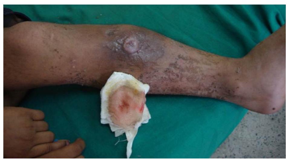
What is the most likely diagnosis for a patient with long history of knee pain and a sinus draining pus?
- Chronic necrotizing osteomyelitis
What are the characteristic findings and diagnosis for a 7-year-old with painful swollen lower thigh and recurrent fever? 
Q1: Abnormal x-ray findings:
- Cavitation
- Periosteal reaction
- Involucrum
- Sequestration
Q2: Diagnosis:
- Chronic osteomyelitis
What is the diagnosis for this acute osteomyelitis case?
- Acute osteomyelitis
What neonatal infection source can cause osteomyelitis?
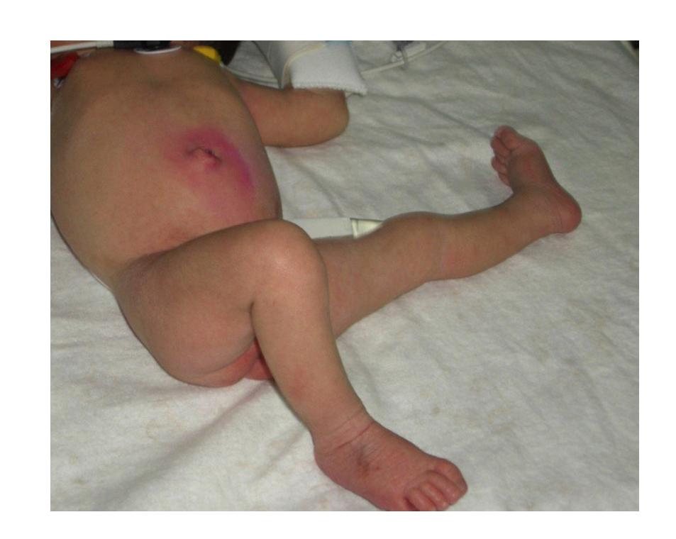
- A source of infection at the umbilicus
- Could cause osteomyelitis in neonates
Acute Osteomyelitis
Clinical Features
- Fever
- Tenderness
- Acute onset
- Often history of trauma or infection source
Diagnosis
- Clinical presentation
- X-ray findings (may be normal early)
- Laboratory studies
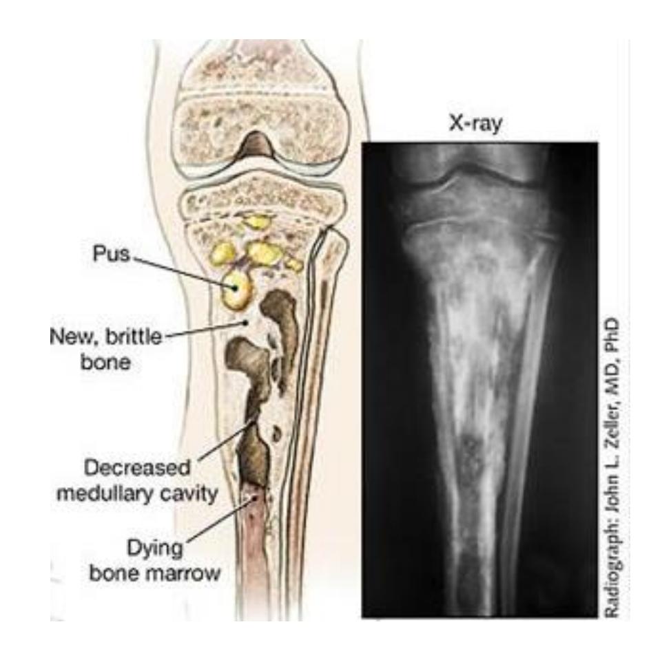
Chronic Osteomyelitis
Radiological Features
- Rarefaction surrounded by sclerosis
- Cavities and sequestra
Key Pathological Features
- Sequestrum: Dead bone fragment that becomes separated from healthy bone
- Involucrum: New bone formation surrounding the sequestrum
- Cavitation: Bone destruction forming cavities
- Sinus formation: Draining tracts to skin surface
Differential Diagnosis
- Tumor
- Infection
Treatment
Medical Management
- Antibiotics (IV initially, then oral based on cultures)
Surgical Management
- Sequestrectomy - removal of dead bone fragments
- Surgical debridement - removal of infected tissue
- May use local antibiotic beads for targeted delivery
Diagnostic Tools
- Sinogram - to show extent of sinus tracts
- Imaging studies (X-ray, CT, MRI)
Clinical Features for Diagnosis
- History: fever, tenderness
- Physical: swelling, warmth, draining sinuses (chronic)
- Laboratory: elevated inflammatory markers
Osteo Infections (Osteomyelitis, Septic Arthritis, TB)
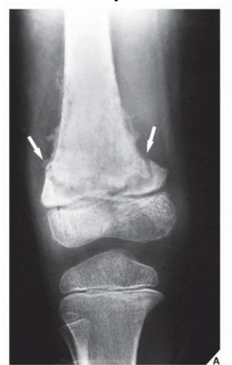
Q8/ An adult male presented to the clinic complaining of Right thigh pain & fever for 6 weeks. He didn’t seek any medical advice. On examination, He is febrile 38 with swelling around the proximal tibia. WBC was 12500, ESR 40 & CRP 5. The osteolytic lesion seen in the x-ray is known as:
- Brodie’s abscess. - IV antibiotics and surgical drainage, with oral antibiotics 4-6 weeks
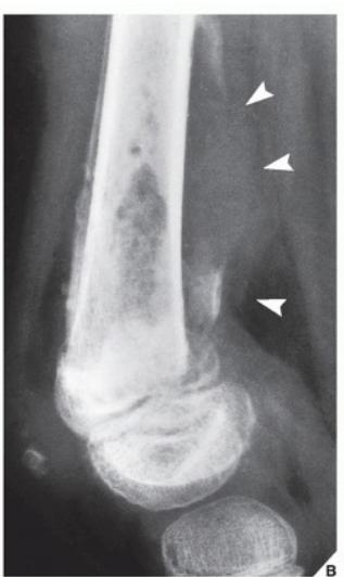
Q16/ An 8-year-old girl presented with a limp. Two years earlier, she had had mild trauma followed by a “bone infection”. Examination revealed a small, pus-secreting wound on the anterior aspect of her left thigh. Her blood count was normal, but her erythrocyte sedimentation rate was 48 mm. According to this radiograph, What do The White Arrows indicate?
- C- Sequestrum
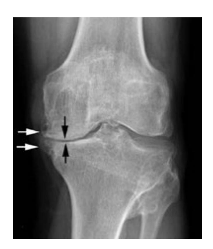
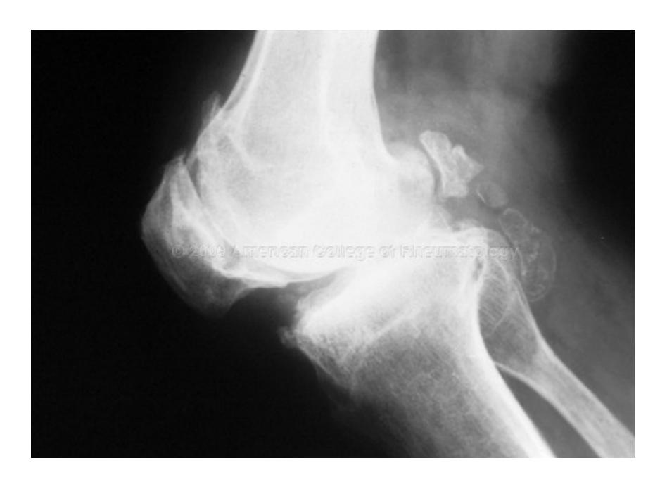
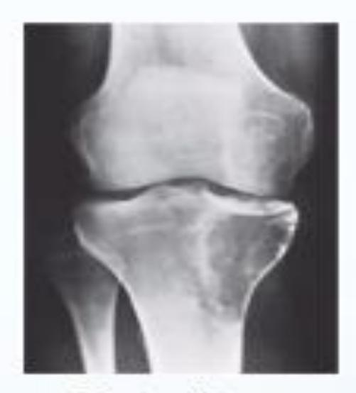
Osteoarthritis
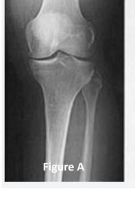
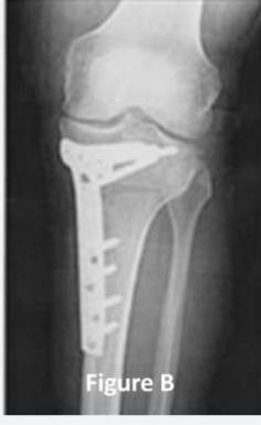
Q9: A 45-year-old male complained of left knee pain for 3 years that didn’t improve with conservative treatment. His x-ray is shown in Figure A. He underwent the procedure, as seen in Figure B. What is the aim of this procedure?
- B. Redistribute the patient’s weight.
Q22: A 55-year-old male was diagnosed with right knee osteoarthritis. He was asking about partial (Unicompartmental) knee arthroplasty. What is the absolute contraindication for this procedure?
- D. Rheumatoid Arthritis
(It may be relative contraindication not absolute? CC)
Q67: A 70-year-old male came with a history of knee pain, and his x-ray in the picture. What is appropriate treatment for him?
- A. Total knee replacement
Radiographic Findings and Diagnosis
Describe the findings:
- Joint space narrowing
- Subchondral sclerosis
- Osteophytes formation
Diagnosis: Osteoarthritis
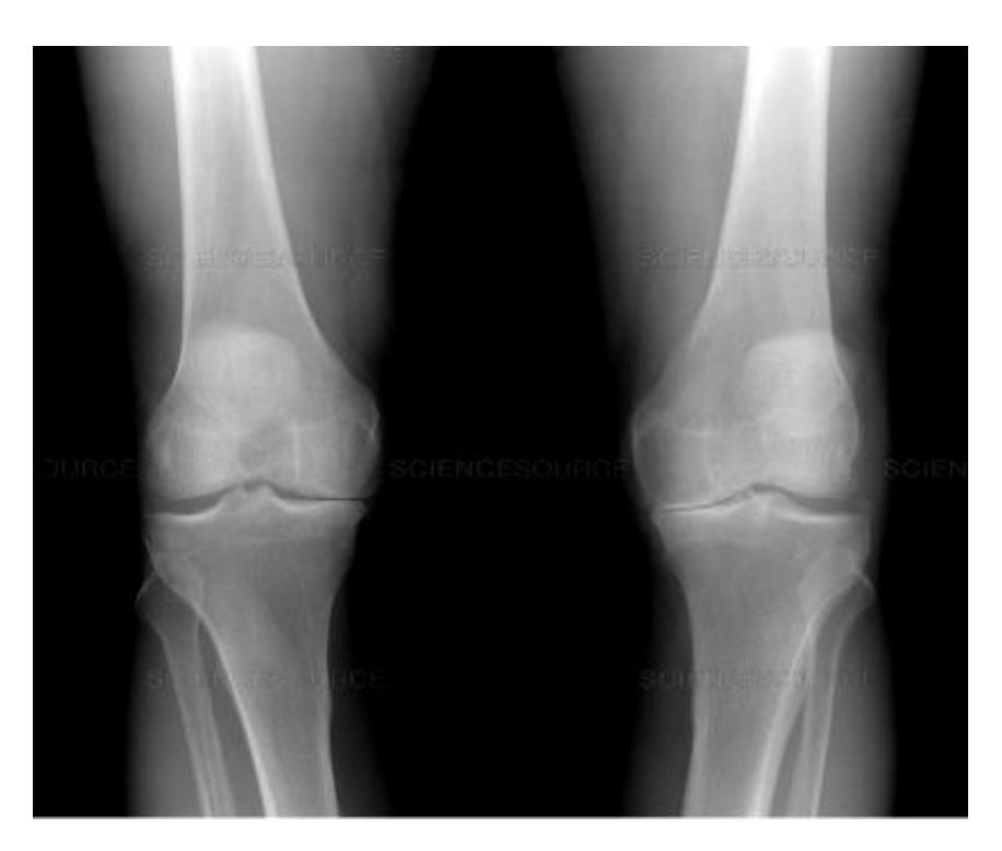
Clinical Case Analysis
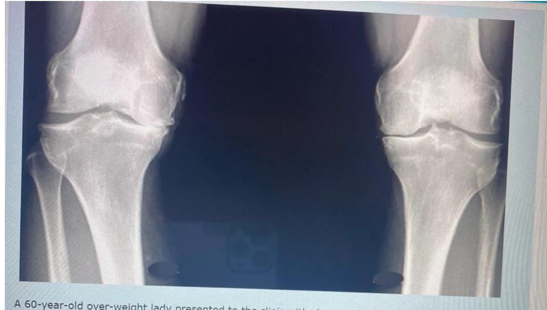
A 60-year-old overweight lady presented to clinic with chronic pain in both knees. A standing AP x-ray of both knees is shown.
Q1: Mention TWO findings on this X-ray
- Narrowing of the joint space
- Osteophytes
Q2: What is the diagnosis?
- Bilateral knee osteoarthritis
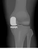
X-ray Interpretation and Management
AP & Lateral x-ray of the knee joint showing narrowing of joint space & sclerosis
Diagnosis?
- Knee Osteoarthritis
Management?
- Exercise & analgesics
- Intra-articular injection
- Knee arthroplasty (depends on the case)
Metabolic Bone Disorders
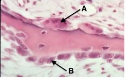
Bone Cell Identification
Which cells are multinucleated with a rough border facing the bone?
- Osteoclasts - These are the bone-resorbing cells characterized by their multinucleated appearance and ruffled border
Osteoporosis and Pathological Fractures
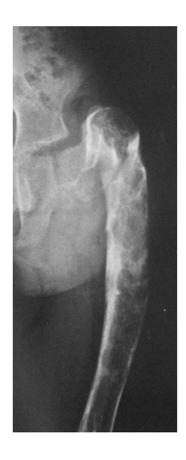
Clinical Case Study
A 50-year-old male tripped over the edge of carpet at home. He heard a crack and is unable to walk. He gave history of progressive weight loss. On clinical examination, he was in pain with a deformity at upper right thigh. He looks pale.
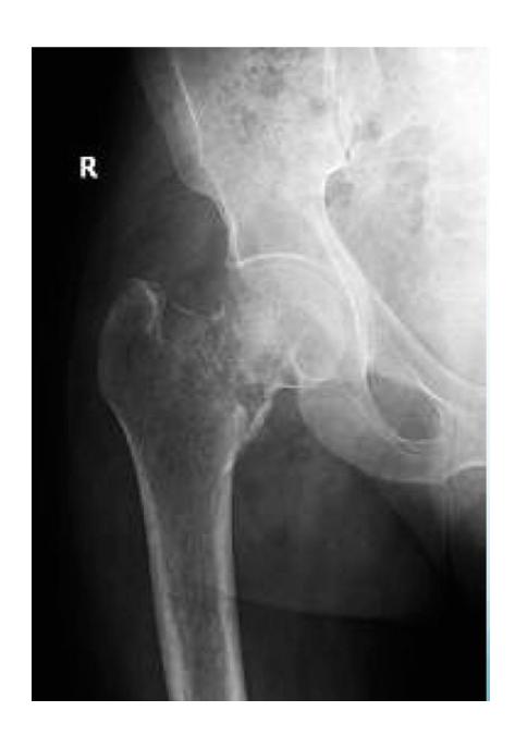
What are the key x-ray findings?
- Decreased bone density in the right proximal femur
- Fracture in the neck of the femur
- AP view of the right proximal femur
What is the diagnosis?
- Pathological fracture due to osteoporosis
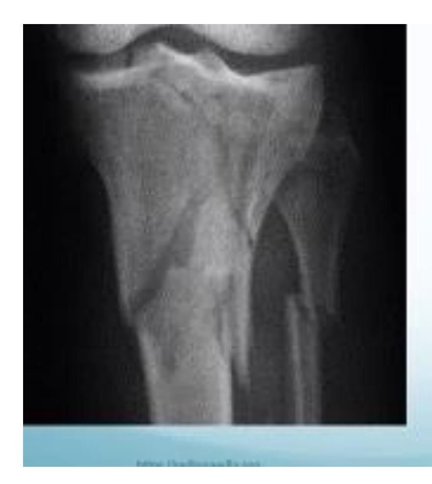
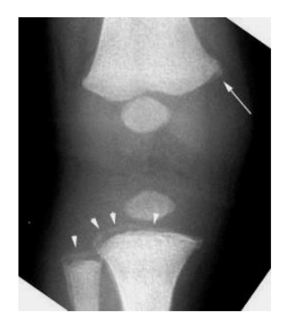
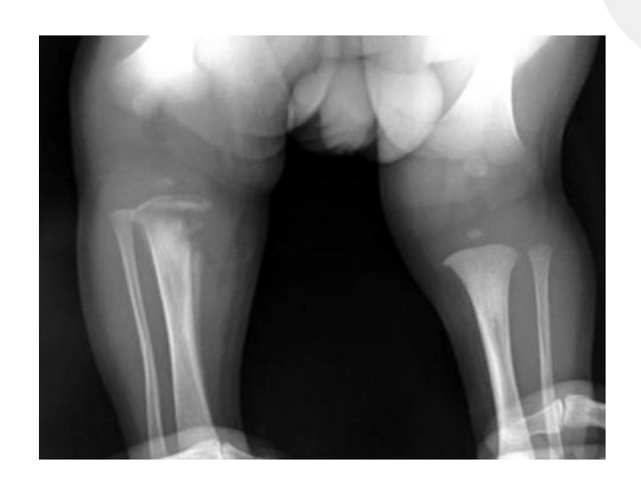
Rickets
Radiological Features
What are the characteristic x-ray findings in rickets?
- Widening of the epiphyseal plate
- Cupping of the metaphysis
- Irregular metaphyseal ends
- Widened physis
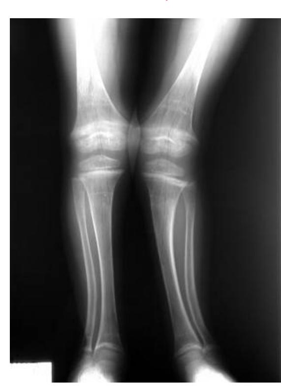

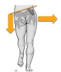
Pediatric Case Study
A 5-year-old boy was brought to the clinic because of a progressively increasing deformity in his left leg for 6 months.
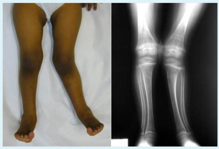
What are the abnormal x-ray findings?
- Widening of epiphyseal plate
- Cupping of metaphysis
How will you treat the underlying cause?
- Vitamin D supplementation
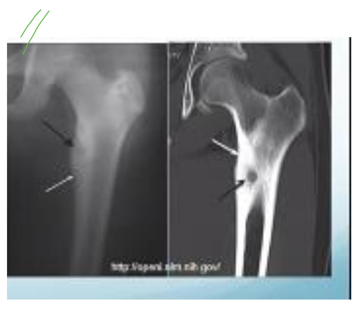
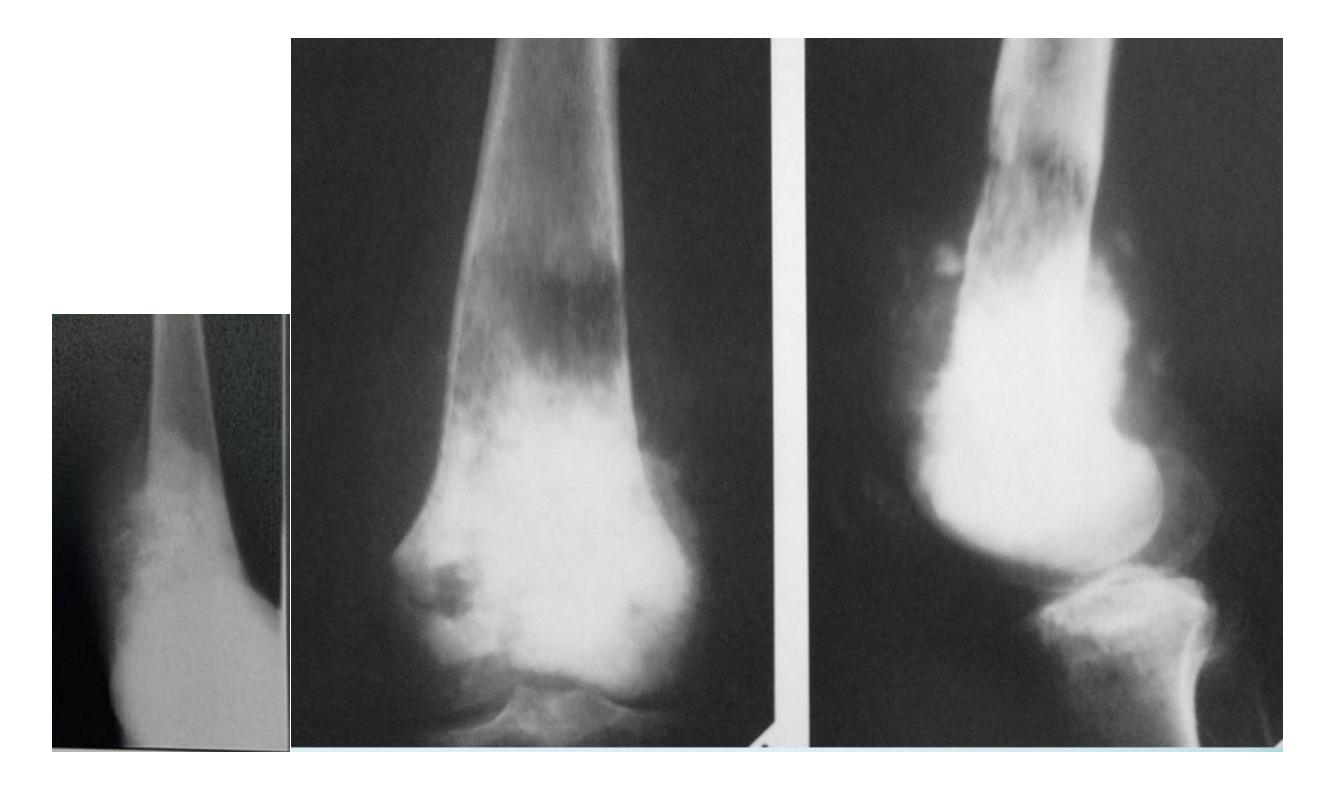
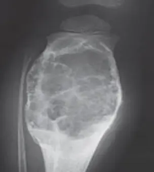
Bone Tumors
Benign Bone Tumors
Non-Ossifying Fibroma
What does this picture show?
- A- Non-ossifying fibroma
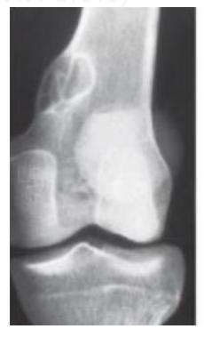
Clinical Features:
- Found in children
- Incidentally discovered
- Asymptomatic
- Cortical defect
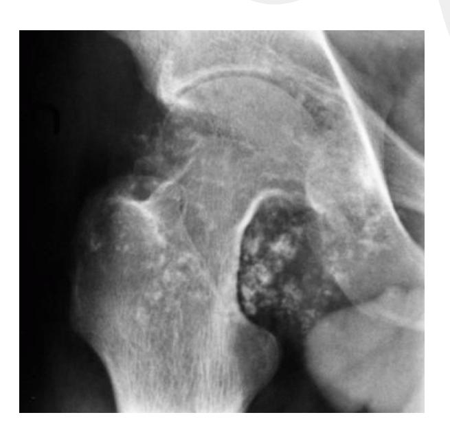
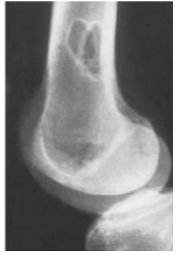
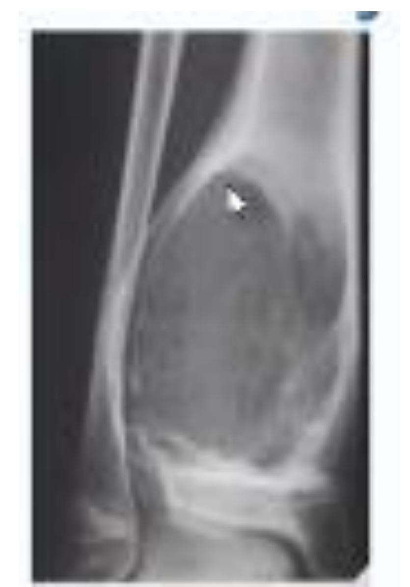
Osteochondroma
What is the diagnosis?
- X-ray showing bony tumor growing away from the epiphysis
- Diagnosis: Osteochondroma
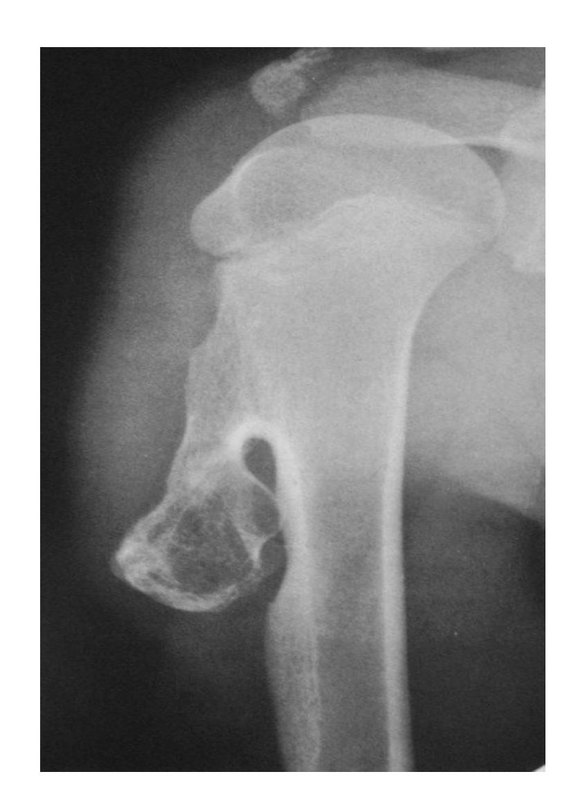
Clinical Features:
- Pain since 2 years in proximal humerus
- Malignant transformation is rare (<1%)
- Treatment: Monitor with ESR, follow-up every 6 months-1 year
- Surgery considered if severe pain (suggesting malignant transformation)
- Surgical risk: Drop hand due to radial nerve injury
Osteoid Osteoma
Clinical Features:
- Night pain
- Bone forming tumor
- Pain relief with aspirin or ibuprofen
- Common in spine
- X-ray may not show anything initially
Radiological Features:
- Sclerotic lesion inside osteolytic lesion (nidus)
- CT shows nidus better
Treatment:
- Excision
- Radio-ablation for spinal lesions
Fibrous Dysplasia
Clinical Scenario:
- Young patient presented with limping and leg length discrepancy
- Diagnosis: Fibrous dysplasia
Malignant Bone Tumors
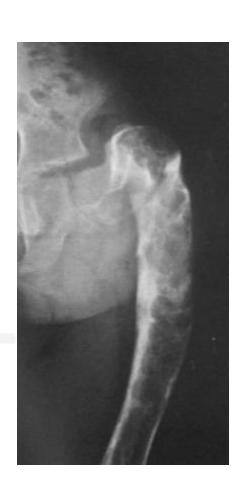
Osteosarcoma
Typical Presentation:
- Age: 10-20 years
- Pain and mass
- Location: Metaphysis of long bone
Characteristic Radiological Findings:
- Sunburst (Sunray) appearance
- Codman’s triangle
- Periosteal reaction
- Bone eating appearance
Clinical Cases:
-
14-year-old patient with pain and swelling at lower right thigh
- Most important X-ray findings: Sun ray appearance, Codman’s triangle
- Diagnosis: Osteosarcoma
-
14-year-old patient complaining of pain and swelling at lower R thigh
- Findings: Codman’s triangle, Sun burst appearance
- Diagnosis: Osteosarcoma

Codman trinagle
Ewing Sarcoma
Clinical Features:
- Occurs in children
- Throbbing pain
- Can be confused with chronic osteomyelitis
- Poor prognosis
Clinical Cases:
-
11-year-old presented with symptoms - diagnosis on imaging
- Diagnosis: Ewing sarcoma
-
Case scenario provided with picture
- Diagnosis: Ewing sarcoma
Giant Cell Tumor
Clinical Features:
- Benign but aggressive
- Eccentric and close to the joint line
- Behavior patterns: Benign, local aggressive, high aggressive
- Occurs after fusion of physis
- Extends to sub-articular region

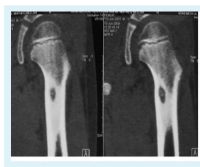
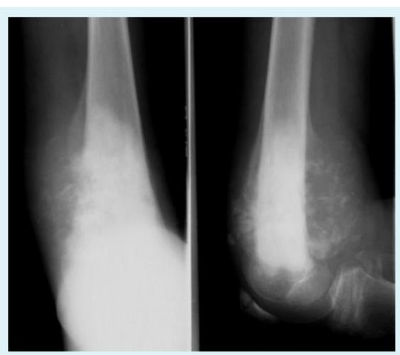
Bone Cysts
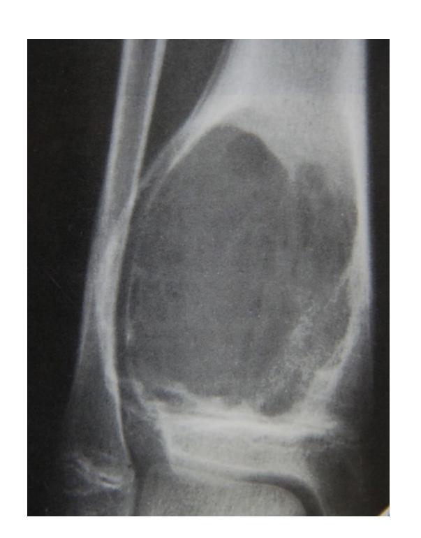
Simple Bone Cyst
Clinical Features:
- Benign
- Children
- Fracture is the first symptom
- Aspiration yields clear fluid
- Surgery for recurrent cases or very large fractures in lower limb
Radiological Features:
- Fills medullary cavity
- Does not expand bone
Clinical Case:
- 12-year-old boy fell and developed severe thigh pain
- AP view of femur showing well demarcated radiolucent lesion without fracture
- Diagnosis: Simple (solitary) bone cyst
- Treatment: Curettage and grafting
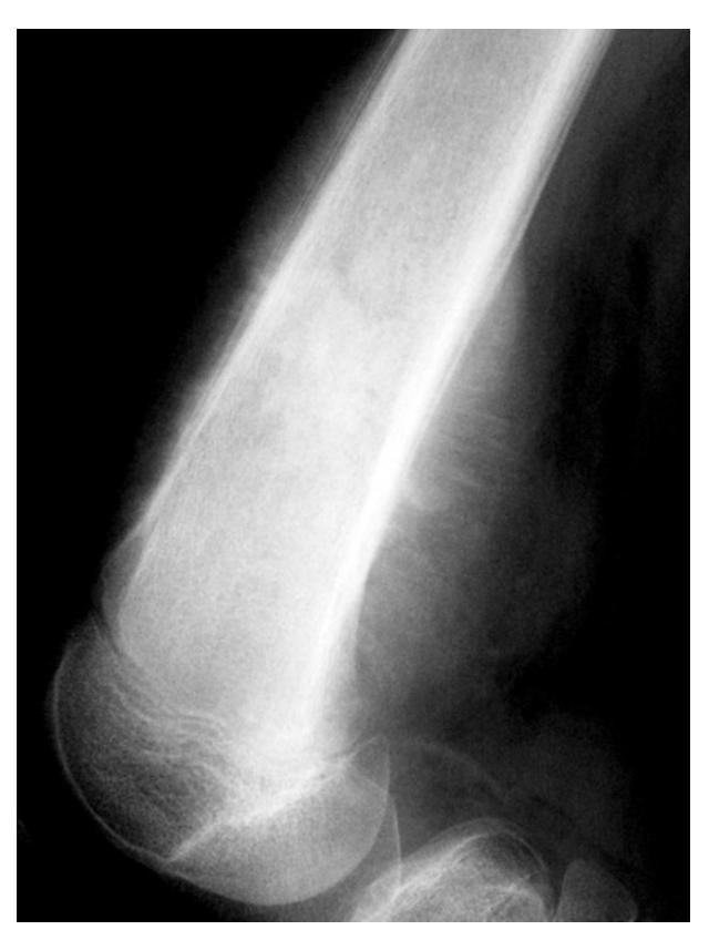
Aneurysmal Bone Cyst
Clinical Features:
- Eccentric, ballooning appearance
- Bloody content
- Found in young adults
Radiological Features:
- Located at metaphyseal side of physis
- Expansile
Metabolic and Other Conditions
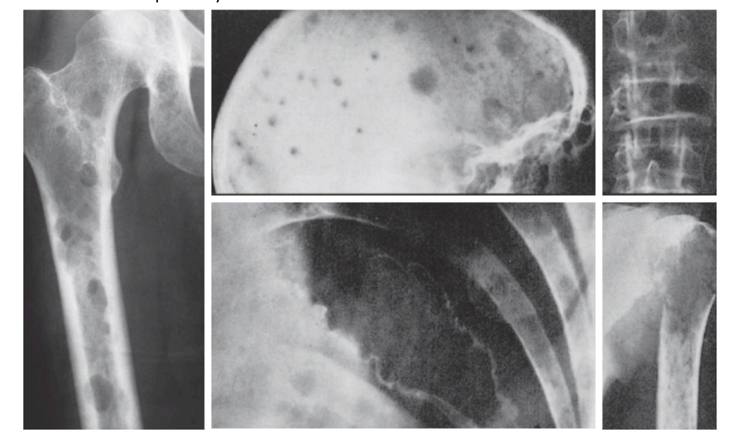
Multiple Myeloma
Clinical Features:
- Age: 45-65 years
- Generalized bone pain
- Involves bone marrow
Radiological Features:
- Multiple punched-out lesions
- Osteopenia, osteoporosis
Clinical Cases:
-
60-year-old man complaining of generalized bone pain, mostly in left thigh
- Sign: Multiple punched out lesions
- Diagnosis: Multiple Myeloma
-
Radiological description
- Lesion: Multiple punched-out lesions
- Associated finding: Osteoporosis
- Diagnosis: Multiple myeloma
Brown Tumor
Clinical Features:
- Associated with hyperparathyroid disease
- Check PTH levels
- Differential diagnosis: infection, malignancy, hyperparathyroidism
Metastatic Bone Disease
Clinical Features:
- Above 50 years old
- Metastasis is differential diagnosis
- Can present as pathological fracture
Radiological Features:
- Osteolytic lesion
- Moth eaten appearance
General Radiological Features
Zone of Transition
Which of the following is correct regarding this image?
- C. Narrow zone of transition or cortical thinning not sure?
Differential Diagnosis by Age
- Children: Ewing sarcoma (when sun-ray appearance and Codman’s triangle present)
- Adults: Osteosarcoma (when sun-ray appearance and Codman’s triangle present)
- Elderly: Consider metastatic disease
Common Presentations
- Many bone tumors present as injury
- Pain is a common symptom across most tumor types
- Location and age are key diagnostic factors
- Radiological appearance helps narrow differential diagnosis