Benign Bone Tumors
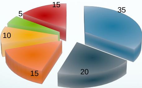
Distribution
- Osteochondroma 35%
- Enchondroma 20%
- Giant Cell 15%
- Osteoid Osteoma 10%
- Fibrous Dysplasia 5%
- Other 15%
Types of Benign Bone Lesions
- Osteochondroma
- Chondroma / chondroblastoma
- Osteoid osteoma / osteoblastoma
- Non-ossifying fibroma
- Simple (Unicameral) Bone Cyst
- Aneurysmal Bone Cyst
- Giant Cell Tumor
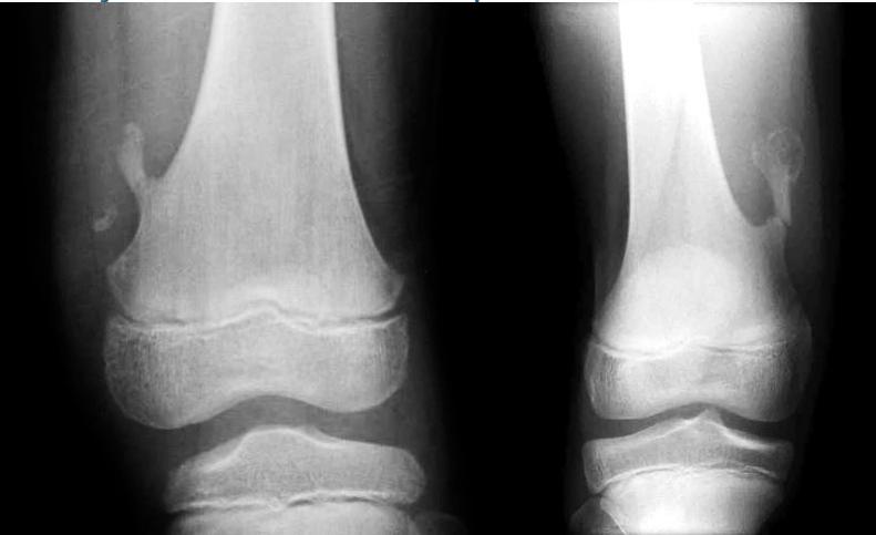
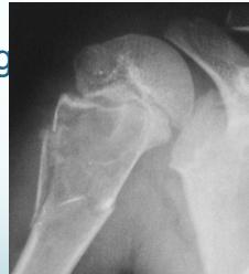
Osteochondroma (Exostosis)
- A common lesion
- Ends of long bone
- Bony overgrowth:
- Away from epiph. plate
- Covered by cartilage
- Growth:
- Stops when epiphysis close
- If continues later:
- ? Malignant transformation
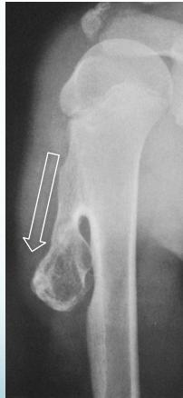
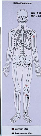
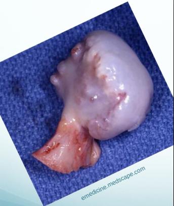
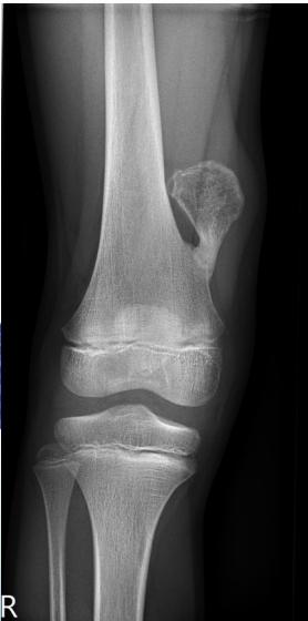
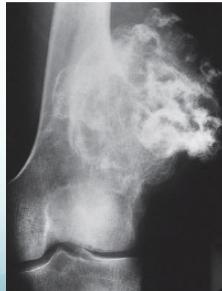
Source: radiopaedia.org, Apley’s System of Orthop. And Fractures
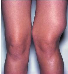
Complications
- May fracture: becomes painful
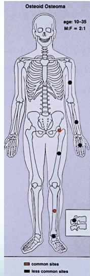
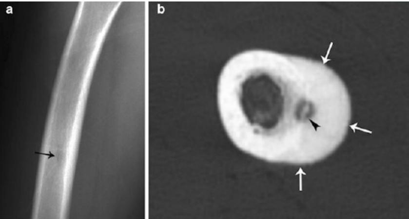
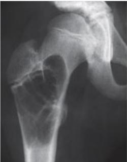
Osteoid Osteoma
- Small tumor (<1 cm)
- Young adults
- Pain, pain, pain:
- Typically relieved by Salicylates
- Sites: Femur, tibia, spine
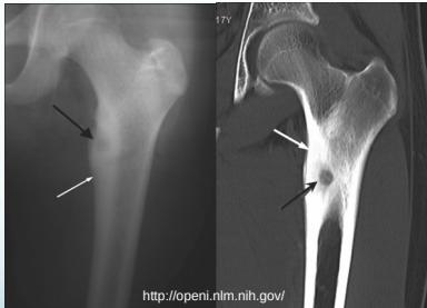
- X-ray:
- Small radiolucent “nidus”
- Surrounded by sclerotic bone
- CT: Shows “nidus” better
- scan: hot
- Treatment: surgical excision, or thermal ablation
Source: Orthopedic Radiology. A Greenspan. Lippincott-Raven
Sources: Apley’s System of Orthop. And Fractures, http://openi.nlm.nih.gov/
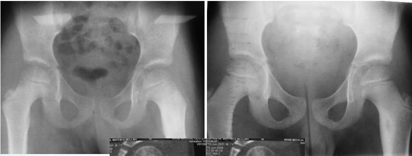
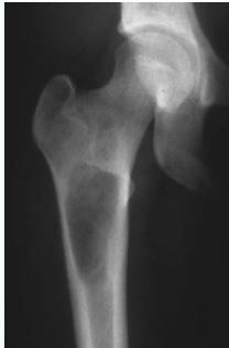
Clinical Example
- 11 year old boy: Pain in left hip
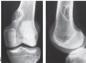
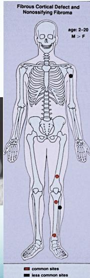
Non-Ossifying Fibroma
- Another name: Fibrous cortical defect
- The commonest benign lesion of bone
- Asymptomatic:
- Incidentally discovered
- Children:
- Disappears later
- Common site:
- Metaphysis of long bones
- Treatment:
- Observation
- Surgery if very large
Source: Apley’s System of Orthop. And Fractures
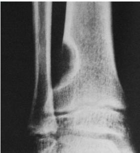
Non-ossifying fibroma …Fibrous cortical defect
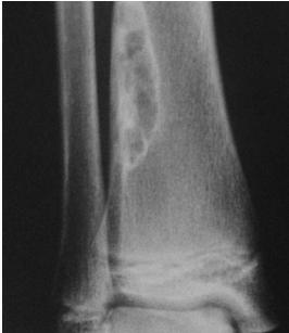
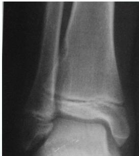
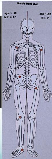
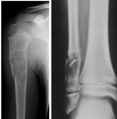
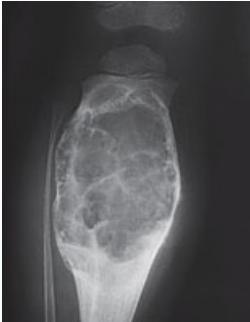
Simple Bone Cyst
- Solitary – unicameral
- Children
- Metaphysis:
- Prox. Humerus and Femur
- Not a tumor:
- Not seen in adults
- Heals spontaneously
- Pathological fracture / or incidental
- Aspirate is clear straw-colored
Sources: www.juniorbones.com, Orthopedic Radiology. A Greenspan. Lippincott-Raven
Source: https://radiopaedia.org
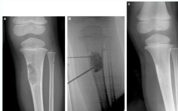
Treatment
- Small, reducing in size:
- Leave alone
- Increasing in size, active:
- Moderate trial of bone marrow injection
- Large (risk of fracture) Curettage & grafting
- Pathological fracture:
- Treat fracture
- Cyst might heal
- Recurrent / injection failed:
- Surgical curettage and bone grafting
Sources: https://www.sciencedirect.com/science/article/pii/S1877056814003338, Orthopedic Radiolgy. A Greenspan. Lippincott-Raven
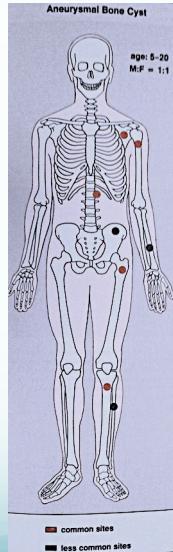
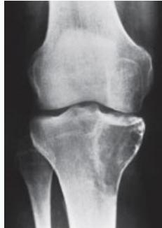
Aneurysmal Bone Cyst
- Child - young adult
- Metaphysis of long bone
- X-ray:
- Well-defined cyst
- Trabeculated
- Eccentrically placed
- Ballooning
- Bloody content
- Treatment:
- Curettage and bone graft
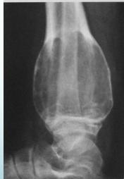
- Metaphysis, Eccentric
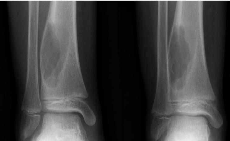
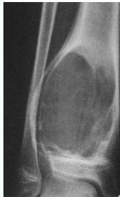
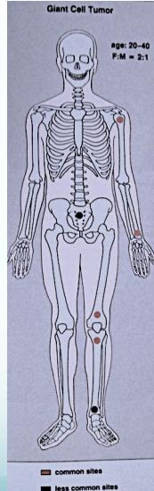
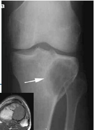
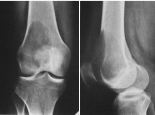
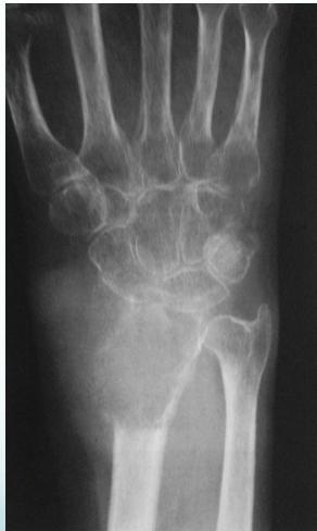
Giant-Cell Tumor
- Unknown origin:
- Giant cells abundant
- Behavior:
- One third benign
- One third locally aggressive
- One third (less) with distant metastasis
- Young adults
- Common sites:
- Around knee
- Proximal humerus
- Distal radius
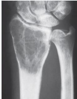
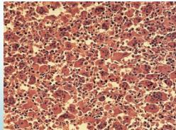
Radiological Features
- Eccentric lesion:
- Radiolucent
- Soap bubble
- Abuts (adjacent) against the joint
- Thin cortex
- Margins may be clear / unclear:
- Depends on aggressiveness
- Treatment:
- Curettage & bone grafting
- More wide excision in recurrent and aggressive lesions
Source: Bone Tumors A Practical Guide to Imaging
Comparison of Cyst-Like Lesions in Bone
Simple Bone Cyst
- Fills medullary cavity
- Does not expand bone
Aneurysmal Bone Cyst
- At metaphyseal side of physis
- Expansile
Giant-Cell Tumor
- After fusion of physis
- Extend to sub-articular