Classification – Predominant Tissue
| Tissue of Origin | Benign | Malignant |
|---|---|---|
| Bone forming | Osteoma Osteoid Osteoma Osteoblastoma | Osteosarcoma |
| Cartilage forming | Chondroma Osteochondroma Chondroblastoma | Chondrosarcoma |
| Fibrous tissue | Fibroma | Fibrosarcoma |
| Giant-cell tumor | Benign Osteoclastoma | Malignant Osteoclastoma |
| Marrow tumors | Ewing’s Sarcoma Myeloma | |
| Vascular | Haemangioma | Haemangiosarcoma |
| Other connective tissue | Fibrous histocytoma Lipoma | Malignant fibrous histocytoma Liposarcoma |
| Other tumors | Neurofibroma | Adamantina |
Clinical Presentation
History
Duration
- Prolonged history:
- In most benign lesions
- Some malignant:
- Slow growing (chondrosarcoma) / in pelvis (expandable)
Age Distribution
- Childhood and adolescence
- Most benign, and some malignant (e.g. Ewing’s sarcoma)
- 4th – 5th decade:
- Chondrosarcoma
- Sixth decade:
- Myeloma (the commonest primary malignant bone tumor)
- Over 50 yrs.:
- Metastatic lesions are the commonest
Symptoms
- Pain:
- In both malignant and benign (usually more in malignant)
- May be caused by:
- Rapid expansion – stretching of tissues
- Central hemorrhage or degeneration
- Insipient pathological fracture
- Tense encapsulation in bone (e.g. osteoid osteoma)
- Swelling
- H/O Trauma
- Neurological symptoms:
- Pressure on nerve / stretching the nerve
- Pathological fracture: minor trauma / strange fracture line
Clinical Examination
- A mass (lump):
- Location
- Discrete (separated) or ill-defined
- Tenderness
- Temperature
- Pulsatile
- Mobility
- …etc
- Range of motion
- LN, pelvis, abdomen, chest, spine
Diagnostic Approaches
Imaging Studies
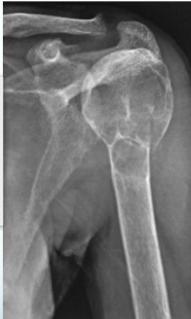
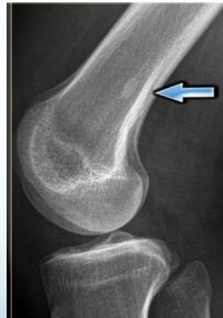
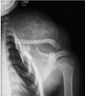
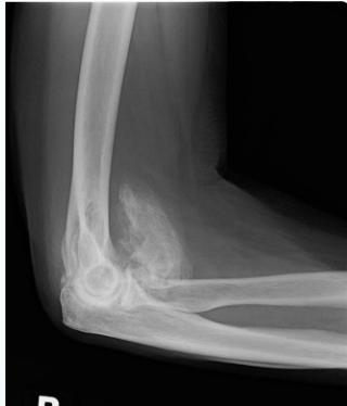
X-rays
- Which bone, and which site in bone?
- Solitary or multiple?
- Bone forming or bone eating?
- Margins: well-defined or ill-defined?
- Calcifications in the lesion?
- Is cortex eroded or destroyed?
- Is there periosteal new bone formation?
- Soft tissue extension?
Note: Shall be discussed separately
Other Imaging Modalities
- Bone scan ():
- Shows the site of lesion / skip lesions / metastasis
- CT:
- Shows Intra and extra-osseous structure and extension
- Good in deep bones (pelvis, spine)
- MRI:
- Tumor spread:
- Within bone, into joints, into soft tissue
- Relation to vessels
- Soft tissue and cartilage tumors
- Tumor spread:
Laboratory Investigations
- Look for infection:
- CBC, differ WBC, CRP, blood c/s
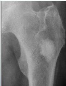
- Look for metabolic disease:
- Brown tumor in hyperparathyroid disease
- Anemia, raised ESR
- S. Alkaline phosphatase
- Tumor markers:
- Bence Jones protein in urine: Myeloma
- S. Acid phosphatase & PSA: Prostatic carcinoma
- Raised serum Calcium in metastasis
Biopsy
- Diagnostic
- Needle biopsy:
- CT-guided
- In the line of further surgical incision
- Open biopsy:
- After all imaging techniques completed
- More reliable
- Site: considering further surgery
- From boundaries
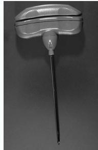
Jamshidi needle
Differential Diagnosis
Soft Tissue and Traumatic Conditions
- Soft tissue hamartomas
- Myositis ossificans
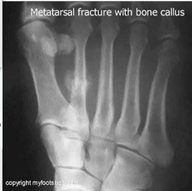
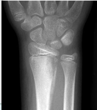
- Stress fracture:
- Histopathology may be confused with osteosarcoma?
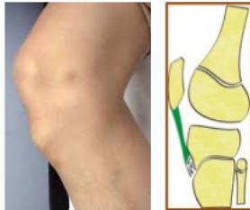
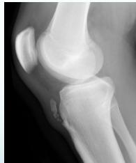
- Tendon avulsion injuries:
- Near hip and knee (e.g. Osgood-Schlatter)
- Infection
Metabolic and Other Bone Lesions
- Hyperparathyroid disease (Brown tumor)
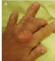
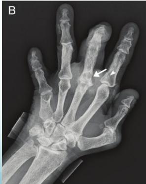
- Gout: Large gouty typhus
- Other bone lesions:
- Cortical defects, bone infarcts, “bone islands”