Imaging Modalities
- X-Ray - initial detection and characterization
- CT Scan - detailed bone architecture and characterization
- MRI - local staging, soft tissue involvement
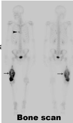
- Bone Scan - detection of bone metastases
- PET-CT - metabolic activity and systemic staging
Imaging Strategy
- Detection & Characterization (X-rays, CT scan)
- Local staging (MRI)
- Systemic staging - evaluation for regional and distant metastases (CT chest, bone scan, PET scan)
X-Ray Interpretation: The 7 Key Features
- Site - bone and location within bone
- Size - dimensions of the lesion
- Matrix - internal composition and calcification patterns
- Pattern/Margins - including zone of transition
- Effect of the lesion on bone - cortical changes
- Reaction of bone to the lesion - periosteal response
- Soft tissue mass - presence or absence
Site Analysis
- Which bone is affected (femur, radius, etc.)
- Location within bone:
- Diaphysis, metaphysis, epiphysis, or combination
- Centric, eccentric, intracortical, surface
Matrix Analysis
- Fibrous, fluid, fat (radiolucent; dark demarcations)
- Cartilaginous (arcs and rings of calcification)
- Osseous (cloud-like, dense)
- Mixed patterns


 Bubbly
 Cloudy
Benign vs Malignant: Pattern Recognition
Destruction Patterns
Benign Process
- Geographic - uniformly destroyed area with sharply defined border
Likely Malignant Process
- Moth-eaten - areas of destruction with ragged borders
Aggressive/Malignant Process
- Permeative - ill-defined area spreading through marrow space

Zone of Transition
 - Mixed
 - Ill-defined
Lesion Effects on Bone
Cortical Changes
-
Cortical thinning ✓ Lower grade, less aggressive

-
Cortical expansion ✓ Low or high grade, tumor mimickers

-
Cortical destruction ✓ High grade, aggressive
Bone Reaction to Lesion
Periosteal Reaction Patterns
- Absent - typically benign lesions
- Mild – one layer, 1-4 mm thick, adjacent to cortex
- Major - >5mm, multilayered or lamellated:
- “Onion-skimming”
- “Hair-on-end”
- “Sunburst”


 Codman’s triangle- causes include infections / tumours
Soft Tissue Mass Assessment
- Absent - suggests benign process
- Present - raises concern for malignancy

Differential Diagnosis by Bone Location
Diaphysis
Characteristic lesions:
- Adamantinoma
- Osteofibrous Dysplasia
- Osteoid Osteoma
- Stress Fracture
- Chronic Osteomyelitis
- Round Cell Tumors (Ewing sarcoma, Lymphoma)
- Langerhans Cell Histiocytosis
- Fibrous Dysplasia
- Fibrous Cortical Defect / Non-ossifying Fibroma
Metaphysis
Characteristic lesions:
- Osteochondroma
- Osteosarcoma
- Fibrosarcoma
- Chondromyxoid Fibroma
- Aneurysmal Bone Cyst
- Enchondroma / Chondrosarcoma
- Simple Bone Cyst
- Fibrous Dysplasia
- Osteomyelitis (pyogenic)
Epiphysis
Characteristic lesions:
- Articular Osteochondroma (Trevor Disease)
- Chondroblastoma (children)
- Giant Cell Tumor (adults)
- Osteomyelitis (fungal, TB)
- Aneurysmal Bone Cyst
- Clear cell chondrosarcoma
Intra-articular Tumors
- Pigmented Villonodular Synovitis (PVNS)
- Synovial Hemangioma
- Intra-articular Loose Bodies


Epiphyseal Lesions: Detailed Analysis
Common Epiphyseal Tumors
-
Chondroblastoma
-
Clear cell chondrosarcoma
-
Osteomyelitis (occasionally)
-
Chondroblastoma
Apophyseal Lesions
()
Epiphyseal-Metaphyseal Lesions
Benign aggressive lesions:
- Giant Cell Tumor (GCT)
- Aneurysmal Bone Cyst (ABC)
- Osteoblastoma
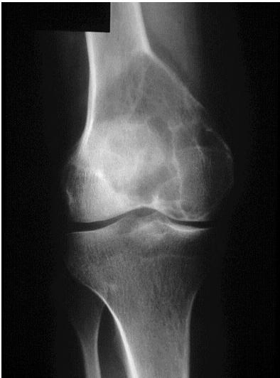
Giant Cell Tumor
Aneurysmal Bone Cyst
/// gce an?
Metaphyseal Lesions: Detailed Examples
Common Metaphyseal Tumors
- Enchondroma
- Simple Bone Cyst (SBC)
- Non-ossifying Fibroma (NOF) / Fibrous cortical defect
- Chondromyxoid fibroma
- Osteosarcoma
- Chondrosarcoma
Simple Bone Cyst
Other Metaphyseal Lesions
 Popcorn, cartilaginous - chondrosarcoma usually - even though theres no perosteal reaction

site: distal femur size: Most (involving how much? / cm?) matrix: mixed - mainly radiopaque , wide zone of transition, cortical destruction, resulted periosteal reaction, and codman’s triangle soft tissue involvement:
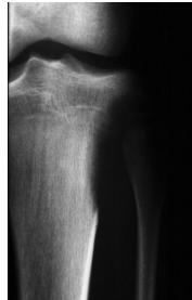
Non-ossifying Fibroma

site: Diaphyseal, metaphysal size: partial matrix: Mixed - Narrow zone of transition, well defined, syndosmosis soft tissue involvement: No tissue involvement
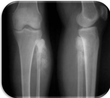
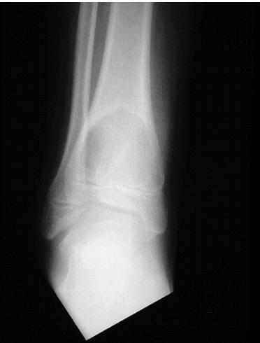
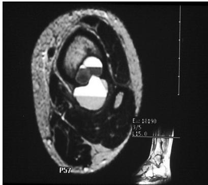
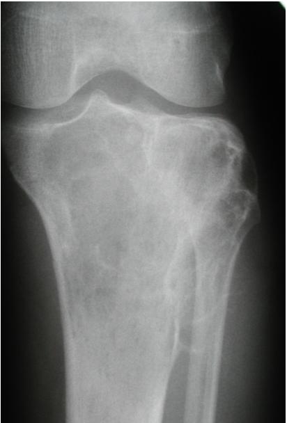
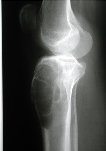
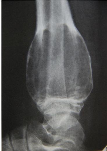
Aneurysmal Bone Cyst

site: Distal tibial
size: Involving most of distal part
matrix: Cortical expansion, radiolucent matrix, well defined, narrow zone of transition
soft tissue involvement: no peristeal reaction
site: Distal shaft of femur size: matrix: Opaque, well defined, ossifying fibroma soft tissue involvement: no peristeal reaction

site: Distal femur size: matrix: soft tissue involvement:
Diaphyseal Lesions: Detailed Examples
Common Diaphyseal Tumors
- Osteoid osteoma
- Fibrous dysplasia
- Osteofibrous dysplasia
- Adamantinoma
- Ewing’s sarcoma
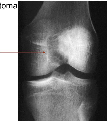
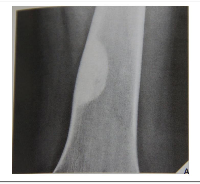
Osteoid Osteoma
Thick cortex

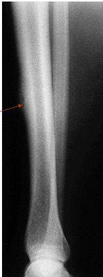
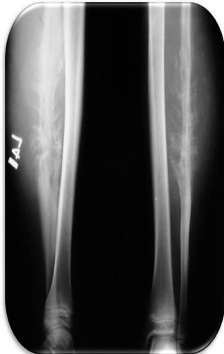
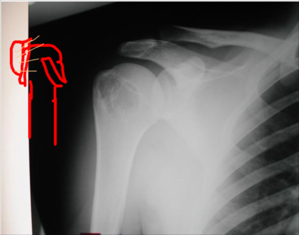
Other Diaphyseal Lesions
Could be malignant, even though no periosteal reaction, may be due old fracture
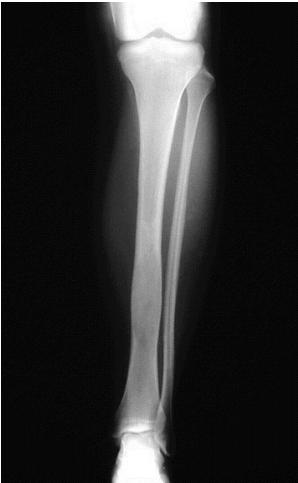

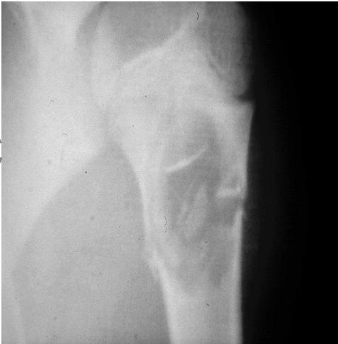
Pathological fracture, after metastasis

Important Diagnostic Note
Always remember infection as differential diagnosis
