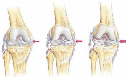
Collateral Ligament Injuries
Clinical Presentation
History:
- Acute injury during sports or trauma
- Acute pain
- Swelling
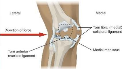
Mechanism of injury:
- Sudden side-way force
- MCL >> LCL (medial collateral ligament is more commonly injured due to anatomical factors)

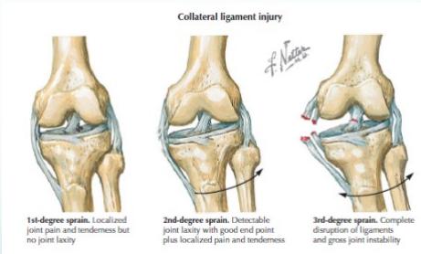
Clinical Signs
- Swelling
- Tenderness (in collaterals / origin / insertion)
- Stressing ligament is painful
- (In complete tears, stressing ligament may produce abnormal movement with little pain)
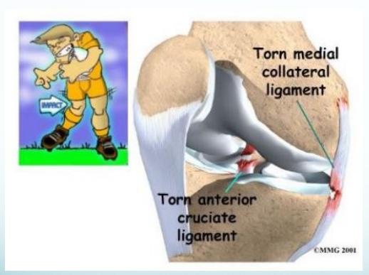
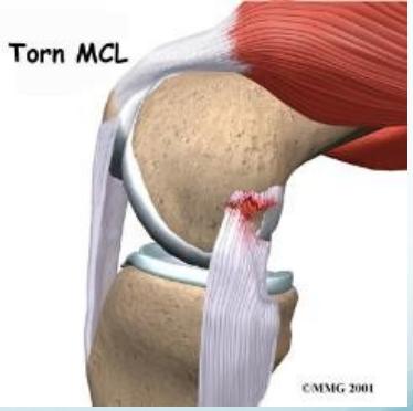
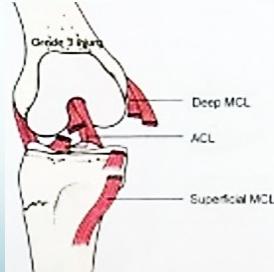
Medial Collateral Ligament (MCL) Injury
Mechanism:
- Valgus stress to the knee
- Most commonly occurs at medial femoral attachment
https://en.wikipedia.org/wiki/Knee
Diagnostic Studies for MCL
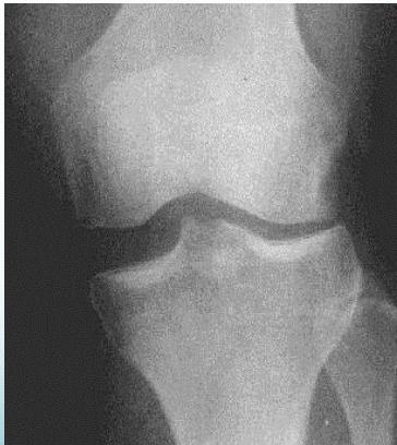
X-ray
- May show avulsion fracture
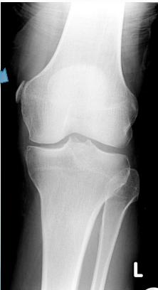
- Stress Film: Shows ligament laxity
MRI
- Best for assessing soft tissue damage


Treatment of MCL Injury
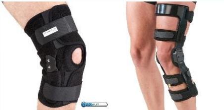
Isolated injuries:
- Hinged Knee Brace for conservative management
Combined injuries:
- Require reconstruction of the respective ligaments (ACL, PCL, posteromedial corner)
Lateral Collateral Ligament (LCL) Injury
Characteristics:
- Isolated LCL injuries are uncommon
- Treatment: Conservative management with brace if grade II
Complete tears with associated ACL/PCL require reconstruction
© Martin Dunitz Ltd. 2001