Dr. Majed Alasbali
Reading an X-Ray of Fractured Bone
Prerequisites
- At least 2 views (True AP and lateral views)
- Entire bone length included
- Appropriate joint views
- Joint above and joint below for trauma cases
Use the Following
- For trauma >> OLD ACID
- For tumor >> 7Q
- For osteoarthritis >> 5 things
Comment On
- Adequacy of the views
- Bone, joints and soft tissue
- Need for previous x-rays

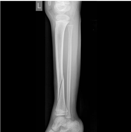
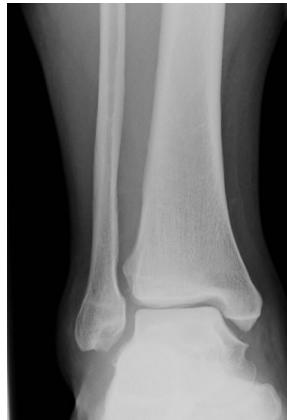
X-ray ap lat closed ,distal tibia extra-articular fracture, complete (two cortex), no articulation involvement, spiral (if two views appear broken), oblique (if one view) Oblique fracture, slight gapping (should be displaced if so), cortex appear normal, minimally displacement, angulation, rotation.
TALK ORTHO TO ME
OLD ACID Framework
O - Open vs Closed
L - Location
D - Degree (complete vs incomplete)
A - Articulation involvement
C - Comminution & Pattern (type)
I - Intrinsic bone quality (cortex)
D - Displacement, angulation (distal to proximal), & rotation
@GWEMresidency
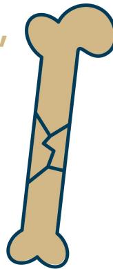
Fracture Types and Characteristics
Displacement Types
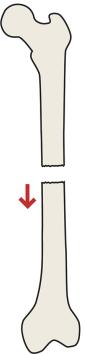
Displaced
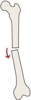
Angulated
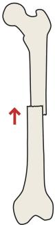
Shortened
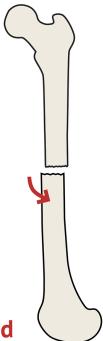
Rotated
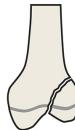
 Bowing
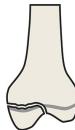
Fissure
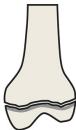
Greenstick
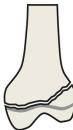
Torus
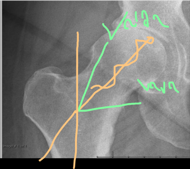
Classification Systems salter harris - (pediatric)
I
II
III (intra-articular)
IV (intra-articular)
V
Tibial platue, linea view -
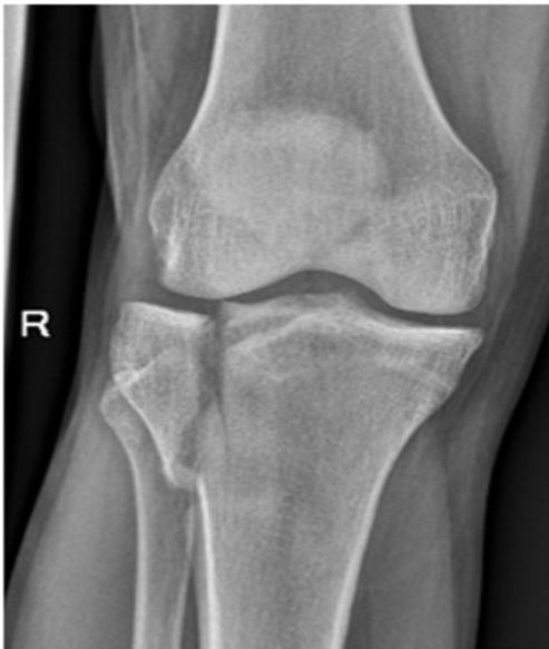
Reading an X-Ray of Osteoarthritic Knee Joint
Five Key Features
- Narrowing of the joint line
- Formation of osteophyte
- Subchondral sclerosis
- Subchondral cyst
- Joint deformity
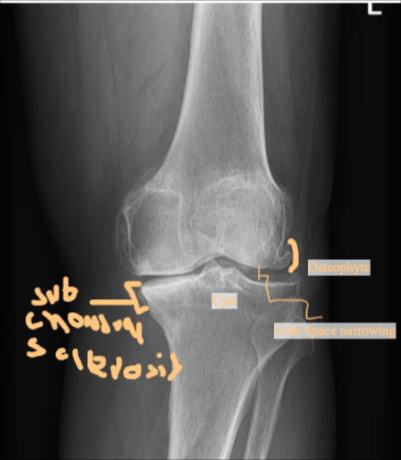
X-ray AP Lateral Osteophyte formation - joint space narrowing - Subchondral cyst adn sclerosis - joint Varus deformity
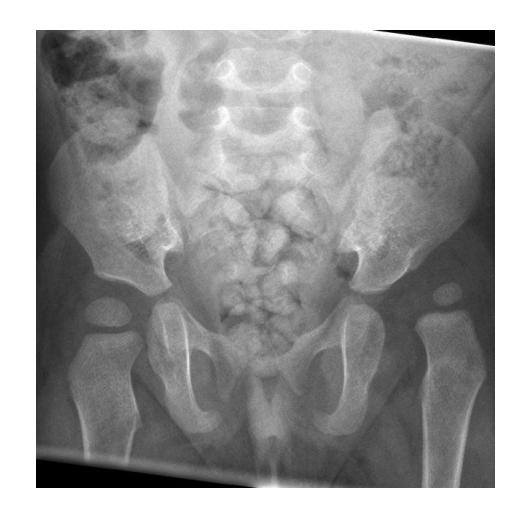
AP HIP Joint line narrowing, osteophyte formation, subchondral sclerosis and cysts, no obvious deformity
Reading an X-Ray of Bone Lesion
7Q Framework
- Site
- Size
- Matrix
- Zone of transition
- Lesion effect
- Bone effect
- Soft tissue
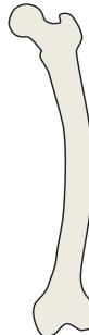
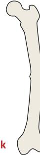
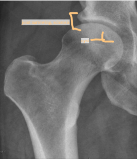
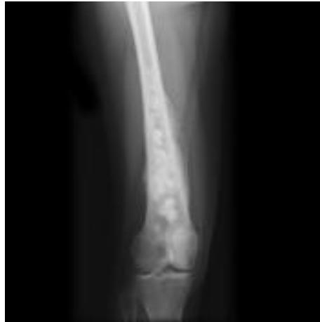
AP/Posterior left femur
- Site: Extra-capsular proximal femur greater trochantric
- Size: 4x3cm apple shaped
- Matrix: Radiolucent,
- Zone of transition: narrow zone of transition
- Bone effect:
- Soft tissue: i cant appreciate soft tissue involvement
most likely benign
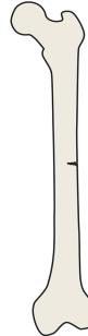
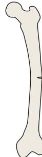
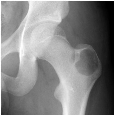
 AP lat x-ray
- Site: multiple small Right femu, metaphysis, ddiaphysis
- Size: varying sizes lesions on humerous
- Matrix: Multiple lesions, mixed
- Zone of transition: wide zone transition,
- Bone effect: cortical destruction, periostea reactions
- Soft tissue: periosteal reaction
malignant
HX EX - Labs - imaging (x-ray ⇒ mri ⇒ CT ⇒ PET) ⇒ Core biopsy (oncology surgeon)
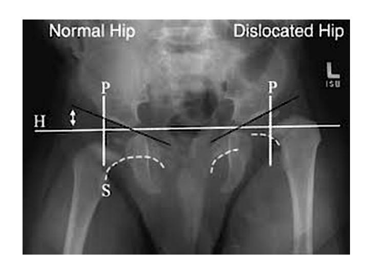
How to Read an X-Rayof Pediatric Hip
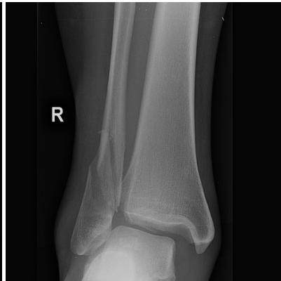
Reading an X-Ray of Ankle Joint Injury
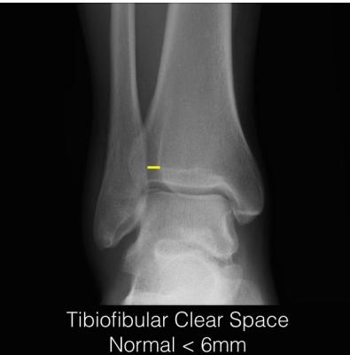
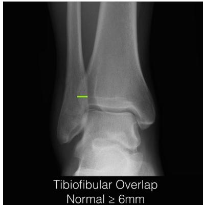
Key Measurements
Tibiofibular Clear Space
- Normal: < 6 mm (AP view)
- Pathological: ≥ 6 mm
Tibiofibular Overlap
- Normal: ≥ 6 mm (AP view)
- Normal: ≥ 2.8 mm (mortise view)
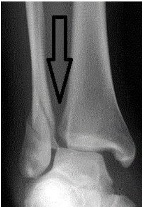
Medial Clear Space
- Normal: < 4-5 mm (mortise view)