Collection from previous batches, made by Abdulmalik Abdulghani, Dania AbuObaid, Faisal Alkharji, Haneen H. Alsultan, Husna Merza, Lina Serhan, Maha Alasmari, Mujahid N Al-muhaydib, Yousef Edress
اللهم يا معلّم موسى علّمني، ويا مفهم سليمان فهّمني، ويا مؤتي لقمان الحكمة وفصل الخطاب آتني الحكمة وفصل الخطاب اللهم اجعل ألستنا عامرة بذكرك، وقلوبنا بخشيتك، وأسرارنا بطاعتك، إنك على كل شيء قدير، حسبنا الله ونعم الوكيل
Any updates of this file would be within https://medatlax.com/Clinical/Level-12/IM-2/Internal-Medicine-2 OSCE & OSPE-CS link.
Contents
- How to Read Chest X Ray
- ECG Rhythms and Arrhythmias
- Chest X-Ray Findings
- Physical Examination Findings
- ECG Localization of Myocardial Infarction
- Pulmonary Edema Management
- Coronary Angiography and Cardiac Surgery
- Pulmonary Edema and Pleural Effusion on Chest X-Ray
- Cerebral Imaging
1. How to Read Chest X Ray
The ABCDE Approach
Remember ABCDE
- A. Airway
- B. Bone
- C. Cardiomegaly
- Cavitation
- Calcification
- D. Density
- E. Effusion
Normal Chest X-Ray
Normal X-Ray

Cardiomegaly: Clinical Case

Wire
-Cardiomegaly
- what drugs should be given
- Aspirin but - warfarin
- follow up monthly for PT/INR
- St of drug + bleeding

2. ECG Rhythms and Arrhythmias
2.1 Supraventricular Tachycardia (SVT)

regular over 160 bpm
Ux: →
- 1st line → Vagal stimulation
- 2nd line → Adenosine or verapamil
2.2 Atrial Fibrillation

irregular
Atrial fibrillation
Ux: →
- Rate control → BB blocker
- Rhythm control → Antiarrhythmic drugs
- Antiarrhythmation (sic, as recorded)
2.3 Ventricular Tachycardia

Ventricular tachycardia
- if Cardiac arrest → CPR & DC shock
- if conscious → Amiodarone
- → wide QRS
- monomorphic
- Regular
2.4 Ventricular Fibrillation

VENTRICULAR FIBRILLATION
- III → CPR & DC shock

2.5 Asystole

Asystole
- check the pt.
2.6 Pulseless Electrical Activity (EMD)

2.7 Complete Heart Block

- in elderly
- complete dissociation between atrium & ventricle
- Profound bradycardia
- III → Pace maker
2.8 Pacemaker

- Pace maker
- in pt. with complete heart block
- on ECG
- with QRS
- Pacemaker spikes (spikes before QRS)
- in dual pace maker
- with QRS
- Pacemaker spikes (spikes before QRS / p-wave)

Patient stoi
Pace maker
2.9 Read any strip in five steps

- Rate · 2. Regularity · 3. P waves · 4. PR interval · 5. QRS duration
2.10 Sinus tachycardia, WPW, torsades, flutter
Sinus tachycardia — 101–160/min, sinus P waves, normal QRS, regular or slightly irregular. Causes: catecholamines, CHF, hypoxia, PE, fever, stress, pain → treat the cause.

WPW — pre-excitation through the bundle of Kent: short PR <0.12, delta wave, wide QRS >0.12. EPS maps the pathway; ablation (surgical or electrical) is curative.

Torsade de pointes — 150–220/min, wide bizarre QRS twisting up and down around the baseline, irregular and paroxysmal. Causes: QT-lengthening drugs (class 1A quinidine), hypokalaemia, ischaemia. Unstable → synchronised cardioversion; also IV magnesium, IV potassium, overdrive pacing.

Atrial flutter — listed by the note among the tachyarrhythmias (rate control as for AF).
2.11 The AV block ladder
| Block | PR | Dropped beats | Clue / cause |
|---|---|---|---|
| 1st degree | >0.20 s, constant | none | Regular, most common conduction disturbance; inferior MI, digoxin, hyperkalaemia, vagal tone, acute rheumatic fever, myocarditis — treat the cause, watch for progression |
| 2nd degree Mobitz I (Wenckebach) | progressively lengthens | a P wave is dropped, repeating | May run 2:1 or 3:1 |
| 2nd degree 2:1 | — | every second P | Block at AV nodal level |
| 3rd degree (complete) | constant P–P, but P not married to QRS; atrial rate always faster | all | Ventricular rate <70, QRS normal or wide (escape site), irregular; MI or conduction-tissue degeneration; atropine + external pacing acutely, permanent pacing chronically (strip in 2.7) |

2.12 Bradyarrhythmias — causes
Sinus bradycardia — hypothyroidism, β-blockers, vomiting or vasovagal syncope, raised intracranial pressure, hypoxia, hypothermia, depression, obstructive jaundice.
Sick sinus — dizziness, fatigue, confusion, syncope and CHF from sinus node dysfunction; atrial tachyarrhythmias may accompany it → brady–tachycardia syndrome.
2.13 VT, VF and asystole — the extras
- Sustained VT = >30 s, or terminated because of haemodynamic collapse; non-sustained = 3 beats to 30 s.
- VT 100–220/min (up to 250), three or more ventricular beats in a row, wide and bizarre QRS, may be regular or irregular. Causes: CAD, acute MI, digitalis toxicity, CHF, ventricular aneurysm. Symptomatic and deteriorating → electrical countershock; drugs: lidocaine (1b), procainamide (1a), bretylium.
- VF causes — CAD, idiopathic cardiomyopathy, hypertrophic cardiomyopathy, long QT, RV dysplasia, rarely WPW.
- Asystole commonly follows termination of atrial, AV-junctional or ventricular tachycardias (an insignificant pause); longer asystole in acute MI/CAD is often fatal. CPR, artificial pacing, atropine.
2.14 Antiarrhythmic classes
Ia quinidine, disopyramide, procainamide · Ib lidocaine, mexiletine · Ic flecainide, propafenone · II atenolol, bisoprolol, metoprolol, sotalol · III amiodarone · IV verapamil, diltiazem.
2.15 When to cardiovert
Haemodynamic instability · CHF · myocardial infarction · angina (stable or unstable) · AF with WPW.
2.16 AF — the extras
Causes: HTN, IHD, cardiomyopathy, valvular disease, thyrotoxicosis, alcohol, pericardial disease, idiopathic.
Rhythm control does not reduce thromboembolic risk and may be proarrhythmic: class 1A can precipitate torsades and enhances AV conduction, so rate first; 1B is useless; 1C (propafenone, flecainide) is a good first choice; amiodarone if post-MI or systolic dysfunction.
Anticoagulate every rheumatic heart disease patient; if <65 with DM, HTN, CHF, CVA, prosthetic valve, thyrotoxicosis, LV dysfunction or LA enlargement → warfarin.
2.17 Three pearls
- See the arrhythmia in the context of the patient, not the ECG alone.
- The patient’s state depends on the rate and underlying heart disease, not the arrhythmia per se.
- Age and associated disease guide the differential before the ECG.
3. Chest X-Ray Findings
Right Upper Lobe Opacity and Pneumonia

- haemorrhous opacity in Rt. upper lobe
- Pneumonia
- Antibiotic
Pulmonary Edema: Clinical Case

Pt. 2 days ago had typical ischemic chest pain. Now he’s having SOB
- Pulmonary edema
- due to Rupture cardiac tamponade or low wall ventricle or Septum or Extensive MI
- imp. investigation to do? echo
TB and Lung Abscess

- consultation
- most imp diagnosis..
- TB
- pt. having fever, ill 3 days
- Lung Abscess
Cardiomegaly: Differential Diagnosis
R

Cardiomegaly DDx
- * Cardiac tamponade “Globose heart”
- Congestive heart failure
- multivascular lesion
- ischemic cardiomyopathy
- dilated cardiomyopathy
most imp investigation? Echo
Pneumothorax

- Left Side pneumothorax
- Shift of trachea
Pt. is known case of Asthma / COPD / Emphysema

- Shift of trachea
- Left Side pneumothorax
- Utr. →
- Health Decompression = Pt. intercostal space (sic, as recorded)
- than chest tube
Intercostal Chest Drainage

Pleural Effusion

- Pleural effusion
- homogeneous opacity
- hll. → Aspiration
Miliary TB

Miliary TB
Fibrocavitary TB

TB, a fibrous body
- cavitation
- fibrosis
TB: • fibrocavitory lesion
Another important finding that you should not (sic, as recorded)
- malignancy
- lung metastasis
Malignancy and Lung Metastasis

L ERECT
malignancy “lung metastasis”

Hilar Lymphadenopathy

- hilar lymphadenopathy
- lymphoma
- TB
3.12 Sarcoidosis

Q1. What is the chest X-ray finding? Bilateral hilar lymphadenopathy (stage 1 — the first sign in this note).
Q2. What is the HRCT finding? Pulmonary infiltrates and fibrosis.

Q3. Which blood tests support it? ↑ serum ACE, ↑ calcium, ↑ ESR.
Q4. How is it confirmed? Biopsy — non-caseating granulomas (diagnostic).
Q5. What must be excluded? TB and fungal infections — bilateral hilar nodes favour sarcoid, unilateral favour TB.
Q6. What does the fibrosis cause? Restrictive lung disease.
Q7. Staging on chest radiograph? Stage 0 no abnormality (5–10%) · stage 1 lymphadenopathy (50%) · stage 2 lymphadenopathy + infiltration (25–30%) · stage 3 infiltration (10–12%) · stage 4 fibrosis (5%).


4. Physical Examination Findings
Physical exam
Xanthelasma and Arcus Senilis


Xanthelasma
- imp. test → lipid profile
- imp drug → Atorvastatin
- SE → myopathy , hepatitis
Arcus Senilis
Xanthoma
Physical exam


diagnosis: 1 kg hyperlipidemia (sic, as recorded)
5. ECG Localization of Myocardial Infarction
Anterior MI

- anterior MI
- left anterior descending artery
Inferior MI

- RT coronary artery
- Suspected PL ventricular infarction
Pericarditis

Universal ST segment elevation with concave upwards PR segment depression
Pericarditis
ttt ⇒ nsaids
Electrical Alterations

Electrical Alterations
- pericardial effusion
- Cardiac tamponade
6. Pulmonary Edema Management

Pulmonary Edema
Treatment (TTT):
- Rest.
- Sitting position.
- Morphine + antiemetic.
- For anxiety and venodilation.
- Diuretics:
- Furosemide.
- Inotropes:
- Dopamine.
- Dobutamine.
Indication for inotropes:
- Hypotension, such as:
- Cardiogenic shock.
- Heart failure (HF) with decreased blood pressure (↓ BP).
7. Coronary Angiography and Cardiac Surgery
Coronary Angiography

- Coronary artery c shape “narrowing”
- used for IHD
- Coronary Angiography
- ttt. →
- Stent
CABG

- midsternal thoracotomy scar (CABG or vascular replacement)
- 2 manuevers bedside should be done
- look at legs for Scar of Saphenous vein
- if no scar → either LIMA or RIMA
- I hear the click sound of valve
8. Pulmonary Edema and Pleural Effusion on Chest X-Ray
Pulmonary Edema: Chest X-Ray

Pulmonary edema
Pleural Effusion: Blunted Costophrenic Angles

Blunted costophrenic angles
9. Cerebral Imaging
9.1 Normal non-contrast CT

Q1. What is this? A normal non-contrast CT head.
Q2. Why is it first-line in the emergency? To exclude haemorrhage; early ischaemic signs may be subtle.
Q3. Which test is most sensitive for posterior fossa and small lacunar infarcts? MRI-DWI.
9.2 Cerebral infarction


Middle cerebral artery (MCA) territory
Q1. What is the diagnosis? Cerebral infarction — here in the MCA territory.
Q2. What is the thrombolysis window? ≤ 4.5 hours, with BP <185/110 and no contraindications; lower the BP first.
Q3. What must be checked before tPA?
- Major head trauma, ischaemic stroke or intracranial/spinal surgery within 3 months
- Previous intracerebral haemorrhage or intracranial neoplasm
- SAH, infective endocarditis or aortic arch dissection
- GI malignancy or recent bleeding within 21 days
- Direct thrombin or factor Xa inhibitor, or warfarin with INR >1.7
- CT showing severe hypoattenuation, hypodensity >1/3 of the hemisphere, or haemorrhage
Q4. Which vessel, and what does it open? MCA or internal carotid — anterior-circulation large-vessel occlusion is the target for mechanical thrombectomy, up to 24 hours in selected patients.
9.3 Cerebral haemorrhage


Right basal ganglia haemorrhage with intraventricular extension (IVH)

Right cerebellar haemorrhage
Q1. What is the diagnosis? Spontaneous bleeding into the brain parenchyma.
Q2. What are the frequent causes? Chronic hypertension (~60%), cerebral amyloid angiopathy, coagulopathy (warfarin, antiplatelets), vascular anomalies (AVM, cavernous malformation), sympathomimetics (cocaine, methamphetamine).
Q3. What is the immediate management? Give mannitol and obtain a neurosurgery consultation; lower the BP intensively to limit haematoma expansion (INTERACT3), reverse anticoagulation (warfarin, DOACs), and evacuate a cerebellar haemorrhage that compresses the brainstem or causes hydrocephalus.
9.4 Subarachnoid haemorrhage


Q1. What is the typical history? Abrupt thunderclap occipital headache — “the worst headache in my life”.
Q2. What is the usual cause? Ruptured berry aneurysm.
Q3. What test confirms it, and how sensitive is it? Non-contrast CT head showing blood in the subarachnoid space — 95% sensitive in the first 24 hours.
Q4. Which investigation separates the causes of the haemorrhage? MRA.
Q5. What are the leading causes of death? Re-bleeding and vasospasm.
Q6. What is the management? Control BP and coagulopathy, secure the aneurysm by coiling or clipping, and give nimodipine to prevent vasospasm.
9.5 Subdural haematoma

Q1. Which patient? Elderly, after minimal trauma.
Q2. What are the features? Headache, weakness on one side, and fluctuation in the level of consciousness.
Q3. What is the treatment? Burr-hole evacuation.
9.6 Meningioma

Q1. What is the diagnosis? Meningioma — an extra-axial mass to be distinguished from haemorrhagic stroke.
Q2. What is the treatment? Resection.
First picture
A 55-year-old man with known cirrhosis comes for routine follow-up. On upper endoscopy, he is found to have small esophageal varices.
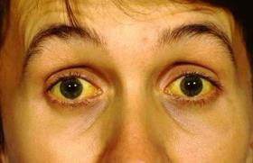
Q1. Treatment of varices? No treatment needed Q2. The cause of Esophegel varices? Portal vein thrombosis Q3. Spider nevi checked by? Diascopy Q4. How to screen for hepatocellular cancer? Serum alpha-fetoprotein (AFP)
Second picture
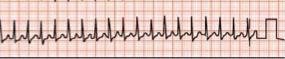
Q1. This is? SVT Q2. Mechanism? Re entry Q3. Bed maneuver to do? Carotid massage Q4. If failed? Adenosine
Third picture
A 35-year-old woman, living in a high-altitude city, comes with headaches and burning pain in her hands. On examination, she has splenomegaly. Laboratory tests reveal elevated hemoglobin, hematocrit, RBCs, WBCs, and platelets
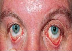
Q1. Diagnosis? Primary polycythemia Q2. Confirm test? Jak 2 test Q3. Initial treatment? Phlebotomy Q4. Treatment of pain in the hand (erythromylalgia)? Aspirin
- [COPD smoker]
- a. Apart from smoking, give 2 Most common causes?
- i. Air pollutions
- ii. Anti-trypsin
- b. What are your investigations, or what would you observe on imaging
- c. How would you treat this case?
- d. What improves mortality?
- a. Apart from smoking, give 2 Most common causes?
- [Stroke] Patient with history of arrythmia, came after losing consciousness,
- a. what the diagnosis based on CT?
- i. Ischemic Stroke
- b. Cause of this stroke
- c. Investigation
- d. Management
- a. what the diagnosis based on CT?
- [Dermatomyositis]
Picture 1 — metallic valve

- What is in the X-ray?
- Metallic valve
- What is the cause?
- Mitral regurgitation
- What medication should be given?
- Warfarin
- If developed fever what is the cause?
- Infective endocarditis
Picture 2 — erythema nodosum

- What is the name of this lesion?
- Erythema nodosum
- What is the cause?
- Sarcoidosis
- How to confirm?
- Lymph node biopsy
- What is the treatment?
- Immunosuppression
Picture 3 — Trousseau’s sign

- What is the name of the sign?
- Trousseau’s Sign
- What is the cause?
- Hypocalcemia
- What is the treatment?
- Calcium gluconate
- What is the passible associated?
- Chronic kidney disease
13. Case Scenario
13.1 Case 1: productive cough, fever, yellowish sputum
30 years old patient with productive cough, fever and yellowish sputum.

Picture similar to this but was veerryyy obvious and clear
- What is the X-ray finding? Whaite opacity in the right lobe
- What is the possible diagnosis? Broncho-pneumonia
- Mention 1 common cause. Strep. Pnuemoniae
- Mention other 2 causes. Mycoplasma and H. influenzae
- Give possible complication. ARDS
- Mention any criteria from CURB 65. Confusion and high urea
13.2 Case 2: 67 years old patient presented with chest pain

- What is the ECG finding? Extensive anterolateral MI
- What are the arteries occlusion? LAD and Circumflex artery
-
- Patient become cyanosed, agitated with palpitations.
- What is the new diagnosis? Acute pulmonary edema OR cardiac tamponade
- What treatment given? Diuretics
- What is the cause of this complication? Septum rupture OR rupture of ventricular wall (Cardiac tamponade)
- What medications to relieve patient’s symptoms? Morphine + antiemetics and Nitroglycerine
Scenario 1
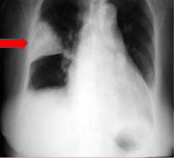
Q1. What you see? Hampton hump sign
Q2. Dx? pulmonary Embolism
Q3. ECG feature? S1, Q3, T3 pattern depression
Q4. Give 3 Risk factors? 1- Prolonged bedrest due to any cause 2- pregnancy 3- long air travel
Q5. Score system name? Wells score
Q6. Give 2 drugs to treat? Heparin & warfarin
Scenario 2
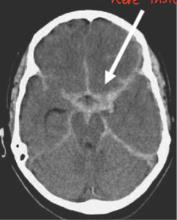
Q1. Dx? Subarachnoid hemorrhage
Q2. Etiology? ruptured berry aneurysm
Q3. Give 3 causes of cerebral hemorrhage? 1- Hypertensive hemorrhage 2- Cerebral amyloid angiopathy 3- Aneurysmal rupture
Q4. Other investigation to confirm diagnosis? MRA
Q5. Give 2 causes of death? 1- Rebleeding 2- vasospasm
Q6. Give one drug and one procedure to prevent the above complication? Drug: Nimodipine – calcium channel blocker Procedure: Endovascular coiling or surgical clipping