ARDSAcute Resp. Distress Syndrome
DR WAQAR
WHAT IS IT?
It is an acute and severe lung injury secondary to some other acute pathology going on either in the lung or anywhere else in the body
It occurs within 1 week of the onset of some other condition (discussed under etiologies)
Mortality is very high in ARDS
Previously called “adult” respiratory distress syndrome
PATHOGENESIS
- There is damage to the blood vessels in the lungs, causing increased capillary permeability. This leads to fluid accumulation in the alveoli and interstitial spaces, just like pulmonary edema, BUT in the absence of CHF( non cardiogenic pulmonary edema).
- Also, it severely reduces gas exchange in the alveoli ( causing hypoxemia)
WHAT HAPPENS AS A RESULT?
-
- Pulmonary edema (non cardiogenic)
-
- Severe hypoxemia ( difficult to improve even with O2)
-
- Stiff lungs (decreased lung compliance)
-
- Diffuse pulmonary infiltrates Bilateral
ETIOLOGIES
-
- Sepsis ( most common cause)
-
- Inhaling harmful fumes chemicals
-
- Severe pneumonia
-
- Body trauma/Fractures
-
- Acute pancreatitis
-
- Burns
-
- Aspiration
S/S
- Acute dyspnea
- Tachypnea
- Cyanosis
- Anxiety
- Fever
- Crackles on auscultation
Symptoms usually occur within a few hours to few days, after the inciting event (eg burns, sepsis etc)
Patient is very critically ill.
BERLIN CRITERIA FOR DIAGNOSIS
According to the “Berlin Criteria”, the diagnosis of ARDS is characterised by the following 4 points:
-
- It is an acute lung injury (occurring within 1 week of another disease)
-
- Bilateral lung infiltrates (not due to any other lung pathology)
-
- Respiratory failure (not explained by any other issue)
-
- Reduced PO2/FiO2 ratio (means severe hypoxia)
In ARDS, there is severe hypoxia in the blood, so PO2 is very low:
eg. If PO2 is 30 & he is on room air, pO2/FiO2 ratio will be 30/0.21 = 142
This is what happens in ARDS
What is PO2 and FiO2?
-
- pO2: Partial pressure of O2 in the blood, or in other words, the O2 level in the blood.
Normal: 75-100mmHg (on ABG)
60 mmHg
-
- FiO2: O2 concentration that you breathe.
Norm. FiO2 on room air: 0.21 (air has 21% O2)
inspired inhaled
So, normal pO2 / FiO2 ratio: 75/0.21 = 357 (or greater, depending on pO2 value)
Normal pO2/FiO2 ratio should be > 300. In ARDS it is less than 300
SEVERITY OF ARDS
According to the Berlin Criteria, ARDS can be divided into 3 severity categories :
- MILD: PO2/FiO2 ratio 200-300
- MODERATE: PO2/FiO2 ratio 100-200
- SEVERE: PO2/FiO2 ratio <100
(normal is > 300)
INVESTIGATIONS
-
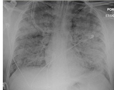
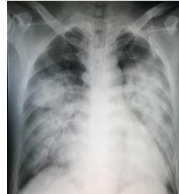
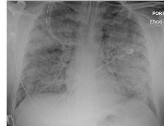
- Chest X ray: Pulmonary infiltrates, usually bilateral & diffuse.
- Infiltrates appear quickly & progress rapidly
- If the patient survives, X ray starts improving after 2 weeks but sometimes may take a long time to normalise
ARDS X-Rays
-
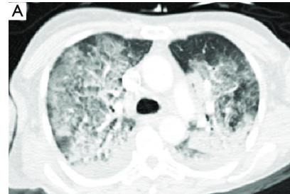
- C.T. scan:
- Gives more details than the X ray
- Not LAAZIM in all patients because diagnosis is clear from overall clinical scenario & X ray
- Before ordering CT, weigh the risk & benefit of doing it (difficult to take a sick ventilated patient for a CT)
CT chest in ARDS
-
- PET scan
-
- Chest ultrasound
-
- ABG: Shows hypoxemia
-
- A-a gradient is high ( what is A-a gradient? →
please listen to me

Alveolar O2 = arterial O2
as can’t diffuse into blood due to
blood, inflammatory infiltrates interstitium
as dist in lungs I alveoli is good but not
diffusing so the difference (A-a gradient is high)
Treatment ICU Admission
The main aim is to give O2 and since many patients are very sick & have very low O2 levels, they are put on a ventilator
-
- Mechanical ventilation with low tidal volume
-
- In some cases, prone position of the patient helps the hypoxemia
the amount of air in going in & out
normal: 500
by face it is stiff to protect from rupture (pneumothorax) → Barotrauma
Corticosteroids & inhaled Nitric oxide have no role in management. ( They were used long time ago)
No drug has shown definitive mortality benefit
Diff. Diagnosis
-
- Cardiogenic pulmonary edema(CHF) Diastolic HF (no Cardiomegaly)
-
- Diffuse pneumonia
-
- Pulmonary hemorrhage
-
- Acute exacerbation of interstitial lung disease
-
- Aspiration Pneumonia
Differences
Cardiogenic pulm. edema
-
- Abnormal 2 D echo
-
- Pleural effusion mostly present
-
- Cardiomegaly
-
- Raised blood levels of a substance called BNP
-
- S3 & S4 on auscultation gallop rhythm
-
- Raised JVP
-
- High wedge pressure
ARDS
-
- 2 D echo normal
-
- Pleural effusion not present
-
- No cardiomegaly
-
- BNP levels normal
-
- No S3 & S4
-
- JVP not raised
-
- Wedge pressure usually normal
what is wedge pressure?
in SMF → heart not pumping → Accumulation of blood
→ Back pressure into lungs → pulmonary BV congested
→ High pressure in lung vessel when measured by Swan Ganz cath (pulmonary capillary wedge pressure)
COMPLICATIONS
-
- Sepsis (most common cause of death)
-
- Oxygen toxicity (due to Oxygen therapy)
-
- Resp failure
-
- Ventilator related complications : motor neuron disease (can’t breathe without ventilator)
Barotrauma, Vent. associated pneumonia, tracheal ulceration (due to the ET tube)
- Ventilator related complications : motor neuron disease (can’t breathe without ventilator)
-
- Lung fibrosis
-
- DVT (bed ridden patient)
- } Later complications
pt. on ventilator, what daily investigation have to be done? Chest X-Ray to make sure ET tube is in the right lung (Above Carina)
PROGNOSIS
- Mortality can be upto 40% in the hospital
- Most people who survive, recover their normal lung function in 6 months to a year.
- Some survivors may develop lung fibrosis later in life
Sepsis is the most common cause of death in ARDS patients