CIRRHOSIS
PART 2
Primary Biliary CHOLANGITIS (PBC)
POINTS:
- Previously called primary biliary cirrhosis
- It is mostly seen in middle aged females.
- There is damage to the “intra hepatic” bile ducts, caused by antibodies. This produces stasis of bile ( intra hepatic cholestasis)
- Finally, it leads to cirrhosis
It is an autoimmune disease
S/S of PBC
- Pruritus : Due to accumulation of bile salts
- Jaundice & fatigue
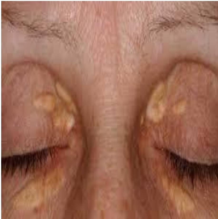
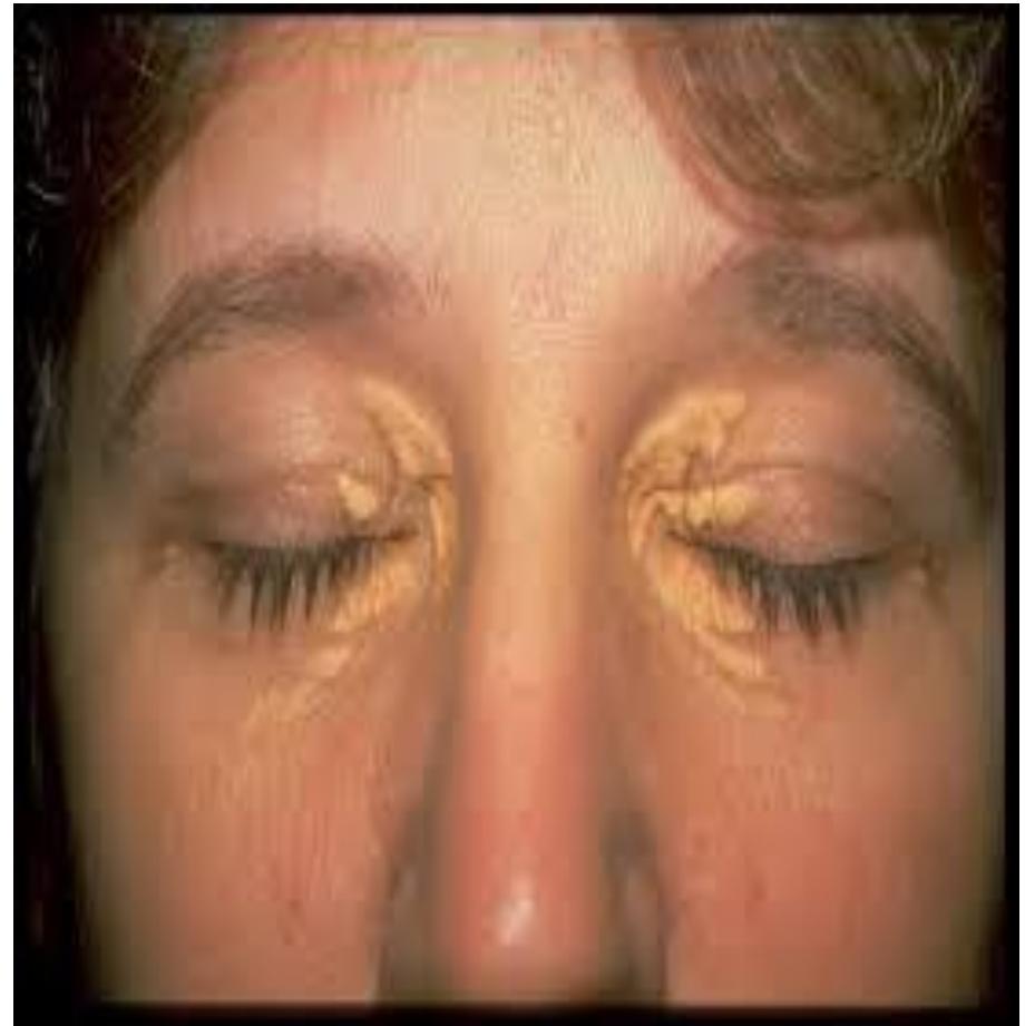
- Xanthelasma: These are cholesterol deposits around the eyes ( Blood cholesterol is very high)
- Fat malabsorption
1) & 2) are the most common symptoms.
Xanthelasma
INVESTIGATIONS
-
Antimitochondrial antibody in the blood ( A.M.A. → Diagnostic)
-
LFTs:
-
Alk.Phos. & bilirubin are very high ( obstructive pattern)
-
AST & ALT minimally high
-
-
High serum Cholesterol
-
Liver Biopsy: Shows destruction of the intra-hepatic bile ducts.
Treatment of P.B.C.
There is no cure of this disease, except liver transplant. Medicines just control the symptoms and slow the progression.
- Urso-deoxy-cholic acid
- Liver transplant ( in advanced disease)
- Symptomatic treatment for pruritus (cholestyramine, antihistamines, Rifampicin)
- Vit A, D & E supplements ( fat soluble vitamins)
It is a progressive disease if not treated
HEMOCHROMATOSIS
- A genetic disease (C282 y gene)
- Too much iron (Fe) absorbed from the intestine & deposited in many organs
- Common organs affected:
| * Liver | * Pancreas | * Skin | * Heart |
| * Joints | * Brain | * Testes |
S/S
- Cirrhosis
- DM
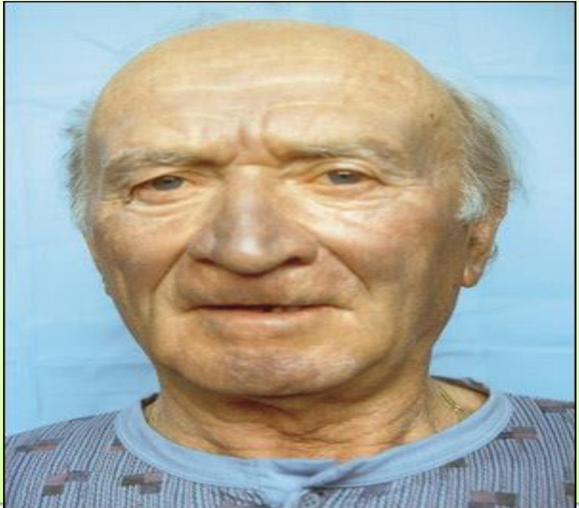
- Skin discoloration
- Cardiomypathy
- Joint pains
If you see these
S/S, think of
hemochromatosis
Note the grey/bronze discoloration on cheeks,
chin & nose
DIAGNOSIS
- Serum ferritin is high. Iron also high.
- Liver biopsy ( gold standard)
- Genetic analysis ( gold standard) → shows C 282 Y gene
TREATMENT OF HEMOCHRO.
- Repeated phlebotomy to remove blood
- Iron chelation therapy with De-feroxamine
First choice treatment is phlebotomy ( forever).
If phlebotomy cannot be done,  chelation therapy with Deferroxamine tablets
Always screen family members of the patient
Autoimmune Hepatitis
Liver damage due to autoantibodies. It can cause acute hepatitis, chronic hepatitis and finally cirrhosis.
SOME GENERAL POINTS
- a) More in females
- b) It is an autoimmune disease, so etiology unknown
- c) Other autoimmune diseases can also be present eg. Grave’s dis. , thyroid disease, celiac disease, rheumatoid arthritis etc.
TYPES OF AIH
Type 1
- more common
- Anti SLA ab +
- ANA +
- Anti smooth muscle ab + (ASMA)
Type 2
- less common
- Anti SLA +
- Anti LKM 1 ab.+
(anti SLA is highly sensitive for autoimmune hepatitis)
S/S
- Can be asymptomatic, incidentally discovered by LFTs
- May present as acute hepatitis
- May present later with S/S of cirrhosis
- Fatigue, jaundice, anorexia, mylagias
INVESTIGATIONS
- LFTs: ALT & AST are much higher than alk. phos.( cell damage pattern)
- Serum globulins are high
- Antibodies: As discussed before
- Liver biopsy: May be done for definite diagnosis :
- shows “piecemeal necrosis” with lymphocytic infiltration
TREATMENT
- Prednisone tabs. or
- Prednisone + Azathioprine
Treat for a year and then taper.
- Liver transplant for end stage liver disease
- Relapse after stopping treatment is frequent,
and may need re-treatment - Steroids + Immunosuppressive (azathioprine)
( standard treatment) - Treatment is usually lifelong in most patients
( disease can relapse after stopping) - Once it has progressed to cirrhosis, liver transplant is the only curative treatment
If cirrhosis has occurred, it will not be reversed
CLINICAL TIP
Long term use of PPIs (omeprazole, pantoprazole etc) can cause :
- Vit B12 deficiency
- Magnesium deficiency
So, anyone taking long term PPIs, check for the above deficiencies
LAST SLIDE IS VERY IMPORTANT