Pulmonary Embolism — High-Yield Revision
Diagnosis → A–E severity → treatment → follow-up. All 61 original image references appear in collapsed, topic-matched figure galleries.
Clinical route
Suspect PE → assess shock → Wells/Geneva → PERC only if very low risk; otherwise D-dimer/YEARS if appropriate, or CTPA directly if high risk → A–E, RV, troponin/BNP → anticoagulation ± reperfusion. CTPA contraindicated → V/Q SPECT. No DVT signs ≠ no PE.
1. Mechanism, causes & presentation
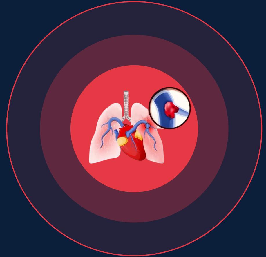
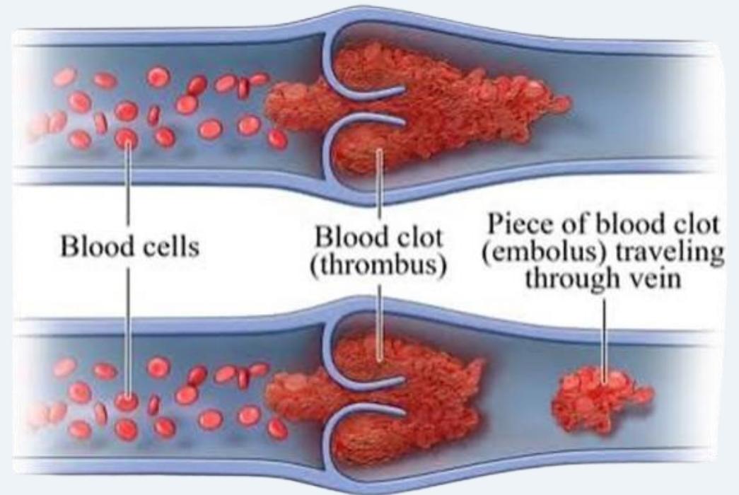
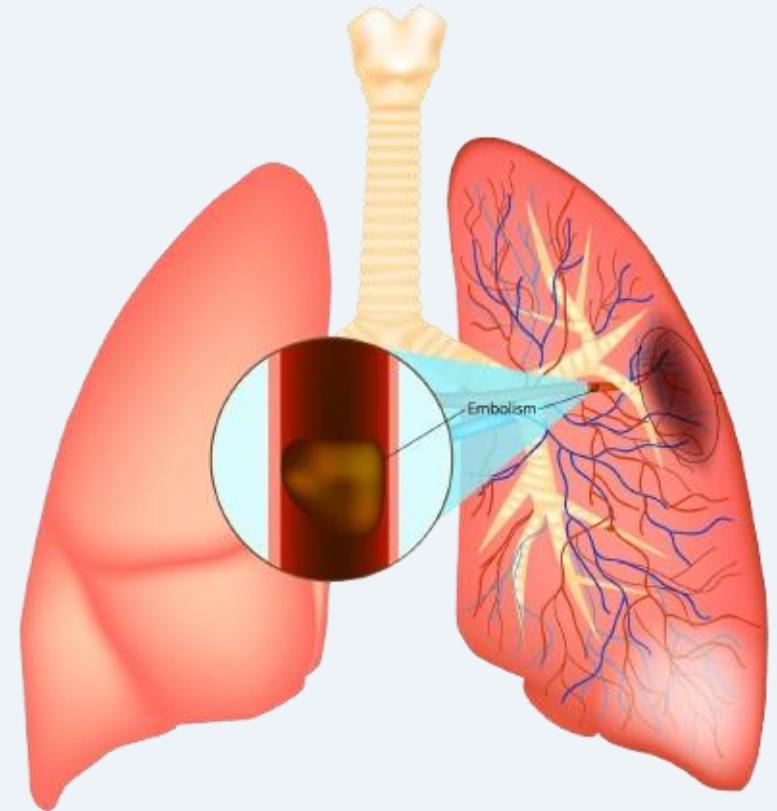
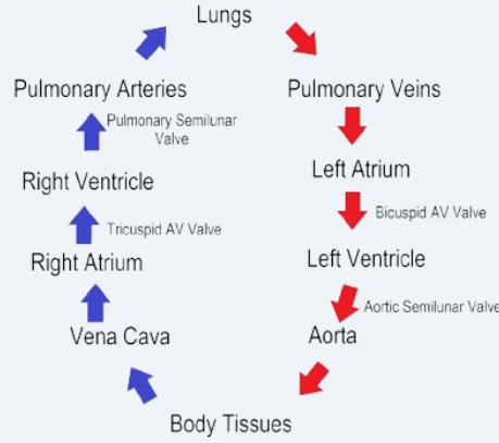
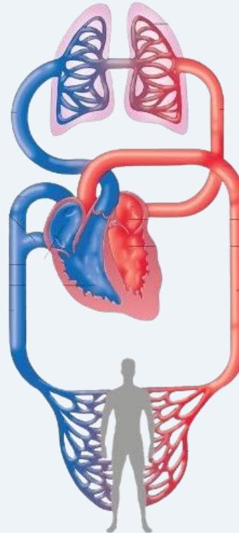
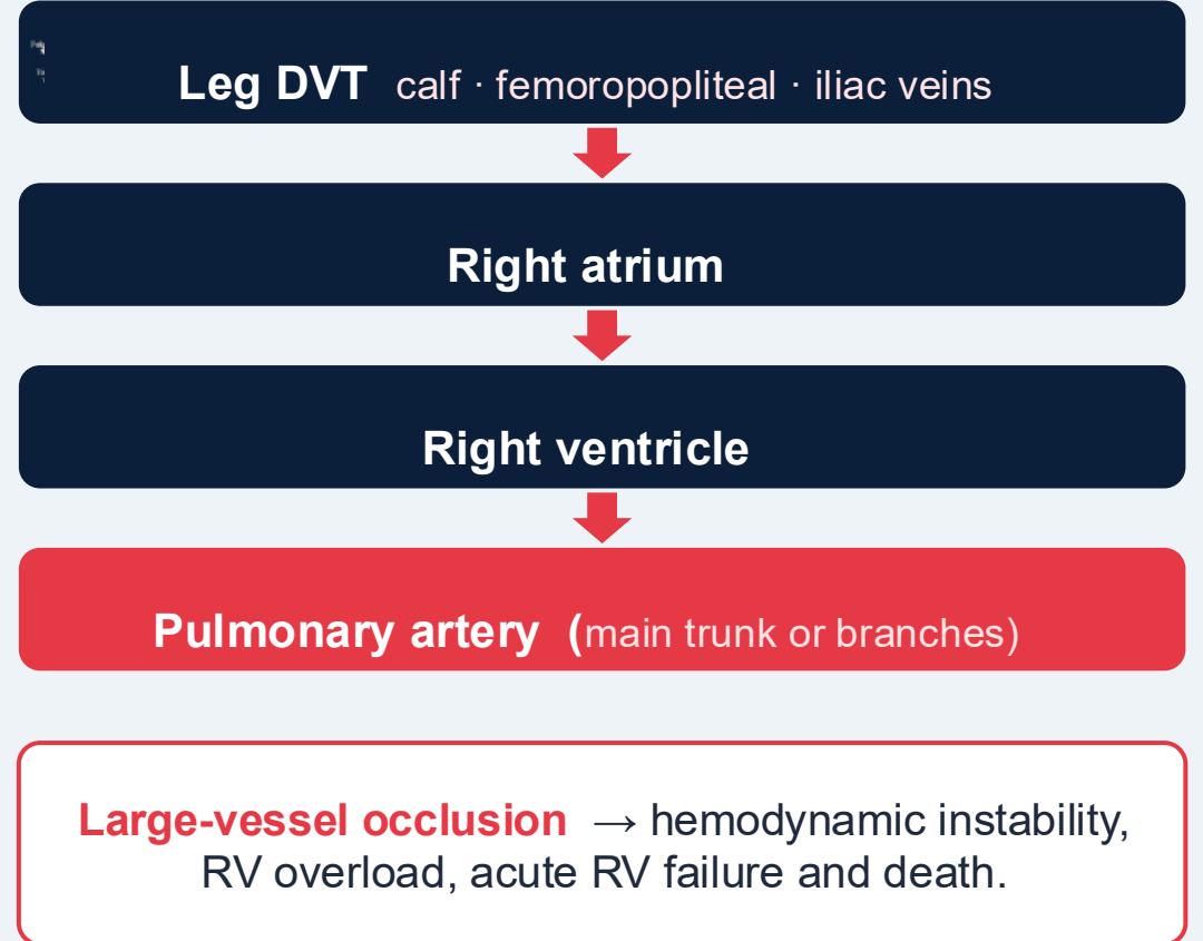
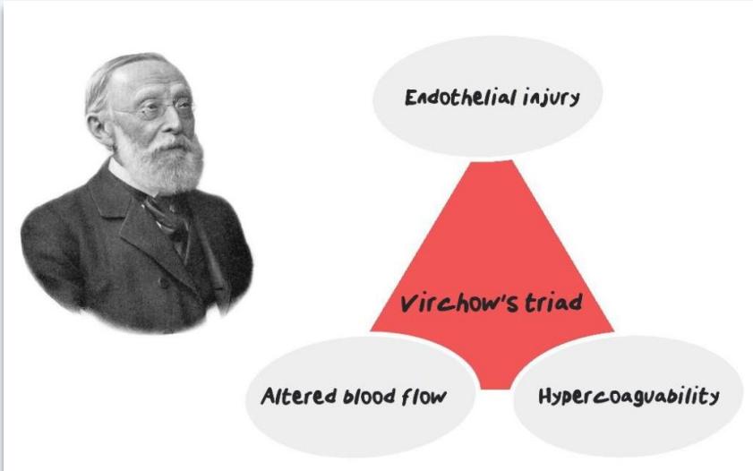
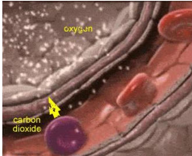
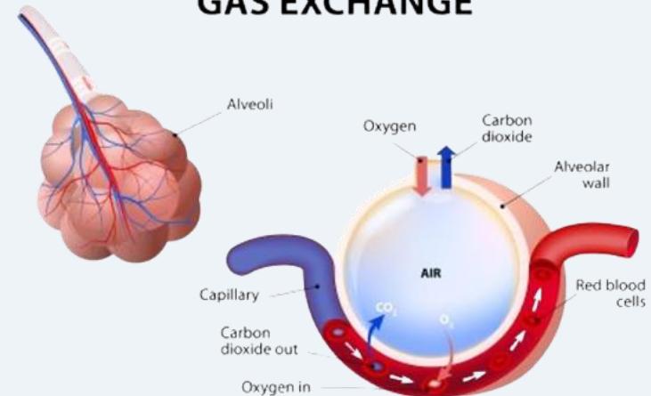
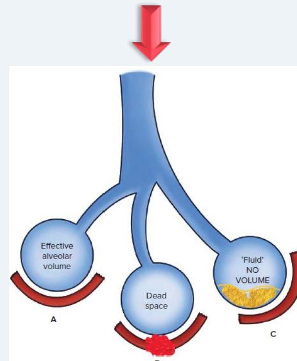
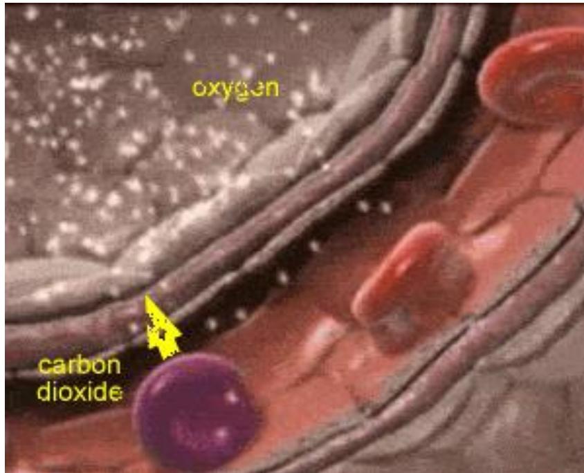
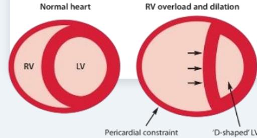
Thrombus = stationary intravascular clot; embolus = dislodged traveling material. Usually leg/pelvic DVT → right heart → pulmonary arteries. Virchow triad: endothelial injury, stasis, hypercoagulability. Normal V/Q ≈1 permits O₂ entry/CO₂ exit. Occlusion creates ventilated/unperfused dead space (high V/Q); redirected blood produces low-V/Q units → hypoxemia. ↑PVR → RV dilation/failure → leftward septal bowing → ↓LV preload/output → hypotension → RV ischemia/arrest.
| Reversible major | Reversible minor | Persistent |
|---|---|---|
| GA surgery >30 min; bedbound admission ≥72 h; cesarean; lower-limb fracture | GA surgery <30 min; hospital admission <72 h; bedbound at home ≥72 h; estrogen, peripartum, trauma with immobility ≥72 h | Active cancer; APS/SLE, thrombophilia; chronic immobility |
| Symptoms | Examination |
|---|---|
| Dyspnea (~70%; commonest); tachypnea; pleuritic pain (sharp on inspiration); cough/hemoptysis (infarction); syncope; DVT leg pain/swelling | HR >100, RR >20, hypoxemia; SBP <90 suggests danger; loud P2, RV heave, JVD; diminished breath sounds, rub, crackles; unilateral warm, tender, red swollen leg. Leg findings may be absent. |
Figures: Pathophysiology (14)


2. Pretest probability & rule-out
| Wells | Pts | Revised Geneva | Pts |
|---|---|---|---|
| DVT signs; PE most likely | 3 each | Age >65; prior VTE | 1; 3 |
| HR >100; immobility ≥3 d or surgery <4 wk; past VTE | 1.5 each | Surgery/fracture ≤1 mo; cancer | 2 each |
| Hemoptysis; active cancer | 1 each | Unilateral leg pain; hemoptysis | 3; 2 |
| — | — | HR 75–94 / ≥95 | 3 / 5 |
| — | — | Leg tenderness + unilateral edema | 4 |
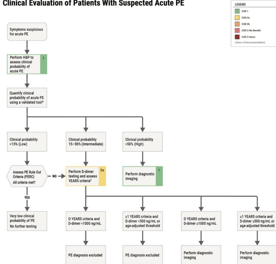
Wells ≤4 → PE unlikely → D-dimer; >4 → PE likely → CTPA. Geneva 0–3 low; 4–10 intermediate; ≥11 high.
| Rule | Exact application |
|---|---|
| PERC | Only gestalt <15%: age <50, HR <100, room-air SpO₂ ≥95%, no unilateral leg swelling, hemoptysis, surgery/trauma <4 wk, prior VTE, or oral estrogen. All 8 negative → no tests; otherwise evaluate further. |
| YEARS | Three items: DVT signs, hemoptysis, PE most likely. 0 → D-dimer <1,000 ng/mL FEU rules out; ≥1 → <500 ng/mL FEU rules out; otherwise imaging. Lecture: ~14% fewer CTPAs. |
| Age-adjusted D-dimer | If >50 years: age ×10 ng/mL FEU (same numeric value in μg/L FEU), in eligible low/intermediate-risk strategies; check assay units. |
| Probability pathway (2026) | <15%: PERC → D-dimer if failed; 15–50%: D-dimer strategy; >50%: image directly (do not exclude with D-dimer). |
Figures: Algorithm (1)
3. Investigations: diagnosis vs prognosis
| Investigation | What to remember |
|---|---|
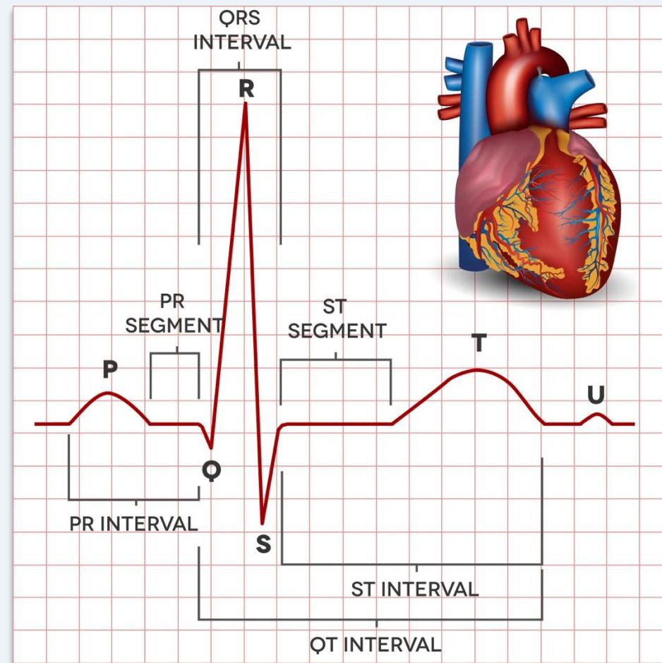
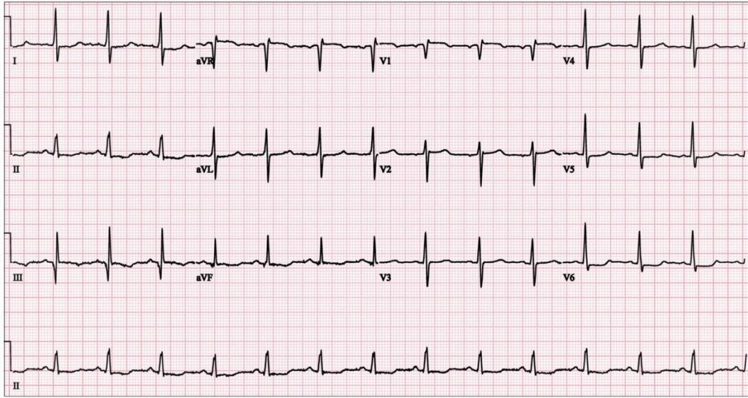
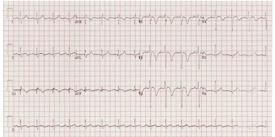
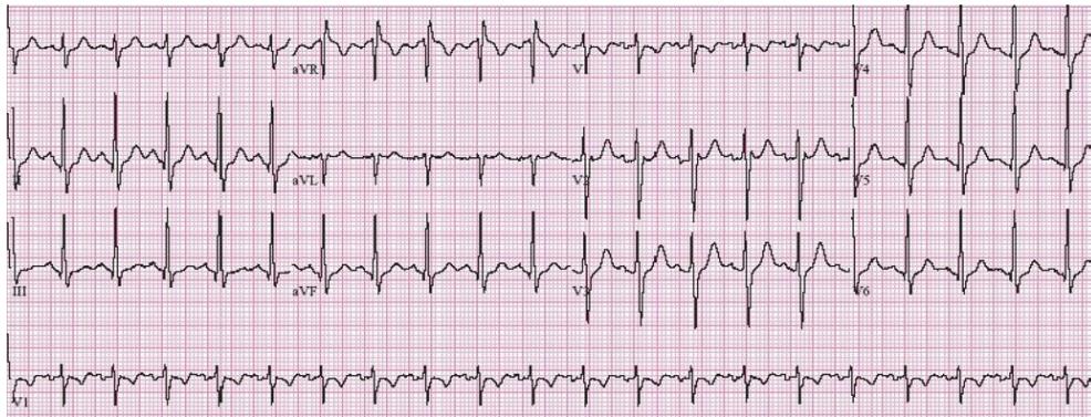
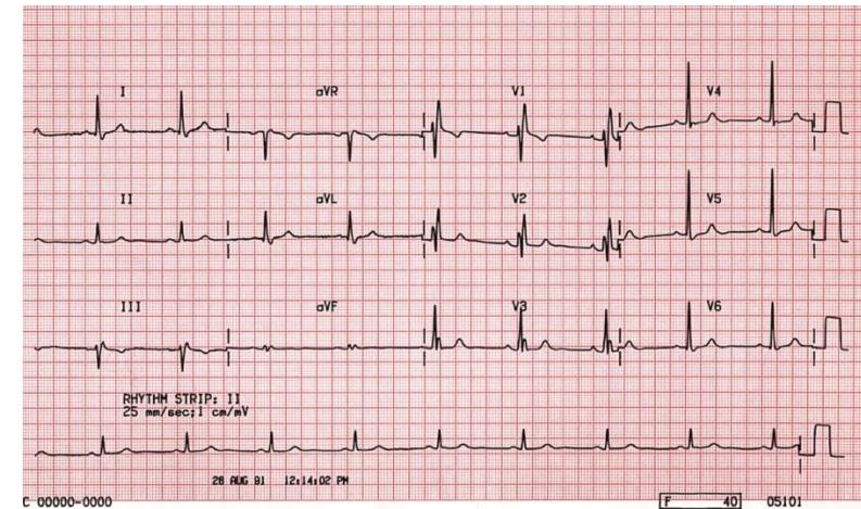
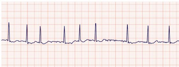
| ECG | Sinus tachycardia = commonest; S1Q3T3 (McGinn–White) = deep S (I), Q + T inversion (III), classic but insensitive; T inversions V1–V4/III/aVF, right axis, RBBB, new AF → RV strain. Not diagnostic. |
| Labs | D-dimer often falsely raised: pregnancy, older age, cardiac disease, malignancy, trauma, infection. Troponin = RV injury; BNP/NT-proBNP = RV stretch (prognosis); ABG often hypoxemia/hypocapnia; normal results do not exclude PE. |
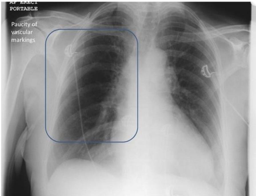
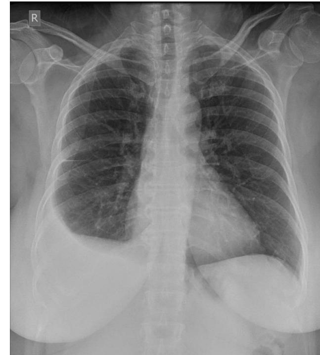
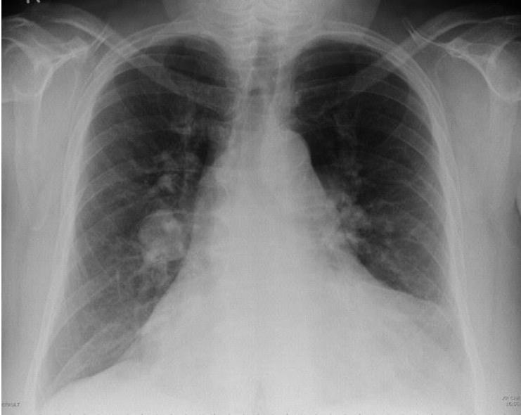
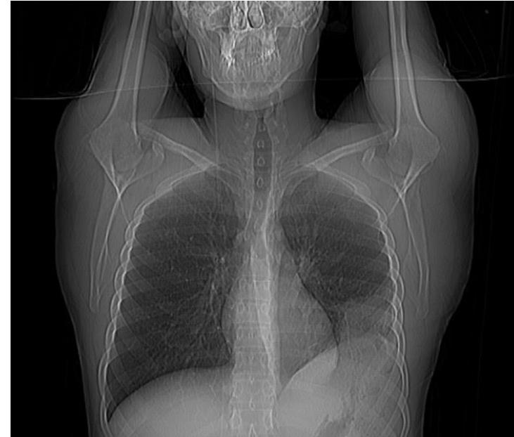
| CXR | Exclude pneumonia/pneumothorax, not PE. Westermark = focal oligemia (~10–15% sensitivity; lecture quiz ~92% specificity); Hampton hump = pleural wedge infarct (~20%); Fleischner = central PA enlargement (~20%); effusion ~35%. |
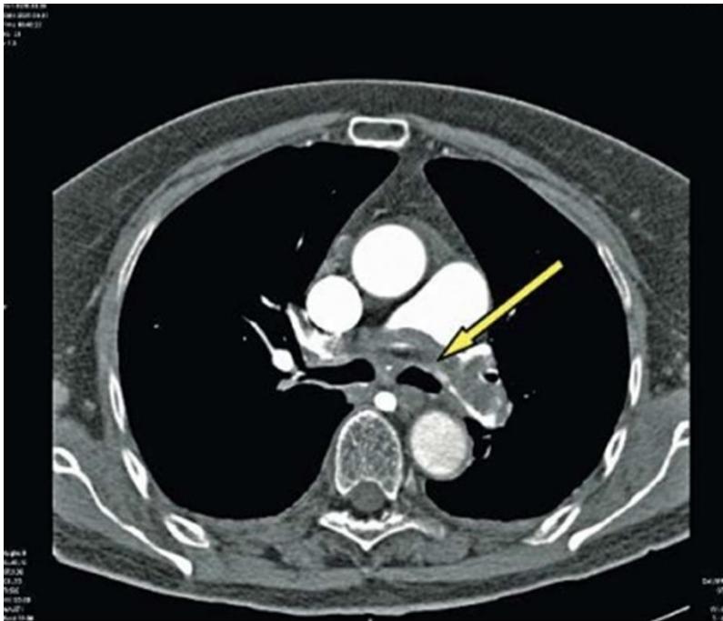
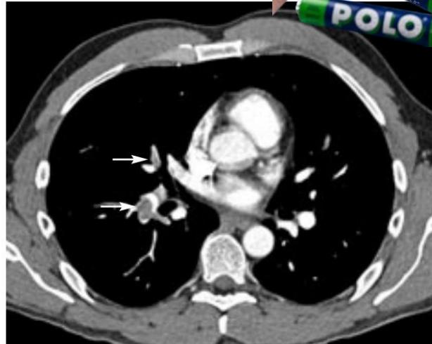
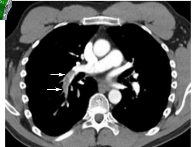
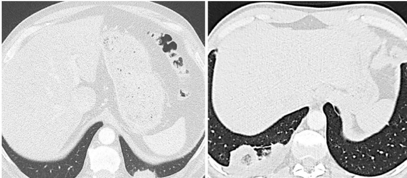
| CTPA | First-line confirmation, specificity ~96% (lecture). Central polo-mint filling defect, saddle clot at PA bifurcation. High suspicion + negative scan → consider further evaluation. Infarct: peripheral wedge/reversed-halo (atoll) ± effusion, especially small branches <3 mm → pleuritic pain/hemoptysis. |
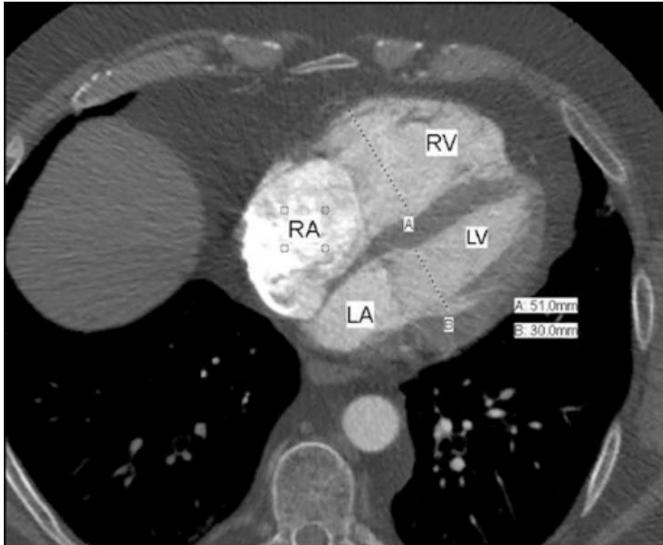
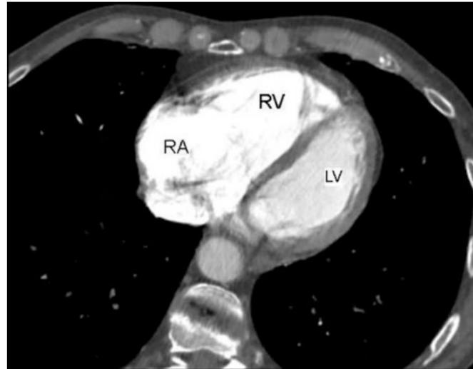
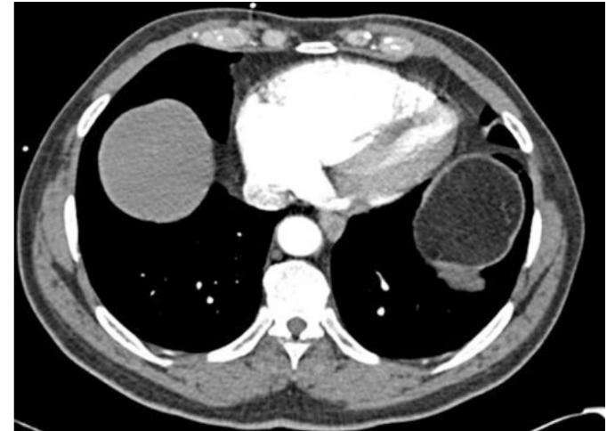
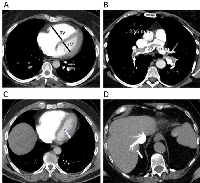
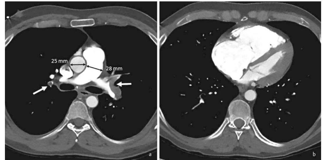
| CT RV strain | RV/LV measured diameter ratio ≥1 (report numerically), RV enlargement, septal bowing, enlarged PA relative to aorta (figure: 28 vs 25 mm), IVC/hepatic contrast reflux → worse short-term prognosis. |
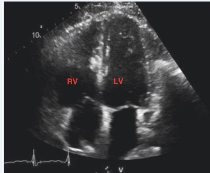
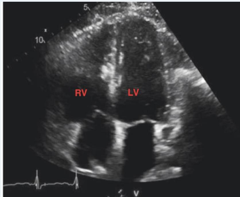
| Echo / triple POCUS | McConnell: RV free-wall hypokinesia, apical sparing. 60/60: PA acceleration <60 ms + TR gradient ≤60 mmHg. TAPSE <16 mm, RV>LV, flat septum, mobile clot-in-transit. Triple POCUS = heart + lung + leg veins (lecture ~90% sensitivity in studied settings; not definitive rule-out). |
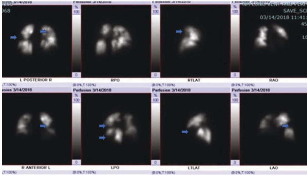
| V/Q SPECT | Segmental perfusion defect with preserved ventilation (mismatch) → PE. Preferred to MRA if CTPA unsuitable (contrast allergy/severe kidney failure). Perfusion tracer ⁹⁹ᵐTc-macroaggregated albumin IV; ventilation inhaled aerosol/carbon tracer; 3-D reconstruction. |
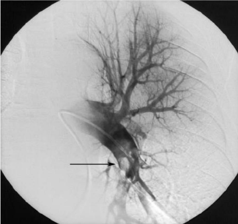
| Pulmonary angiography | Historic invasive gold standard, now chiefly during catheter intervention. |
Selective cause workup: thrombophilia: factor V Leiden, factor II mutation, homocysteine, protein C (chromogenic), protein S (activity), antithrombin III (factor Xa assay); APS: lupus anticoagulant, anticardiolipin, anti-β₂-glycoprotein-I; SLE: ANA, anti-dsDNA, anti-Sm; evaluate cancer when indicated. Not all tests are appropriate routinely or during acute anticoagulation.
Figures: Ecg (6)
Figures: Laboratory (1)
Figures: Cxr (4)
Figures: Ctpa (10)

Figures: Echo (2)
Figures: Vq (2)
4. Severity: 2011 → 2019 → 2026
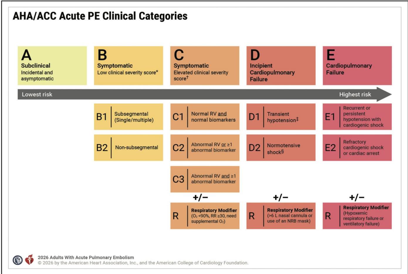
2011 AHA: low / submassive / massive → 2019 ESC: low / intermediate-low / intermediate-high / high → 2026 AHA/ACC: A–E. Wells/Geneva/YEARS predict diagnosis; PESI/sPESI/Bova assess prognosis; Hestia evaluates outpatient suitability. PESI calculator.
PESI score: age in years; male +10, cancer +30, HF +10, lung disease +10, HR ≥110 +20, SBP <100 +30, RR >30 +20, T <36°C +20, altered consciousness +60, SaO₂ <90% +20. Class I ≤65 (0–1.6%); II 66–85 (1.7–3.5%); III 86–105 (3.2–7.1%); IV 106–125 (4–11.4%); V >125 (10–24.5% 30-day mortality, lecture cohorts).
sPESI: 1 point each for age >80, cancer, heart/lung disease, HR ≥110, SBP <100, SaO₂ <90%. 0 ≈1% vs ≥1 ≈10.9% 30-day mortality in cited cohort; only carefully selected low-risk patients qualify for home management.
| Category | Distinguishing criteria |
|---|---|
| A | Incidental + asymptomatic (subclinical). |
| B | Symptomatic, PESI ≤85/sPESI 0 (lecture also Bova ≤4/Hestia 0); B1 single/multiple subsegmental, B2 non-subsegmental. |
| C | Symptomatic, PESI >85/sPESI ≥1 (lecture: Bova >4/Hestia ≥1); C1 RV and biomarkers normal; C2 one abnormal; C3 both RV + biomarker abnormal. |
| D | Impending cardiopulmonary failure: D1 transient hypotension; D2 normotensive shock (hypoperfusion despite preserved BP). |
| E | Failure: E1 recurrent/persistent hypotension with shock; E2 refractory shock or cardiac arrest. |
Shock markers: arrest needing CPR; SBP <90 or drop ≥40 for >15 min (not arrhythmia/hypovolemia/sepsis); pressors despite adequate filling plus hypoperfusion (confusion, clammy skin, oliguria, lactate↑). R respiratory modifier: B/C SpO₂ <90%, RR ≥30 or supplemental O₂; D ≥6 L/min nasal cannula or non-rebreather mask (original lecture says >6 L); E hypoxemic/ventilatory failure. C–E: PERT assessment.
Figures: Classification (2)
5. Acute management
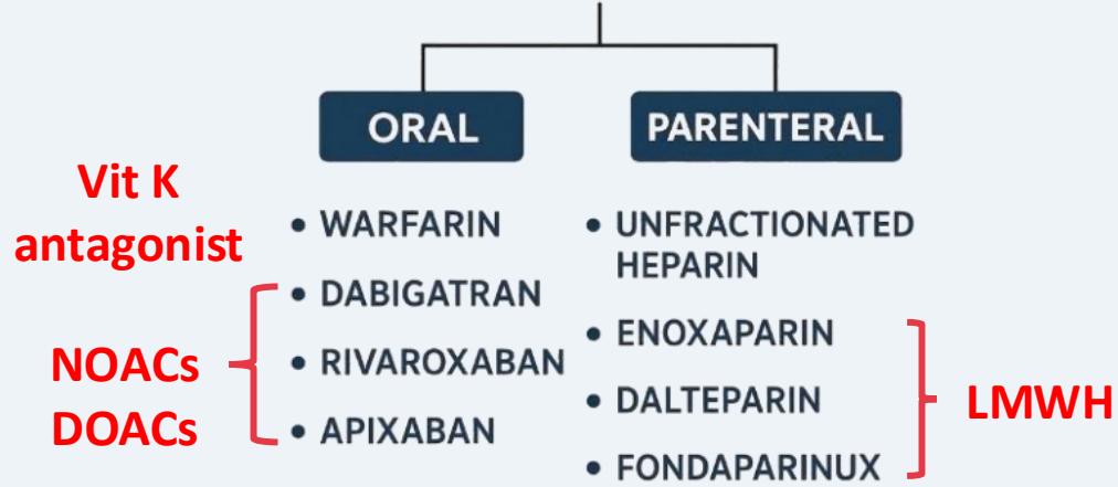
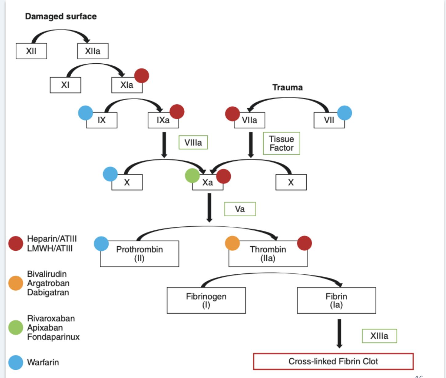
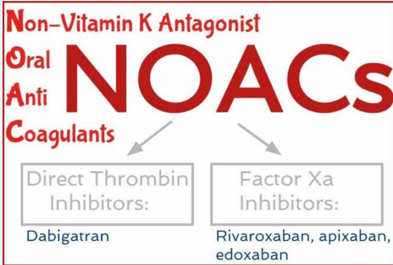
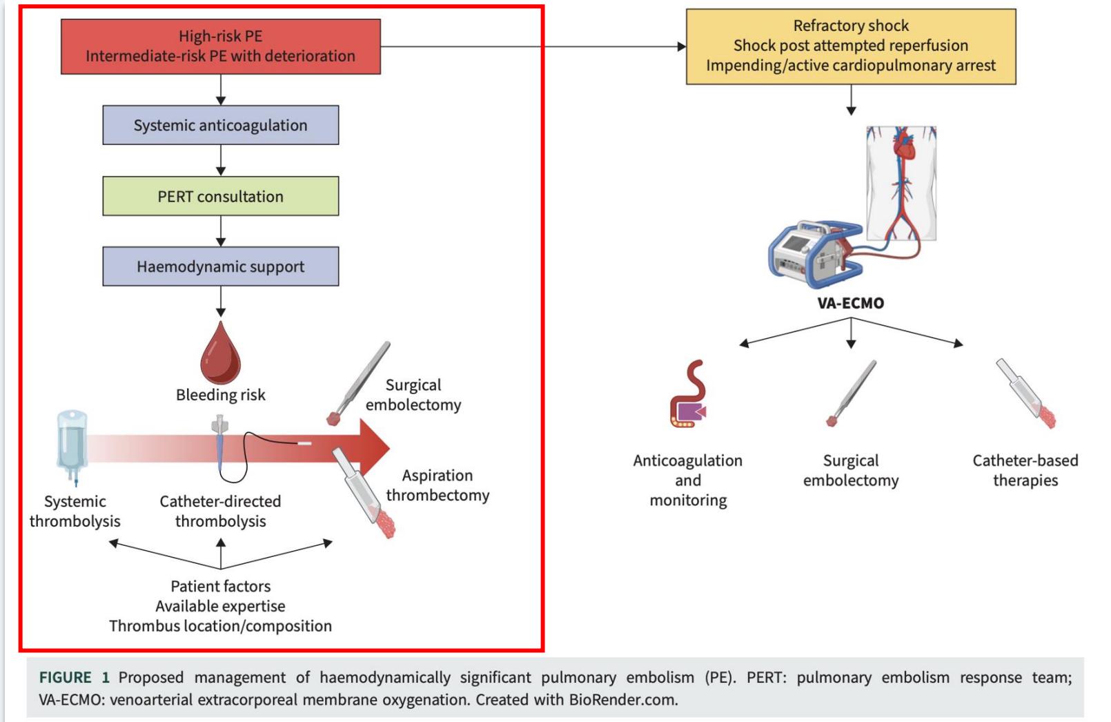
Low-risk: consider early discharge if safe. Intermediate-low: anticoagulate/admit; intermediate-high: monitor, rescue reperfusion if deterioration. AHA/ACC: C3 routine lysis benefit uncertain; D1–D2 advanced reperfusion may be considered; E1–E2 consider urgent systemic lysis when bleeding risk acceptable. O₂/support circulation (avoid RV-overloading fluids); anticoagulate promptly if safe. Parenteral LMWH preferred over UFH when suitable (fondaparinux also an option); DOACs over warfarin in eligible patients (less major bleeding; no INR checks).
| Drug | Acute treatment → maintenance |
|---|---|
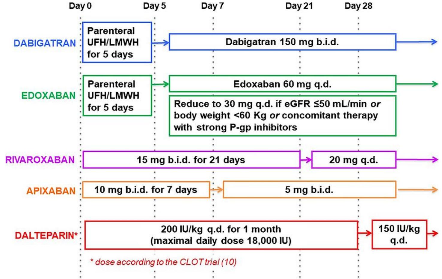
| Apixaban (Xa) | 10 mg BID ×7 d → 5 mg BID; no heparin lead-in. |
| Rivaroxaban (Xa) | 15 mg BID ×21 d → 20 mg daily; no heparin lead-in. |
| Dabigatran (thrombin) | ≥5 d UFH/LMWH → 150 mg BID. |
| Edoxaban (Xa) | ≥5 d UFH/LMWH → 60 mg daily; 30 mg if CrCl 15–50 mL/min, weight ≤60 kg or certain P-gp inhibitors (use CrCl, not eGFR alone). |
| Warfarin | Heparin overlap ≥5 d AND INR ≥2 for ≥24 h; target INR 2–3; e.g. APS/cost barriers. |
| Dalteparin (CLOT trial) | 200 IU/kg daily ×1 mo (max 18,000 IU/d) → 150 IU/kg daily. |
Systemic lysis: alteplase 100 mg IV over 2 h for appropriately selected high-risk/unstable PE. Lecture proposes 50 mg or 0.6 mg/kg to reduce bleeding with similar efficacy; evidence is less secure, so not routinely interchangeable with full dose. Other agents: streptokinase IV/24 h, urokinase IV/12 h (listed as PE-approved in lecture), tenecteplase single bolus, reteplase two boluses (last two not FDA-approved for PE, per lecture). Major danger: intracranial/retroperitoneal hemorrhage.
| Systemic lysis contraindications in lecture | Examples |
|---|---|
| Absolute/major | Active bleeding, prior ICH, ischemic stroke <3 mo, brain/spinal surgery <1 mo, CNS tumor/vascular lesion, head trauma with skull fracture/brain injury, aortic dissection, abdominal surgery <7 d. |
| Relative | BP >180/110, recent bleeding, ischemic stroke >3 mo, current anticoagulant, recent non-CNS surgery/procedure, traumatic/prolonged CPR, pericarditis/effusion, retinopathy, pregnancy, age >75, weight <60 kg. |
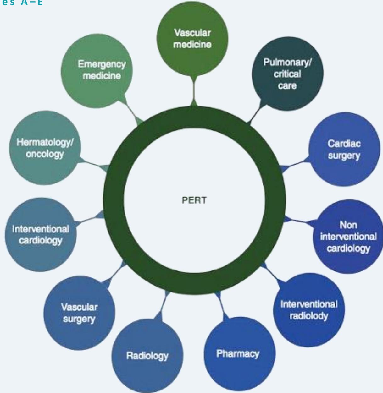
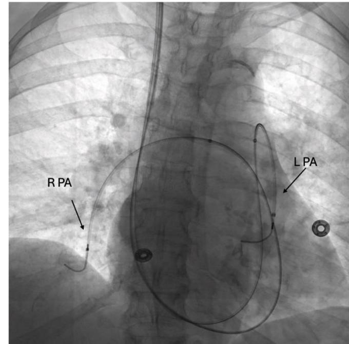
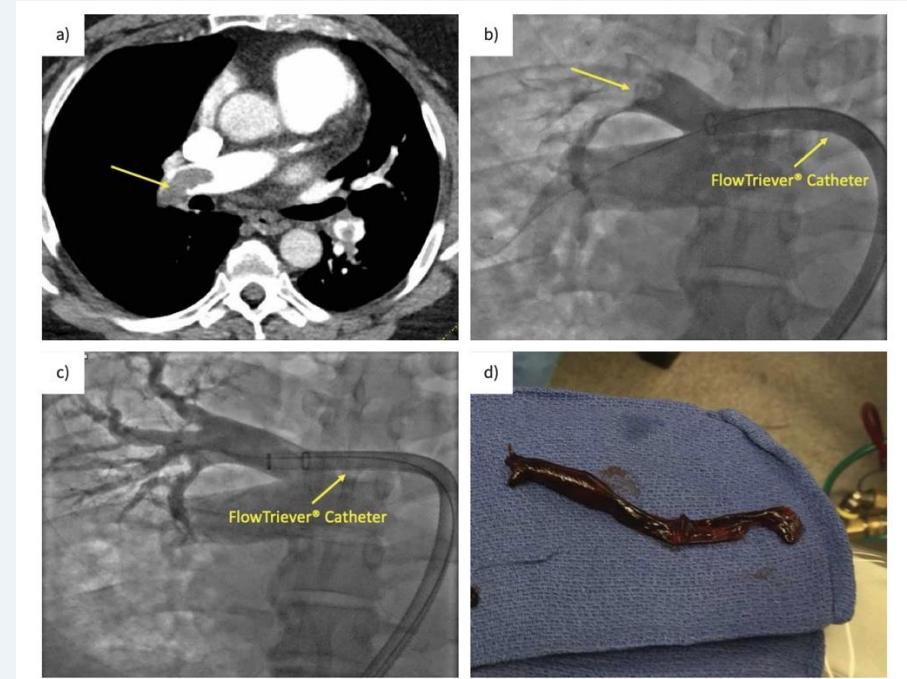
PERT (C–E/deterioration, shock, arrest) chooses anticoagulation, support, systemic lysis, catheter lysis, aspiration, surgery or VA-ECMO by instability/bleeding/anatomy/expertise. Catheter-directed lysis: consider with deterioration when systemic lysis is unsuitable (bleeding risk persists); 4–5 Fr PA catheter, lecture alteplase ~0.5–1 mg/h/catheter ×12–24 h. Mechanical aspiration: clot removal with little/no lytic → improves PA flow, RV/hemodynamics. Surgery: lysis contraindicated/failed; ECMO: bridge in refractory shock/arrest.
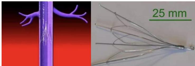
IVC filter: retrievable preferred if anticoagulation absolutely contraindicated (active bleeding); recurrent PE despite therapeutic anticoagulation is a contested exception (verify adherence/dose, consider changing agent first). Remove once anticoagulation resumes/risk subsides (lecture 29–54 d, not a universal deadline). Late risks: DVT, IVC occlusion, fracture, perforation, cardiac migration.
Figures: Treatment (8)
6. Duration, sequelae & special populations
- Major transient provocation: lecture 3–6 mo anticoagulation; commonly 3 mo. Unprovoked/persistent risk: consider extended therapy with periodic benefit–bleeding review.
- After ≥3 mo: post-PE dyspnea/functional limitation up to ~50%; RVO = persistent perfusion defect (~50%, often asymptomatic). CTED/CTEPD = symptomatic thromboembolic disease without resting PH; CTEPH = group 4 pulmonary hypertension. Persistent dyspnea warrants investigation.
| Special case | Agent choice |
|---|---|
| Active cancer | DOAC or LMWH; usually continue while active. |
| Pregnancy | LMWH/UFH; avoid DOACs/warfarin. |
| Breastfeeding | LMWH/UFH/warfarin safe; avoid DOACs. |
| Obesity (BMI >30) | DOACs generally reasonable/preferred. |
| APS | Warfarin > DOACs, especially high-risk APS. |
| Brain tumors | Individualized DOAC vs LMWH balancing bleeding. |
| CKD stage 2–3 | Suitable DOAC if renal dosing permits. |
| Child–Pugh C | Avoid DOACs. |
| HIT | Stop heparin; argatroban, bivalirudin, fondaparinux, or eligible DOAC. |
7. Non-thrombotic pulmonary emboli
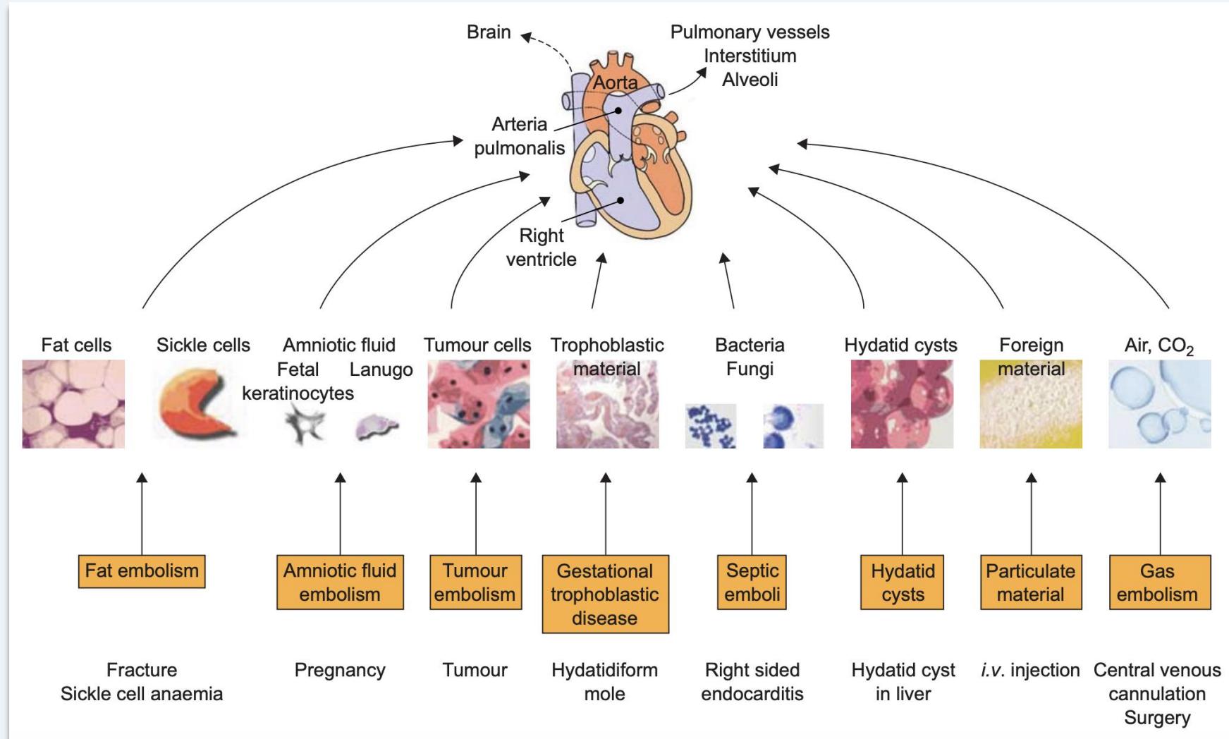
Gas, fat, amniotic fluid, septic, tumor emboli: non-biological material → chiefly mechanical blockage; biological emboli → inflammatory vascular/lung/RV injury. Management differs from thrombotic VTE.
| Type | Sources → signature | Echo | Management |
|---|---|---|---|
| Air/gas | CVC/dialysis/cardiac line, laparoscopy, diving → sudden cardiopulmonary/neurologic deterioration; mill-wheel murmur | Air ± PFO | Stop source, 100% O₂, support, aspirate air if feasible, hyperbaric O₂ for severe neurologic events; lecture left lateral + Trendelenburg when appropriate. |
| Fat | Long-bone/pelvic fracture, orthopedics, liposuction, lipid infusion; latent 6–12 h, onset 24–72 h → respiratory distress + cerebral signs + petechiae | Nonspecific | O₂/ventilation, neuro monitoring ± ECMO; no proven drug. |
| Amniotic | Labor (~70% pre-delivery per lecture), C-section, abortion, amniocentesis → abrupt hypoxemia, collapse ± DIC, seizures/coma | ↑PA pressure, RV failure, TR (TEE) | Resuscitate, correct coagulopathy (blood/FFP/platelets/cryo ± TXA), urgent delivery when indicated. |
| Septic | Tricuspid endocarditis, infected line, IV drugs, periodontal infection → fever, cough/hemoptysis, infection focus | Vegetation (TEE > TTE) | Broad-spectrum IV antibiotics + source control, remove infected line/drain abscess or empyema ± surgery. |
| Tumor | Hepatocellular, breast, renal, gastric, sarcoma, choriocarcinoma → subacute progressive dyspnea/cor pulmonale | Nonspecific | Treat cancer + pulmonary hypertension. |
Figures: Ntpe (11)


Evidence/precision
Lecture-specific prevalence figures, assay cutoffs, doses and relative/absolute lysis restrictions are retained for exam revision; actual management depends on local protocol and the patient. V/Q mismatch = perfusion (not ventilation) defect; avoid combining different D-dimer adjustment rules without a validated pathway.