Radiology and Images of Important Diseases
Dr. Nasr Eldin Yousif Ahmed
Associate Professor of Medicine & Cardiology
Department of Medicine
Al Maarefa University
How to Read Chest X Ray
Remember ABCDE
- A. Airway
- B. Bone
- C. Cardiomegaly
- Cavitation
- Calcification
- D. Density
- E. Effusion
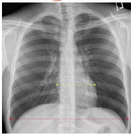
Normal X-Ray
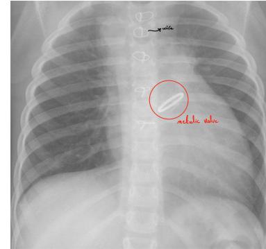
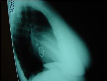
wire
-Cardiomegaly
what drugs should be given
Aspirin but - warfarin
follow up monthly for PT/INR
St of drug + bleeding
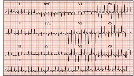
The figure shows a 12-lead ECG tracing on a standard red grid. The leads are labeled as follows:
The tracing shows a series of waves for each lead, with a clear progression of the QRS complex and T waves. A small scale bar is visible in the bottom right corner of the grid.
I aVR V1 V4
II aVL V2 V5
III aVF V3 V6
Regular
160 b/min
2/17/2026
Dr. Nasr Eldin Yousif Ahmed
6
Supraventricular tachycardia (pocket in reentry)
Ux: →
1st line → Vagal stimulation
2nd line → Adenosine or verapamil
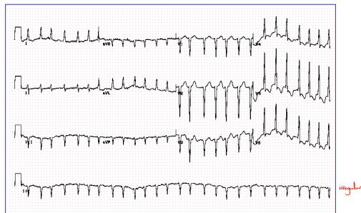
Four ECG leads (I, II, III, aVF) showing a normal sinus rhythm with a clear P wave, QRS complex, and T wave. The leads are stacked vertically.
irregular
Atrial fibrillation
Ux: →
Rate control → BB blocker
Rhythm control → Antiarrhythmic drugs
Antiarrhythmation
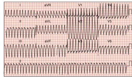
The figure shows a 12-lead ECG tracing on a standard red grid. The leads are labeled as follows:
Ventricular tachycardia
if Cardiac arrest → CPR & DC shock
if conscious → Amiodarone
→ wide QRS
monomorphic
Regular
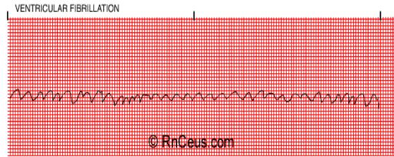
VENTRICULAR FIBRILLATION
© RnCeus.com
Ventricular fibrillation
III → CPR & DC shock
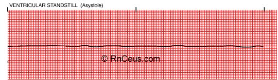
VENTRICULAR STANDSTILL (Asystole)
© RnCeus.com
Asystole
to check the pt.
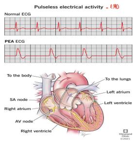
Pulseless electrical activity = EMD
Normal ECG
PEA ECG
To the body
To the lungs
SA node
Left atrium
Right atrium
Left ventricle
AV node
Right ventricle
© 2014
Cengage
Learning
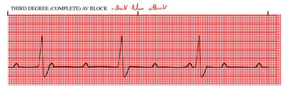
- in elderly
- complete dissociation between atrium & ventricle
- Profound bradycardia
III → Pace maker
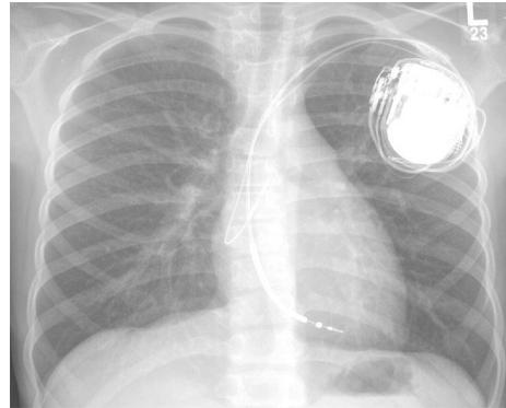
- Pace maker
- in pt. with complete heart block
- on ECG
with QRS
Pacemaker spikes (spikes before QRS) - in dual pace maker
with QRS
Pacemaker spikes (spikes before QRS / p-wave)
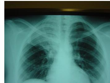
- haemorrhous opacity in Rt. upper lobe
- Pneumonia
- Antibiotic
Pt. 2 days ago had typical ischemic chest pain. Now he’s having SOB
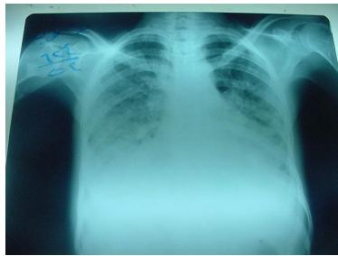
- Pulmonary edema
- due to Rupture cardiac tamponade or
low wall ventricle or Septum
or Extensive MI - imp. investigation to do?
echo
consultation
most imp diagnosis..
TB
pt. having fever, ill 3 days
Lung Abscess
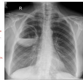
R
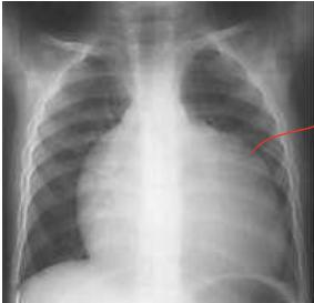
Cardiomegaly DDx
* Cardiac tamponade “Globose heart”
Congestive heart failure
multivascular lesion
ischemic cardiomyopathy
dilated cardiomyopathy
most imp investigation?
Echo
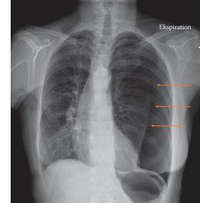
Left Side pneumothorax
Shift of trachea
Pt. is known case of Asthma / COPD / Emphysema
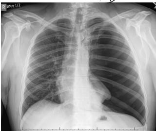
Shift of trachea
Left Side pneumothorax
Utr. →
Health Decompression = Pt. intercostal space
than chest tube
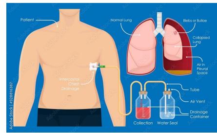
The diagram illustrates the intercostal chest drainage procedure. On the left, a patient’s torso shows the drainage tube inserted into the chest. On the right, a cross-section of a normal lung is shown with labels for the pleura, pleural space, and lung. Below the lung, a diagram shows the drainage container with a water seal and a collection bottle. Labels include: Patient, Normal Lung, Bilets or Bulbous, Collapsed Lung, Air in Pleural Space, Intercostal Chest Drainage, Tube, Air Vent, Drainage Container, Collection, and Water Seal.
Patient
Normal Lung
Bilets or Bulbous
Collapsed Lung
Air in Pleural Space
Intercostal Chest Drainage
Tube
Air Vent
Drainage Container
Collection
Water Seal
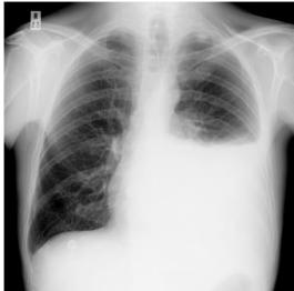
- Pleural effusion
- homogeneous opacity
- hll. → Aspiration
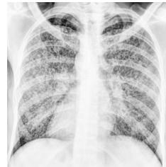
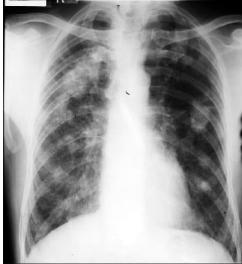
Miliary TB
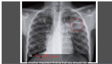
TB, a fibrous body
- cavitation
- fibrosis
TB: • fibrocavitory lesion
Another important finding that you should not
- malignancy
lung metastasis
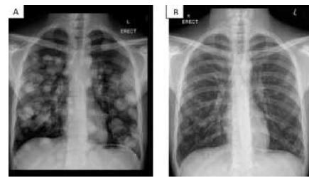
L
ERECT
malignancy
”lung metastasis”
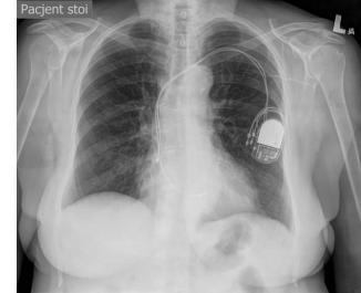
Patient stoi
Pace maker
Physical exam
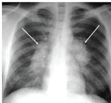
- hilar lymphadenopathy
- lymphoma
- TB
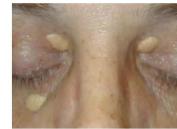
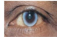
Xanthelasma
imp. test → lipid profile
imp drug → Atorvastatin
SE → myopathy , hepatitis
Arcus senilis
Physical exam
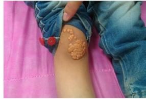
diagnosis: 1 kg hyperlipidemia
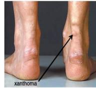
Xanthoma
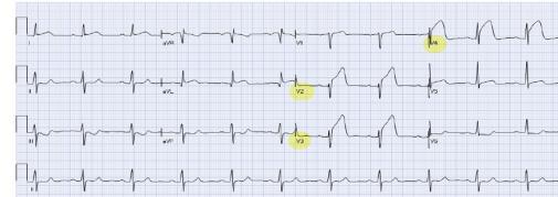
A 12-lead ECG tracing on a standard grid background. The leads are labeled: I, aVR, aVL, aVF, II, III, and V1 through V6. Lead V2 is highlighted with a yellow circle. Lead aVR is also highlighted with a yellow circle.
I aVR aVL aVF
II III V1 V2 V3 V4 V5 V6
- Atheros MI
- left anterior descending artery
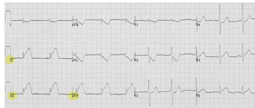
A 12-lead ECG tracing on a standard grid. The leads are labeled: I, aVR, V1, V4, II, aVL, V3, V5, III, aVF, V2, V6. Leads II and aVF are highlighted with yellow circles.
I aVR V1 V4
II aVL V3 V5
III aVF V2 V6
- inferior MI
- PL coronary artery
- Suspected PL ventricular infarction
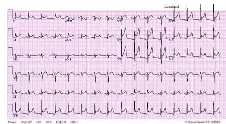
The figure is a 12-lead ECG tracing on a standard pink grid. The leads are labeled: I, II, III, aVR, aVL, aVF, V1, V2, V3, V4, V5, and V6. The tracing shows a regular sinus rhythm with clear P, Q, R, S, and T waves. The paper speed is 25mm/s and the voltage is 1mV. The patient’s name is ‘Shawland’.
Shawland
12-lead ECG tracing with leads I, II, III, aVR, aVL, aVF, V1, V2, V3, V4, V5, and V6.
25mm/s 10mm/mV 400Hz 060C 1206.204 CD: 1
Universal ST segment elevation with concave upwards PR segment depression
Pericarditis
Uk. → WASD
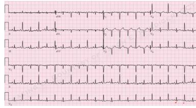
A 12-lead ECG tracing on a standard pink grid. The leads are labeled: I, aVR, II, aVL, III, aVF, V1, V2, V3, V4, V5, and V6. The tracing shows a regular sinus rhythm with clear P, Q, R, S, and T waves. There are no obvious pathological changes such as ST-segment depression or Q-waves.
I aVR II aVL III aVF V1 V2 V3 V4 V5 V6
Electrical Alterations
- Rhythmical affection
- Cardiac temperature
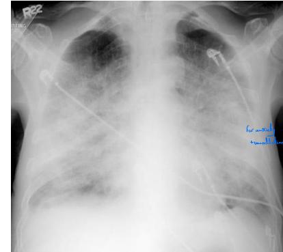
Pulmonary Edema
Ull. →
Put
Sitting position
for anxiety → morphine, automatic
transducer
the only indication for inotropes “dopamine"
"dobutamine”
is hypotensive (cardiogenic shock or
HF with L BP)
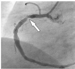
Coronary artery c shape
”narrowing”
used for IHD
Coronary Angiography
Ull. →
Stent
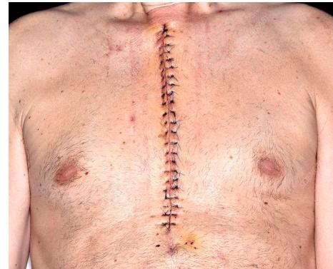
midsternal thoracotomy scar (CABG or
vascular replacement)
2 mmunics bedside should be done
look at legs for Scar of Saphenous vein
if no scar → either LIMA or RIMA
I hear the click sound of valve
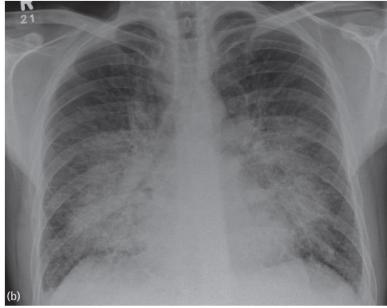
Pulmonary edema
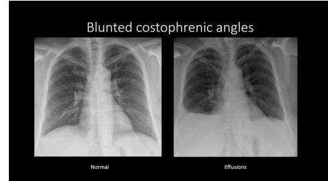
Blunted costophrenic angles
Normal
Effusions
Two side-by-side chest X-rays. The left image is labeled ‘Normal’ and shows clear costophrenic angles. The right image is labeled ‘Effusions’ and shows blunted costophrenic angles with fluid accumulation in the pleural space.
The title at the top reads ‘Blunted costophrenic angles’.
pleural effusion
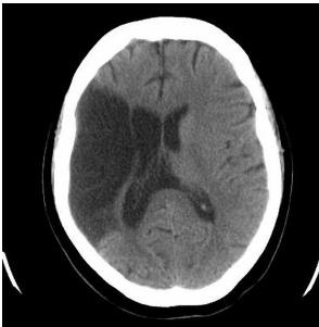
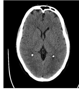
Cerebral infarction
Thrombolytic if
but test
JBP
Have check if there’s CS for TPA
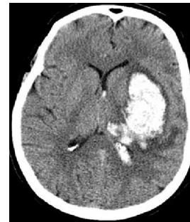
Cerebral hemorrhage
Administer mannitol
then neurosurgery consultation
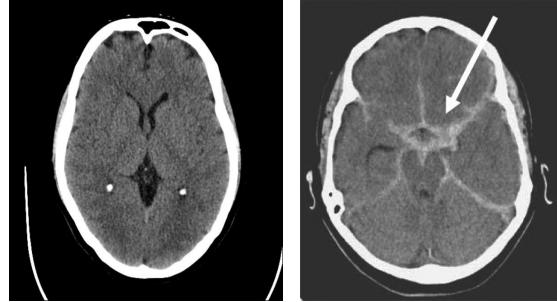
Sudden thunderclap headache
most common cz: Ruptured berry Aneurysm
41
Dr. Nasr Eldin Yousif Ahmed
2/17/2026
MRA to differ between causes of hemorrhage
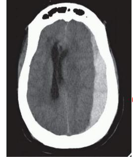
Elderly
headache
weakness
minor trauma
fluctuating level of conciousness
ttt →
Burr hole evacuation
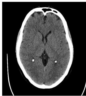
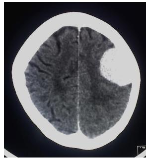
meningioma
ttt. → Resection