Collected from Basim Alhomida, , Faisal Alkharji, Leenah Turjoman, Prof. Alam, Tassnim Hakeem, Yasser Alkhudairy
اللهم يا معلّم موسى علّمني، ويا مفهم سليمان فهّمني، ويا مؤتي لقمان الحكمة وفصل الخطاب آتني الحكمة وفصل الخطاب اللهم اجعل ألستنا عامرة بذكرك، وقلوبنا بخشيتك، وأسرارنا بطاعتك، إنك على كل شيء قدير، حسبنا الله ونعم الوكيل
Table of Contents
- Station 1: Surgical Anatomy of the Abdominal Wall & Common Abdominal Incisions
- Station 2: Acute Abdomen & Intestinal Obstruction (History & Physical Exam)
- Station 3: Stoma & Colostomy Bedside Physical Examination & Assessment
- Station 4: Biliary Colic, Chronic Cholecystitis & Acute Cholecystitis
- Station 5: Obstructive Jaundice, Choledocholithiasis & Acute Ascending Cholangitis
- Station 6: Gallstone Ileus & Mechanical Bowel Obstruction
- Station 7: Acute & Chronic Pancreatitis, Complications & Surgical Site Infections
- Station 8: Acute Appendicitis (Clinical Signs, Alvarado Score & Management)
- Station 9: Groin & Abdominal Wall Hernias Examination & Surgical Repair
- Station 10: Breast Lump Clinical Scenario, Physical Examination & Triple Assessment
- Station 11: Thyroid Gland & Solitary Neck Mass Physical Examination & Assessment
- Station 12: Lower Limb Peripheral Vascular Physical Examination & Arterial Disease
- Station 13: Diabetic Foot Ulcer Examination & Surgical Management (Amputations)
- Station 14: Digital Rectal Examination (DRE / PR Exam) & Proctoscopy
- Station 15: Nasogastric Tube (NGT) Insertion Procedural Station
Station 1: Surgical Anatomy of the Abdominal Wall & Common Abdominal Incisions
A. Core Clinical Objectives & Examination Overview
- The 4 Cardinal Chief Complaints in General Surgery ():
- Pain (Acute abdomen, colic, peritonitis, ischemia)
- Lump / Swelling (Thyroid, Breast, Hernia, Lymph node)
- Ulcer (Diabetic, arterial, venous, malignant)
- Stoma / Bleeding / Fistula / Jaundice
- Master anterior abdominal wall layers to ensure safe surgical entry, anatomic closure, and prevention of incisional herniation.
- Differentiate somatic versus visceral peritoneal neuroanatomy to explain localized versus referred abdominal pain.
- Distinguish colonic morphology from small intestine during diagnostic laparotomy and laparoscopy.
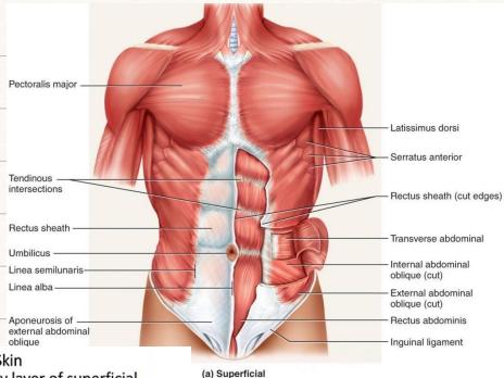
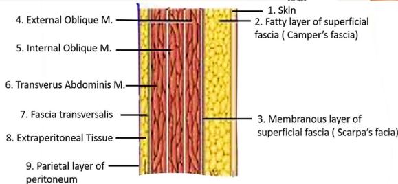
B. Anterior Abdominal Wall Layers (Superficial to Deep)
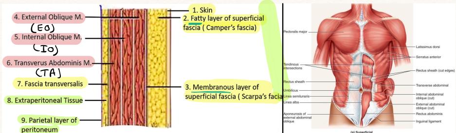
- Skin: Cleavage lines (Langer’s lines) run horizontally; transverse incisions produce optimal cosmetic healing and minimal wound tension.
- Superficial Fascia:
- Camper’s Fascia: Fatty, thick outer layer continuing into thigh and perineum.
- Scarpa’s Fascia: Membranous, dense inner layer attached to fascia lata of thigh; continues into perineum as Colles’ fascia (confines extravasated urine or hematoma).
- External Oblique Muscle: Fibers run inferomedially (“hands in pockets” — directed downwards, forwards, and medially). Aponeurosis terminates medially in linea alba and inferiorly rolls under to form the Inguinal Ligament (Poupart’s).
- Internal Oblique Muscle: Fibers run superomedially (upwards and forwards), perpendicular to external oblique.
- Transversus Abdominis Muscle: Deepest flat muscle layer; fibers run strictly transversely / horizontally.
- Fascia Transversalis: Continuous fascial lining of internal abdominal cavity; forms posterior wall of inguinal canal and deep inguinal ring.
- Extraperitoneal Fat
- Parietal Peritoneum: Innervated by somatic sensory intercostal nerves (T7–L1) sharp, well-localized somatic pain and muscular guarding upon inflammation.
C. The Rectus Sheath & The Arcuate Line
- Above the Arcuate Line (Upper 3/4):
- Anterior Sheath: External Oblique aponeurosis + Anterior lamina of Internal Oblique aponeurosis.
- Posterior Sheath: Posterior lamina of Internal Oblique aponeurosis + Transversus Abdominis aponeurosis + Fascia Transversalis.
- Below the Arcuate Line (Lower 1/4 — halfway between umbilicus and pubic symphysis):
- Anterior Sheath: All three aponeuroses (EO + IO + TA) pass entirely anterior to the rectus abdominis.
- Posterior Sheath: Deficient; rectus abdominis rests directly against Fascia Transversalis and peritoneum. (Site of Spigelian hernia emergence).
- Rectus Sheath Contents:
- Rectus abdominis muscle, Pyramidalis muscle.
- Superior Epigastric vessels (from Internal Thoracic) and Inferior Epigastric vessels (from External Iliac; anastomose within sheath).
- Terminal branches of intercostal nerves T7–T12 (travel between IO and TA before piercing sheath).
D. Peritoneal Spaces & Embryological Divisions
- Parietal vs. Visceral Peritoneum:
- Parietal: Somatic innervation (T7–L1); exquisitely sensitive to cut, stitch, ischemia, and localized inflammation.
- Visceral: Autonomic sympathetic/parasympathetic innervation; insensitive to touch, burning, or cutting; sensitive exclusively to stretch, distension, and ischemia (produces dull, poorly localized midline visceral pain).
- Intraperitoneal Organs: Suspended from abdominal wall by mesentery (stomach, jejunum, ileum, transverse colon, sigmoid colon, appendix, liver, spleen).
- Retroperitoneal Structures Mnemonic — “SAD PUCKER”:
- S: Suprarenal (Adrenal) glands
- A: Aorta & Inferior Vena Cava
- D: Duodenum (2nd, 3rd, and 4th parts)
- P: Pancreas (head, neck, and body; tail is intraperitoneal in splenorenal ligament)
- U: Ureters
- C: Colon (Ascending and Descending colon; fixed retroperitoneally via Toldt’s fascia)
- K: Kidneys
- E: Esophagus (distal abdominal segment)
- R: Rectum (middle and lower thirds; upper third is retroperitoneal / subperitoneal)
- Embryological Gut Divisions & Neurovascular Pedicles:
- Foregut (Celiac Trunk): Distal esophagus to ampulla of Vater in 2nd duodenum; liver, gallbladder, pancreas, spleen. Parasympathetic: Vagus (CN X); Sympathetic: Greater Splanchnic (T5–T9) Epigastric visceral pain.
- Midgut (Superior Mesenteric Artery [SMA]): Ampulla of Vater to proximal 2/3 of transverse colon. Parasympathetic: Vagus (CN X); Sympathetic: Lesser Splanchnic (T10–T11) Periumbilical visceral pain.
- Hindgut (Inferior Mesenteric Artery [IMA]): Distal 1/3 of transverse colon to upper anal canal (pectinate line). Parasympathetic: Pelvic Splanchnic (S2–S4 / nervi erigentes); Sympathetic: Lumbar Splanchnic (L1–L2) Hypogastric/suprapubic visceral pain.
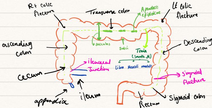
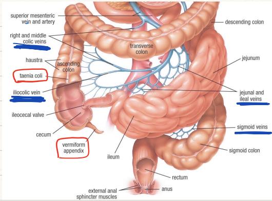
E. Surgical Anatomy of the Large Intestine
- Dimensions & Surgical Calibers:
- Cecum: Width 9 cm, Length 6 cm (widest segment; highest Laplace tension highest perforation risk in distal LBO).
- Ascending Colon: Diameter 6.35 cm, Length 20 cm (retroperitoneal).
- Transverse Colon: Diameter 6 cm, Length 46 cm (intraperitoneal with transverse mesocolon).
- Descending Colon: Diameter 7 cm, Length 25 cm (retroperitoneal).
- Sigmoid Colon: Diameter 7 cm, Length 35–60 cm (intraperitoneal with V-shaped sigmoid mesocolon; redundant prone to volvulus).
- The Three Distinct Morphological Features of the Colon (Distinguish from Small Bowel):
- Teniae Coli: Three longitudinal bands of smooth muscle ( wide) converging at the appendiceal base:
- Taenia libera: Anterior, free surface (free of mesenteric or omental attachment).
- Taenia mesocolica: Posteromedial, along the mesenteric border.
- Taenia omentalis: Posterolateral, along attachment of greater omentum.
- Haustrations / Sacculi (Sacculations): Sac-like pouches formed because teniae are shorter than underlying circular muscle. Interrupted by transverse sulci / creases (do not cross full lumen circumference, unlike circular valvulae conniventes of small bowel).
- Appendices Epiploicae: Peritoneal fat tags along the colon surface (absent on cecum, small intestine, and rectum).
- Teniae Coli: Three longitudinal bands of smooth muscle ( wide) converging at the appendiceal base:
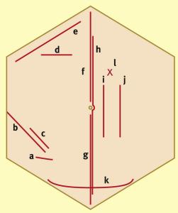
F. Surgical Incisions & Indications
| Incision | Anatomical Orientation & Location | Clinical & Operative Indications |
|---|---|---|
| Midline (Upper / Lower / Full) | Linea alba from xiphoid process to pubic symphysis | Emergency exploratory laparotomy, major trauma, widespread peritonitis, AAA repair. Rapid entry, avascular, excellent universal exposure. |
| Right Subcostal (Kocher) | inferior and parallel to right costal margin | Open Cholecystectomy, biliary tract reconstruction. (Left subcostal used for Splenectomy). |
| McBurney’s (Gridiron) | Oblique incision at junction of lateral 1/3 and medial 2/3 of ASIS-umbilicus line | Open Appendectomy (muscle-splitting along natural fiber lines). |
| Lanz Incision | Transverse skin crease incision below McBurney’s point | Open Appendectomy in pediatrics/cosmetic preference; superior wound healing. |
| Rutherford-Morison | Curvilinear muscle-cutting incision in right or left lower quadrant | Open Right or Left Hemicolectomy, renal transplantation, sigmoid resection, extraperitoneal access to iliac vessels. |
| Pfannenstiel | Transverse convex-downward incision above pubic symphysis along Langer lines | Cesarean section, abdominal hysterectomy, open prostatectomy, urological and gynaecological procedures. (Rectus muscles separated without cutting). |
| RUQ Transverse | Horizontal incision extending laterally from midline across rectus | Pediatric open laparotomy, biliary atresia repair (Kasai), infant bowel surgery. |
| Palmer’s Point | Left upper quadrant, below left costal margin in midclavicular line | Primary Veress needle / laparoscopic optical trocar insertion in non-virgin abdomen (avoids periumbilical adhesions from previous surgery). |
Station 2: Acute Abdomen & Intestinal Obstruction (History & Physical Exam)
A. Systematic Clinical History Taking
- Clinical History Taking Questions (Handwritten Ward Notes):
- Onset: Sudden / instantaneous (“Did the pain start completely suddenly like an explosion?”) perforated peptic ulcer, ruptured AAA, acute mesenteric embolus.
- Relation to Meals: Timing & duration relative to food (“Does the pain start before or after eating, and how long does it last?”) duodenal ulcer relieved by food; gastric ulcer / biliary colic provoked by food ( postprandially).
- Chronological Timeline: Detailed sequential onset (“Walk me through the exact day and hour the pain first started up until now.“)
- Pain Severity & Impact: Numerical scale 1–10; sleep disruption, inability to straighten up.
- Past Medical & Surgical History (Essential for Differential Diagnosis):
- Prior abdominal surgeries (virgin vs non-virgin abdomen adhesions vs malignancy).
- Chronic medical illnesses (diabetes, cardiovascular disease, renal failure).
- Constitutional symptoms: appetite loss, severe unintentional weight loss (
sever Diet / unhealthy / surgery / malignant disease). - Past similar attacks: biliary colic, recurrent subacute obstruction, diverticular flare-ups.
- Past Medications & Allergies: NSAIDs/steroids (peptic ulceration), anticoagulants/warfarin, antibiotics, known drug allergies.
- Social & Environmental History: City vs rural residence, alcohol consumption history, smoking pack-years.
- Cardiovascular & Visceral Secretion Kinetics:
- Daily digestive secretions enter GI tract: Saliva , Gastric juice , Bile , Pancreatic juice , Small intestinal secretions .
- Totaling ; 98% reabsorbed by jejunum, ileum, and right colon. Proximal obstruction rapidly produces massive fluid third-spacing, severe dehydration, and hypokalemic hypochloremic metabolic alkalosis.
B. The Cardinal Tetrad of Bowel Obstruction
| Cardinal Sign / Symptom | Small Bowel Obstruction (SBO) | Large Bowel Obstruction (LBO) |
|---|---|---|
| Abdominal Pain | Early, intense, colicky, periumbilical; spasms every | Late, lower abdominal / hypogastric, continuous dull aching |
| Vomiting | Early, frequent, profuse; bilious progressing to foul feculent | Late, infrequent, or absent if competent ileocecal valve |
| Abdominal Distension | Minimal in high proximal SBO; moderate in distal ileal SBO | Marked, prominent, generalized peripheral distension |
| Absolute Obstipation | Late (residual gas/stool in colon evacuated over first 12–24h) | Early and complete (failure to pass both flatus and feces) |
C. Etiology: Virgin vs. Non-Virgin Abdomen
- “Virgin Abdomen” (No prior abdominal scars):
- Colorectal Malignancy is the #1 cause of LBO (), followed by diverticular stricture, volvulus, and incarcerated virgin external hernia.
- SBO in virgin abdomen: incarcerated primary groin/femoral hernia, gallstone ileus, Crohn’s stricture, intussusception.
- “Non-Virgin Abdomen” (Prior laparotomy scars):
- Postoperative Adhesions are the #1 cause (), followed by incisional hernia, recurrence, volvulus.
D. Physical Examination of the Acute Abdomen
- Inspection: Symmetry, surgical scars (non-virgin abdomen), visible peristalsis (ladder pattern in thin patients), cough impulse at hernial orifices.
- Palpation:
- Guarding (Involuntary Muscular Rigidity): Reflex contraction of abdominal wall muscles peritoneal inflammation.
- Rebound Tenderness (Blumberg’s sign): Sharp pain upon rapid release of slow deep palpation parietal peritonitis.
- Rigid “Board-Like” Abdomen: Generalized involuntary spasm indicating catastrophic chemical peritonitis (perforated ulcer).
- Percussion: Tympanitic over gas-distended bowel loops (hyper- or hyporesonant); shifting dullness in concomitant ascites; loss of liver dullness indicates free intraperitoneal air.
- Auscultation: Hyperactive, high-pitched, metallic “tinkling” bowel sounds in early mechanical obstruction; silent abdomen in paralytic ileus or generalized peritonitis. Auscultate for aortic/renal bruits.
E. Imaging & Laboratory Diagnostic Workup
- Essential Laboratory Workup:
- CBC: Hemoglobin/hematocrit (hemoconcentration vs occult bleeding), leukocytosis with left shift, platelet count.
- Urea, Creatinine & Electrolytes (U&E): Dehydration, prerenal azotemia, hypokalemia from prolonged vomiting.
- Liver Function Tests (LFTs): Necroinflammatory (AST, ALT) vs Cholestatic (ALP, Bilirubin).
- Coagulation Profile: PT/INR, PTT (essential pre-op).
- Blood Gas Analysis: ABG / VBG (metabolic acidosis, elevated serum lactate indicates bowel ischemia/strangulation).
- Plain Abdominal Radiograph (AXR):
- Small Bowel Obstruction: Central dilated loops (), multiple air-fluid levels in “step-ladder” pattern, visible valvulae conniventes (plicae circulares) crossing the complete transverse width of the lumen.
- Large Bowel Obstruction: Peripheral dilated loops (, cecum ), haustrations crossing only partial width of the lumen.
- Erect Chest X-ray (CXR): Mandatory to rule out pneumoperitoneum (free crescent of air under right hemidiaphragm = perforated hollow viscus / niche perforated duodenum).
- Contrast-Enhanced CT (CECT) of Abdomen & Pelvis (Gold Standard):
- Identifies transition point, demonstrates “whirl sign” in volvulus, detects closed-loop obstruction, and evaluates bowel ischemia (thick, hypo-enhancing wall, pneumatosis intestinalis, portal venous gas).
- Oncologic Pearl: CT can underestimate tumor size by due to microscopic submucosal spread.
- Staging: CT CAP (Chest, Abdomen, Pelvis) is mandatory in suspected colorectal malignancy to screen for pulmonary and hepatic metastases.
- Colonoscopy: Indicated in partial or early obstructing tumors; contraindicated in acute complete mechanical obstruction with threatened cecal perforation.
F. Surgical Management & Operative Strategies
- Resuscitation: IV fluid resuscitation, NPO, NGT decompression, Foley catheter, broad-spectrum IV antibiotics.
- Surgical Pathways for Colonic Obstruction:
- Resection with Primary Anastomosis: In stable, non-septic patients with viable bowel.
- Hartmann’s Procedure (Resection & Diversion): Resection of obstructing rectosigmoid tumor; the distal rectal stump is oversewn and left closed within the pelvis (Hartmann’s pouch), and the proximal bowel is exteriorized through the left rectus muscle as an end colostomy in LIF (standard in elderly, septic, or hemodynamically unstable patients).
- Proximal Diversion Loop Stoma: Palliative stoma without tumor resection in locally unresectable/fixed tumors.
- Subtotal / Total Colectomy with Ileorectal Anastomosis: For obstructing left-sided tumors with ischemic, severely distended, or perforated cecum.
- Self-Expanding Metal Stent (SEMS): Palliative decompression or bridge-to-surgery; carries perforation risk and inferior oncological outcomes if curative resection is planned.
- Prognostic Pearl: Stage IV sigmoid adenocarcinoma carries an estimated risk of recurrence even following palliative surgical intervention.
Station 3: Stoma & Colostomy Bedside Physical Examination & Assessment
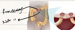
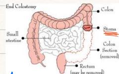
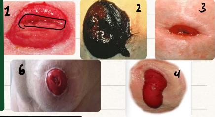
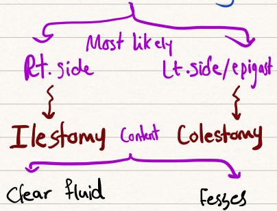
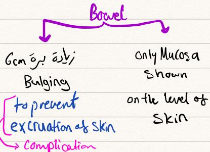
A. Surgical Classification of Stomas
| Characteristic | Ileostomy | Colostomy |
|---|---|---|
| Typical Anatomical Site | Right Iliac Fossa (RIF) | Left Iliac Fossa (LIF) |
| External Stoma Contour | Spouted (Everted ) | Flush with Skin (or minimally raised ) |
| Lumen Wall Characteristics | Thin, velvety mucosal wall; continuous flow | Thick, rugose mucosal wall; intermittent output |
| Effluent Consistency | Liquid, semi-solid, green-yellow bile; corrosive proteolytic enzymes | Formed, solid, dark brown feces; non-corrosive |
| Bag Appliance Type | Drainable pouch with bottom clip/outlet | Closed-end disposable filter pouch |
- Surgical Configurations:
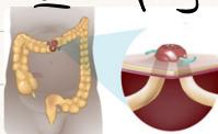
- End Stoma: Single functional lumen brought through rectus muscle (e.g., permanent end colostomy in APR; end colostomy in Hartmann’s; end ileostomy in total proctocolectomy). Produces formed feces (colostomy) or liquid/semi-solid effluent (ileostomy).
- Loop Stoma: Intact bowel loop exteriorized over a plastic bridge/rod:
- Proximal Limb: Functional; discharges stool/effluent.
- Distal Limb: Non-functional; discharges small volumes of mucus (“mucous fistula”). Used for temporary upstream defunctioning/protection of distal anastomosis.
- Supporting Bridge / Rod: An artificial plastic rod or bridge is placed beneath the exteriorized bowel loop to prevent slipping back (retraction) into the peritoneal cavity until secure peritoneal adhesions develop (removed around day 5–7).
- Double-Barrel Stoma: Bowel transected completely; proximal functional stoma and distal defunctioned mucous fistula brought out as two separate distinct stomas on the abdominal wall.
- Mucous Fistula: Distal lumen flush with skin, producing small amounts of clear or cloudy mucus.
B. Step-by-Step Bedside Physical Examination (‘s Protocol)
- Preparation: WIPES, exposure from nipples to mid-thigh, supine, chaperone present.
- Inspect the Appliance (Stoma Bag):
- Location: Right vs. Left Iliac fossa (temporary loop colostomies occasionally placed in transverse colon / right hypochondrium).
- Pouch type: One-piece vs. two-piece; drainable vs. closed-end.
- Contents: Volume, color, consistency (liquid bilious vs. formed feces vs. cloudy mucous urine in ileal conduit / urostomy).
- Maintenance Note: Empty or change bag when to full to avoid excess weight dragging on peristomal seal.
- Inspect the Uncovered Stoma:
- Remove stoma bag gently using adhesive remover wipe.
- Number of Lumens: Single lumen (End) vs. Two lumens (Loop or Double-barrel).
- Height: Spouted ( above skin Ileostomy) vs. Flush with skin ( Colostomy).
- Mucosa Quality: Healthy shiny pink/red (viable) vs. Pale (ischemia/anemia) vs. Dark purple/black (gangrene/necrosis).
- Mucocutaneous Junction (Transitional Border between Bowel and Skin):
- Carefully inspect the circumferential seam uniting bowel mucosa to skin.
- Look for healthy healing, mucocutaneous separation, suture line infection, hyperpigmentation, or purulent breakdown.
- Peristomal Skin: Peristomal erythema, chemical excoriation (enzymatic irritation from leaking ileostomy fluid), pyoderma gangrenosum (IBD), candidiasis (satellite lesions).
- Dynamic Maneuver & Palpation:
- Ask patient to turn head away and cough: observe for Parastomal Hernia (bulge around or beneath stoma aperture).
- Palpate around stoma for fascial defects, tenderness, or subcutaneous abscess collection.
- Digital Examination of the Stoma (Gloved Lubricated Index Finger):
- Gently insert finger into stoma lumen to assess fascial ring caliber (rules out fascial stomal stenosis), confirm direction of bowel, and verify absence of obstructing intraluminal bolus.
C. Stoma Complications & Management
- Early Complications ():
- Ischemia / Necrosis: Inadequate mesenteric blood supply or tight fascial defect; dark purple/black mucosa. If necrosis extends below fascial level emergency surgical revision.
- Retraction: Tension on mesenteric pedicle causes stoma to sink below skin level severe leakage and excoriation.
- Mucocutaneous Separation: Disruption of suture line between mucosa and skin; heals by secondary intention with risk of stricture.
- High-Output Stoma (): Dehydration, hypokalemia, hypomagnesemia, and prerenal failure; treat with loperamide, codeine, and oral rehydration solutions (St. Mark’s).
- Late Complications ():
- Parastomal Hernia: Incisional hernia through stoma trephine defect; managed with support belt or surgical mesh repair (Sugarbaker technique).
- Prolapse: Intussusception of bowel loop through stoma; reduced conservatively or revised surgically.
- Stomal Stenosis: Cicatricial scarring at skin or fascial level producing mechanical obstruction; managed with gentle digital dilatation or surgical refashioning.
Station 4: Biliary Colic, Chronic Cholecystitis & Acute Cholecystitis
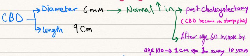
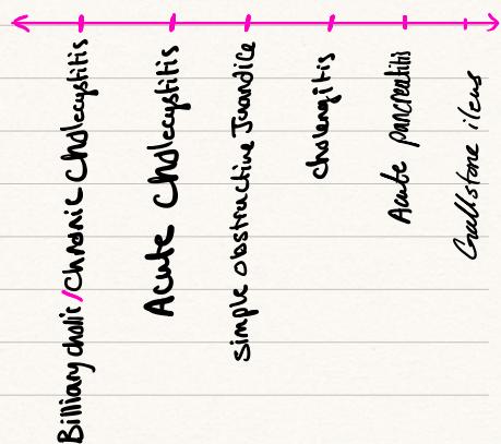
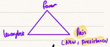
A. Surgical Pathophysiology & Gallstone Disease
- Biochemical Origins of Gallstones:
- Bile Salt synthesis starting molecule: Cholesterol.
- Bilirubin synthesis starting molecule: Heme (breakdown product of senescent red blood cells).
- Bile Composition: Water, electrolytes, Cholesterol, Bilirubin, and Lecithin (phospholipids). Disruption of bile salt/phospholipid to cholesterol ratio leads to precipitation and stone crystallization.
- Radiographic Pearl: Only of gallstones are radiopaque / calcified visible on plain AXR; are radiolucent requiring ultrasound.
- Risk Factors (The 5 F’s & High-Yield Demographics): Female, Forty, Fat (), Fertile (multiparity, estrogen), Fair.
- Ethnicity: Highest prevalence in Native Americans (Pima Indians) and Scandinavian populations.
- Rapid Weight Loss: Severe caloric restriction, starvation diets, bariatric surgery cholesterol mobilization and impaired gallbladder emptying.
- Hormonal: Progesterone acts as a smooth muscle relaxant gallbladder hypomotility and stasis; estrogen increases cholesterol saturation in bile.
- Pediatric & Hematologic Pearl: In children and young adults presenting with gallstones, rule out Sickle Cell Disease (chronic hemolysis generates calcium bilirubinate pigment stones; clinical confusion frequently occurs between a sickle cell vaso-occlusive crisis and acute cholecystitis).
- Gallstone Types:
- Cholesterol Stones (80%): Yellow-green, crystalline, radiolucent.
- Black Pigment Stones: Calcium bilirubinate; associated with chronic hemolysis (sickle cell, thalassemia, hereditary spherocytosis) and cirrhosis.
- Brown Pigment Stones: Calcium salts of unconjugated bilirubin; associated with chronic biliary infection, stasis, and Clonorchis sinensis.
- The Clinical Spectrum:
- Asymptomatic Cholelithiasis: Incidental finding; no surgery indicated except in porcelain gallbladder (malignancy risk), stone , sickle cell anemia, or planned bariatric surgery.
- Biliary Colic: Transient impaction of calculus in cystic duct; severe episodic RUQ/epigastric pain radiating to right shoulder and right infrascapular region, lasting , resolving spontaneously; normal inflammatory markers; completely normal LFTs.
- Acute Cholecystitis: Persistent cystic duct impaction chemical then bacterial inflammation continuous RUQ pain, fever, leukocytosis, and localized peritonitis.
- Medical Dissolution Therapy: Ursodeoxycholic acid (UDCA) can dissolve small () radiolucent cholesterol stones in functioning gallbladders, reserved exclusively for high-risk surgical candidates unfit for anesthesia.
B. Bedside Signs & Special Pathologies
- Murphy’s Sign: Examiner palpates deeply below right costal margin at midclavicular line (gallbladder bed) and asks patient to take a deep inspiration. Breath is suddenly arrested by sharp pain as the inflamed gallbladder contacts examining fingers. (Must be absent on left side to be positive).
- Sonographic Murphy’s Sign: Maximal focal tenderness elicited directly over the sonographically identified gallbladder with the ultrasound transducer (sensitivity ).
- Scleral Icterus Pearl (Mild Scleral Icteric Tinge in Cholecystitis):
- In uncomplicated acute cholecystitis, overt jaundice is absent. However, mild scleral icterus (“mild yellowish change in the sclera”) may occur due to local inflammatory edema tracking along common bile duct.
- Threshold Pearl: If total bilirubin , suspect concomitant Choledocholithiasis or Mirizzi Syndrome (stone impacted in cystic duct Hartmann’s pouch compressing the common hepatic duct).
- Acalculous Cholecystitis: Acute gallbladder inflammation without gallstones. Occurs in critically ill, septic, burn, or major trauma ICU patients on prolonged TPN. High gangrene and perforation rates; managed with emergent percutaneous cholecystostomy tube drainage.
- Ceftriaxone Pseudolithiasis: High-dose Ceftriaxone forms insoluble calcium-ceftriaxone salts excreted into bile, precipitating reversible biliary sludge and pseudolithiasis.
- Emphysematous Cholecystitis: Gas within gallbladder lumen and wall caused by gas-forming anaerobes (Clostridium perfringens, E. coli); affects elderly diabetic males (
DM Pt); high perforation rate; requires emergency cholecystectomy.
C. Diagnostic Workup & Imaging
- Transabdominal Ultrasound (First-Line Modality of Choice):
- Ultrasound Triad: 1) Gallstones with posterior acoustic shadowing (anechoic shadow), 2) Gallbladder wall thickening (), 3) Pericholecystic fluid halo.
- Differential Diagnosis of Gallbladder Wall Thickening (): Acute cholecystitis, ascites, acute viral hepatitis, hypoalbuminemia, congestive heart failure, adenomyomatosis.
- HIDA Scan (Cholescintigraphy — Gold Standard Functional Test):
- Indicated when ultrasound is equivocal.
- Technetium-99m labeled hepatic iminodiacetic acid excreted into biliary tract:
- Positive Test (+): Non-visualization of gallbladder with prompt visualization of duodenum (cystic duct is occluded by impacted stone confirms Acute Cholecystitis).
- Negative Test (-): Prompt gallbladder visualization within 1 hour (cystic duct is patent excludes Acute Cholecystitis).
D. Surgical Anatomy: Calot’s Triangle & Safe Cholecystectomy
- Triangle of Calot (True Anatomical Boundaries):
- Superior: Inferior surface of the liver.
- Medial: Common Hepatic Duct.
- Lateral / Inferior: Cystic Duct.
- Contents: Cystic Artery, cystic lymph node of Lund (Mascagni’s), autonomic nerves, and loose connective tissue.
- Strasberg’s “Critical View of Safety” (Mandatory to Prevent Bile Duct Injury):
- Complete clearance of all fat and fibrous tissue from the hepatocystic triangle.
- Dissection of the lower third of the gallbladder off the liver bed (cystic plate visualized).
- Exactly TWO (and only two) structures seen entering the gallbladder: the Cystic Duct and the Cystic Artery.
- Timing of Surgery:
- Early Laparoscopic Cholecystectomy (Index Admission, within 72 hours of symptom onset): Standard of care; reduces total hospital stay, complication rates, and avoids recurrent biliary attacks.
- Conservative therapy reserved for frail, high-risk surgical candidates.
Station 5: Obstructive Jaundice, Choledocholithiasis & Acute Ascending Cholangitis
A. Pathophysiology of Jaundice
- Normal Serum Bilirubin: ().
- Clinical Detection:
- Sclera & Under Tongue (Sublingual frenulum): Detectable when bilirubin () due to high elastin content with affinity for bilirubin.
- Skin Icterus: Detectable when bilirubin ().
- Classification of Jaundice & LFT Patterns:
- Pre-Hepatic (Hemolytic): Excess unconjugated bilirubin production exceeding hepatic conjugating capacity (hemolytic anemia, transfusion reaction); normal AST/ALT/ALP; acholuric jaundice (dark stool, normal urine color because unconjugated bilirubin is water-insoluble).
- Intra-Hepatic (Hepatocellular / Necroinflammatory): Hepatocyte injury (viral hepatitis, paracetamol toxicity, alcohol); marked elevation of AST & ALT (); mixed conjugated/unconjugated hyperbilirubinemia; dark urine.
- Post-Hepatic (Obstructive / Surgical / Cholestatic): Mechanical obstruction to bile outflow downstream of hepatic ducts; Direct (conjugated) bilirubin of total; markedly elevated Alkaline Phosphatase (ALP) and GGT ( ULN) with mild AST/ALT elevation.
- Parasitic Biliary Obstruction:
- Ascaris lumbricoides: Roundworm can migrate through the ampulla of Vater into the CBD, causing biliary colic, strictures, and cholangitis.
B. Clinical Features of Extrahepatic Biliary Obstruction
- Cardinal Symptoms:
- Dark “Tea-Colored” Urine: Water-soluble conjugated bilirubin filtered by renal glomeruli.
- Pale, Clay-Colored (Acholic) Stools: Absence of stercobilinogen reaching the intestinal lumen.
- Pruritus (Intractable Itching): Cutaneous deposition of systemic bile salts / bile acids; associated scratch marks and excoriations over limbs and trunk.
- Fat Malabsorption & Steatorrhea: Absence of bile salts in duodenum impairs micelle formation bulky, foul-smelling, floating stools and deficiency of fat-soluble vitamins (A, D, E, and K).
- Vitamin K Deficiency: Impairs hepatic -carboxylation of clotting factors II, VII, IX, and X (mnemonic
"1972"[Dependent clotting factor 1972]), producing prolonged Prothrombin Time (PT) / elevated INR. - Pre-Operative Management: Administer Vitamin K IV/SC pre-operatively to correct coagulopathy before surgical or endoscopic intervention.
C. Differential Diagnosis & Courvoisier’s Law
| Clinical Feature | Choledocholithiasis (Gallstones) | Periampullary / Pancreatic Carcinoma |
|---|---|---|
| Pain Profile | Severe, episodic, colicky RUQ/epigastric pain | Painless, progressive, relentless jaundice; dull back pain |
| Course of Jaundice | Fluctuating jaundice (stone acts as ball-valve) | Steadily worsening, deep dark-bronze/green jaundice |
| Systemic Symptoms | Transient chills, fever, nausea | Anorexia, profound unremitting weight loss, cachexia |
| Palpable Gallbladder | NOT Palpable (shrunken, fibrotic, chronically scarred) | Smooth, enlarged, tense, non-tender palpable mass |
- Courvoisier’s Law: “In the presence of jaundice, an enlarged palpable gallbladder is unlikely to be due to gallstones, because previous chronic inflammation has rendered the gallbladder wall fibrotic, contracted, and incapable of dilatation; it is more likely due to malignant obstruction of the common bile duct (e.g., carcinoma of the pancreatic head, cholangiocarcinoma, or periampullary tumor).”
- Exceptions to Courvoisier’s Law: Double stone impaction (stones in cystic duct and CBD simultaneously), Mirizzi syndrome, choledochal cyst, Oriental cholangiohepatitis.
D. Acute Ascending Cholangitis (Surgical Emergency)
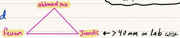
- Charcot’s Triad: 1) Severe RUQ Abdominal Pain, 2) High Spiking Fever with Rigors, 3) Obstructive Jaundice.
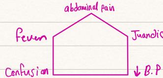
- Reynolds’ Pentad (Severe Suppurative Cholangitis with Shock):
- Charcot’s Triad PLUS:
- 4) Hypotension / Septic Shock
-
- Altered Mental Status / Lethargy / Confusion
- Pathophysiology: Bile duct obstruction elevates biliary pressure above hepatic venous pressure (), forcing bacteria and endotoxins (E. coli, Klebsiella, Enterobacter, Bacteroides) across hepatocyte junctions directly into systemic circulation.
- Complication: Pyogenic liver abscess can develop secondary to ascending biliary sepsis.
- Charcot’s Triad PLUS:
- Emergency Management Protocol:
- Immediate resuscitation: Oxygen, aggressive IV crystalloids, ICU admission.
- Urgent broad-spectrum IV antibiotics: Piperacillin-Tazobactam (Tazocin) or Ceftriaxone + Metronidazole.
- Correct coagulopathy: IV Vitamin K () and Fresh Frozen Plasma (FFP).
- Emergency Decompression of the Biliary Tree (< 24 hours):
- ERCP with Endoscopic Sphincterotomy & Biliary Stent Placement (Gold Standard).
- MRCP indicated if clinically obstructed but ultrasound and labs are equivocal.
- Percutaneous Transhepatic Biliary Drainage (PTBD) if ERCP fails or anatomy altered (Roux-en-Y).
- Surgical CBD exploration with T-tube placement as last resort.
Station 6: Gallstone Ileus & Mechanical Bowel Obstruction
A. Pathophysiology & Fistula Formation
- Mechanism: Mechanical small bowel obstruction caused by intraluminal impaction of a large gallstone ().
- Fistula Pathogenesis: Recurrent acute cholecystitis dense adhesions between gallbladder fundus and adjacent bowel transmural ischemic pressure necrosis cholecystoenteric fistula:
- Cholecystoduodenal Fistula (70% — Most common): Gallbladder erodes into duodenal bulb; stone travels downstream to impact at the narrowest site: terminal ileum adjacent to the ileocecal valve (60–70% of cases).
- Cholecystocolic Fistula: Gallbladder erodes into hepatic flexure / right colic flexure; stone usually passes per rectum with feces without obstruction unless colonic stricture exists.
- Cholecystogastric Fistula: Gallbladder erodes into stomach gastric outlet obstruction (Bouveret Syndrome).
- Site of Impaction: Stone travels through jejunum and ileum to impact at the narrowest small bowel segment: terminal ileum adjacent to ileocecal valve.
B. Clinical Presentation
- Patient Profile: Elderly female (), frail, comorbidity, chronic biliary disease history.
- “Tumbling” Obstruction: Intermittent subacute SBO (colicky pain and vomiting alternating with symptom-free intervals as stone temporarily lodges, dislodges, travels, and finally impacts at terminal ileum).
- Established Obstruction: Severe colicky pain, profuse vomiting (becoming feculent), marked distension, obstipation, severe dehydration, prerenal azotemia.
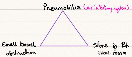
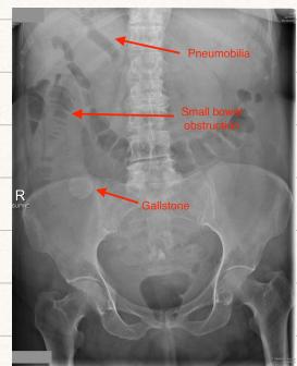
C. Diagnostic Hallmark — Rigler’s Triad
| Finding | Radiographic Features | Clinical Significance |
|---|---|---|
| 1. Pneumobilia | Branching air lucencies over liver hilum / porta hepatis | Pathognomonic of cholecystoenteric communication |
| 2. Small Bowel Obstruction | Centrally dilated loops (), multiple air-fluid levels, collapsed colon | Confirms mechanical obstruction |
| 3. Ectopic Gallstone | Calcified radio-opaque calculus in RIF / pelvis (30–35% of AXRs) | Identifies the offending intraluminal calculus |
- Diagnostic Modality of Choice: Contrast-Enhanced CT (CECT) (sensitivity , specificity ). Accurately demonstrates fistula, confirms stone impaction site, rules out secondary synchronous stones, and evaluates bowel viability.
D. Surgical Management & Decision-Making
- Resuscitation: Aggressive IV crystalloid resuscitation, NGT decompression, IV broad-spectrum antibiotics.
- Operative Procedure — Enterolithotomy:
- Midline exploratory laparotomy; run entire bowel from Treitz to ileocecal valve (exclude synchronous proximal/distal stones; present in ).
- Incision Site: Longitudinal enterotomy on antimesenteric border in healthy, pliable bowel PROXIMAL to the impacted stone.
- Never incise over the stone: Direct pressure creates ischemic, ulcerated mucosa prone to breakdown.
- Closure: Transverse 2-layer closure of enterotomy to prevent luminal stricture.
- One-Stage vs. Two-Stage Decision:
- Enterolithotomy Alone (Preferred standard in emergency): Mortality . Fistula frequently undergoes spontaneous closure once distal pressure is relieved.
- One-Stage (Enterolithotomy + Cholecystectomy + Fistula Division): High mortality () in elderly septic patients with obliteration of Calot’s triangle; reserved strictly for young, stable patients with minimal local inflammation.
Station 7: Acute & Chronic Pancreatitis, Complications & Surgical Site Infections
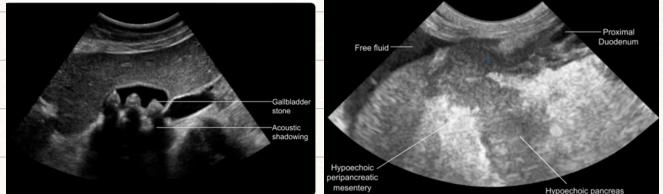
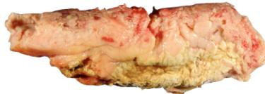
A. Etiology & Mnemonic “I GET SMASHED”
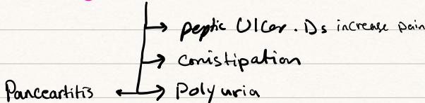
- Top 2 Causes (> 80%): Gallstones (40–50%) (transient ampullary occlusion ductal hypertension/bile reflux) and Ethanol (30–40%) (acinar toxicity, protein plugging, sphincter of Oddi spasm).
- Mnemonic Breakdown:
- I: Idiopathic.
- G: Gallstones.
- E: Ethanol abuse.
- T: Trauma (blunt abdominal trauma / bicycle handlebar injury — #1 cause in children).
- S: Steroids.
- M: Mumps and other viruses (Coxsackie B, CMV, HIV).
- A: Autoimmune pancreatitis (IgG4-related systemic disease, occasionally).
- S: Scorpion sting (Tityus trinitatis venom).
- H: Hyperlipidemia (Type I, IV, V hypertriglyceridemia, serum TG ) / Hypercalcemia.
- E: ERCP (iatrogenic mechanical/thermal injury).
- D: Drugs (Azathioprine, 6-MP, Thiazides, Furosemide, Didanosine, Valproic acid, Tetracyclines).
- Hypercalcemia Workup (“Bones, Stones, Groans, Moans”):
- Primary hyperparathyroidism, Vitamin D toxicity, osteolytic bone metastases, PTHrP paraneoplastic secretion, immobilization, factitious (tourniquet stasis during venous extraction), and Sarcoidosis (macrophages express 1-alpha-hydroxylase elevated ; bilateral hilar lymphadenopathy).
B. Clinical Presentation & Physical Examination
- Pain Profile: Sudden, severe, stabbing/boring epigastric pain radiating to mid-back (T10–L1); aggravated supine, relieved by sitting upright and leaning forward with knees drawn up. Associated with intractable nausea, profuse vomiting, diaphoresis, and tachycardia.
- Signs of Retroperitoneal Hemorrhage (Appear at 48–72h; severe necrotizing disease):
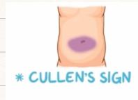
- Cullen’s Sign: Periumbilical ecchymosis (blood travels via falciform ligament to subcutaneous tissue).
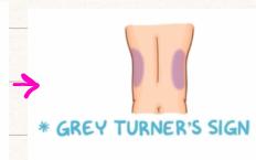
- Grey Turner’s Sign: Bilateral flank ecchymosis (retroperitoneal blood tracks via posterior pararenal space to abdominal wall).
- Fox’s Sign: Ecchymosis over anterior-superior thigh below inguinal ligament.
C. Diagnostic Criteria (Revised Atlanta Criteria)
- Diagnosis confirmed by of the following 3 criteria:
- Characteristic Pain: Severe, acute epigastric pain radiating to the back.
- Biochemical Marker: Serum Lipase or Amylase Upper Limit of Normal (ULN):
- Lipase (Gold Standard): 95% sensitivity, 98% specificity; rises at 4–8h, peaks at 24h, remains elevated for 8 to 14 days (ideal for late presentations).
- Amylase: Rises at 2–12h, normalizes at 3 to 5 days; less specific; absolute level does NOT correlate with disease severity. Urinary amylase is more specific than serum.
- Cross-Sectional Imaging: CECT, MRI, or transabdominal ultrasound demonstrating acute pancreatic inflammation/peripancreatic fluid.
D. Consultant Clinical Q&A Pearls & Differential Diagnosis of Hyperamylasemia
- Causes of Hyperamylasemia:
- Pancreatic: Acute pancreatitis, pancreatic trauma, pseudocyst, pancreatic carcinoma.
- Non-Pancreatic GI: Posterior Duodenal Ulcer perforation (erodes Gastroduodenal Artery severe hemorrhage and peritoneal leak), acute mesenteric ischemia, intestinal obstruction, acute cholecystitis.
- Salivary Glands: Mumps, parotitis, salivary gland calculi or trauma.
- Benign / Non-Pathological:
- Macroamylasemia: Normal/benign variant where amylase complexes with serum immunoglobulins (IgA/IgG), preventing renal glomerular filtration persistently elevated serum amylase, completely normal lipase, and normal/low urine amylase.
- Pregnancy: Amylase secreted by the placenta into maternal circulation.
- Q: Timing of Cholecystectomy in Biliary Pancreatitis?
- Perform Index Admission Laparoscopic Cholecystectomy once acute attack settles and enzymes normalize (48–72h, prior to discharge). Delaying carries a 30–60% recurrence risk within 6 weeks and a 10% mortality risk from recurrent severe pancreatitis.
- Q: Indications for Urgent ERCP (< 24–72h)?
- Concomitant Acute Ascending Cholangitis (fever, jaundice, shock).
- Persistent CBD obstruction (worsening jaundice, progressive bilirubin rise, dilated CBD on US/MRCP, impacted stone).
- Severe biliary pancreatitis in frail patients failing conservative therapy.
- Q: Clinically improved patient with persistently elevated amylase weeks later?
- Suspect Pancreatic Pseudocyst (active enzymes sequestered in fibrous cavity continue leaking into circulation).
- Q: Discharged patient returns 3–4 weeks later with upper abdominal fullness, pain, and vomiting?
- Suspect Pancreatic Pseudocyst obstructing gastric antrum or duodenal sweep.
- Vascular Complication — Splenic Vein Thrombosis:
- Pancreatic tail/body inflammation spreads to adjacent splenic vein thrombosis Left-Sided (Sinistral) Portal Hypertension isolated gastric varices, splenomegaly, and normal liver architecture (curative treatment is Splenectomy).
E. Prognostication: Ranson, Glasgow & APACHE II Criteria
- Ranson’s Criteria:
Timing Variable Threshold At Admission (“GALAW”) Glucose () Age LDH AST WBC At 48 Hours (“C-HOBIS”) Calcium (serum) () (fat saponification) Hematocrit drop Oxygen () (acute lung injury / ARDS) Base Deficit (metabolic acidosis) Increase in BUN () (despite fluids) Sequestration of Fluid (third-spacing) - Scoring: 0–2: Mild (mortality ); 3–4: Moderate (mortality ); 5–6: Severe (mortality ); : Catastrophic (mortality ).
- Glasgow (Imrie) Score: Assessed within 48 hours for both biliary and alcoholic pancreatitis ( indicates severe pancreatitis): Age , WBC , Glucose , Urea , , Calcium , Albumin , LDH .
- APACHE II Score: Evaluates 12 physiological parameters; score predicts severe attack and can be updated continuously.
F. Imaging Pearls & CT Timing
- Abdominal Radiograph (AXR):
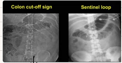
- Sentinel Loop: Focal localized ileus in jejunum adjacent to inflamed pancreas.
- Colon Cut-off Sign: Abrupt cutoff of colonic gas at splenic flexure due to inflammatory spasm of phrenicocolic ligament.
- Timing of Contrast-Enhanced CT (CECT):
- Optimal Window: 72 to 96 hours (3–4 days) post-onset.
- Why not early on admission? Pancreatic microvascular perfusion takes 48–72h to establish; early CT significantly underestimates extent of pancreatic necrosis.
- Revised Atlanta Classification of Pancreatic Collections:
- : Acute Peripancreatic Fluid Collection (APFC) (interstitial; no wall) vs. Acute Necrotic Collection (ANC) (necrotizing; contains liquid and solid debris; no fibrous wall).
- : Pancreatic Pseudocyst (fibrous wall, no solid necrosis; lined by granulation tissue, NOT epithelium) vs. Walled-Off Necrosis (WON) (fibrous wall containing necrotic parenchyma and infected/sterile debris).
- Antibiotic Strategy in Severe Pancreatitis:
- Prophylactic antibiotics are NOT indicated in mild or sterile pancreatitis.
- In documented infected necrotizing pancreatitis (gas on CT or FNA positive): Carbapenems (Imipenem / Meropenem) due to superior pancreatic tissue penetration.
G. Respiratory Complications: ARDS & Pulmonary Toilet
- Acute Respiratory Distress Syndrome (ARDS) (AECC Definition):
- Acute onset, bilateral fluffy pulmonary infiltrates on CXR, (ALI ), with no clinical evidence of left atrial hypertension (wedge pressure ).
- Pathogenesis: Systemic release of pancreatic phospholipase damages alveolar type II pneumocytes surfactant destruction microatelectasis and capillary leak.
- Mechanical Ventilation Strategy (MCQ Pearl): Lung-protective ventilation with low tidal volume (, target ideal body weight) and high PEEP up to to prevent alveolar barotrauma.
- Postoperative Pulmonary Toilet:
- Incentive spirometry, chest physiotherapy, deep breathing exercises, early ambulation to clear secretions and prevent postoperative atelectasis and pneumonia.
H. Surgical Site Infections (SSIs) & CDC Classification
- CDC Surgical Site Infection Classification:
- Superficial Incisional SSI (): Involves skin and subcutaneous tissue only. Treatment: Open suture line, drain, clean, change dressings; antibiotics usually not required unless surrounding cellulitis.
- Deep Incisional SSI (, or with implant): Involves deep soft tissues (fascia and muscle layers). Treatment: Open wound down to fascia, debride non-viable tissue, pack wound, tertiary/delayed primary closure, systemic antibiotics.
- Organ / Space SSI (, or with implant): Involves any anatomical space manipulated during surgery (pelvic abscess, subhepatic abscess, interloop abscess). Treatment: Percutaneous ultrasound/CT-guided drainage, targeted IV antibiotics.
- Surgical Wound Contamination Classification:
- Class I (Clean): Uninfected operative wound; no entry into respiratory, alimentary, genital, or urinary tract (e.g., elective hernia repair, thyroidectomy). SSI risk . Prophylactic antibiotics generally not indicated unless prosthetic mesh/implant used.
- Class II (Clean-Contaminated): Operative wound entering respiratory, alimentary, genital, or urinary tract under controlled conditions without unusual contamination (e.g., elective cholecystectomy, elective colectomy). SSI risk . Prophylactic antibiotics indicated.
- Class III (Contaminated): Open, fresh accidental wounds; major breaks in sterile technique; gross spillage from GI tract (e.g., perforated diverticulum with purulent peritonitis). SSI risk . Prophylactic/therapeutic antibiotics mandatory.
- Class IV (Dirty / Infected): Old traumatic wounds with retained devitalized tissue; existing clinical infection or perforated viscera (e.g., fecal peritonitis from perforated stercoral ulcer). SSI risk . Full therapeutic antibiotics course.
- Foreign Body SSI Pearl:
- In patients with prosthetic implants (hernia mesh, cardiac pacemakers, prosthetic valve replacements), SSI can present 3 to 6 months post-operatively; the classic causative organism is Staphylococcus epidermidis (deep skin flora forming protective biofilm).
Station 8: Acute Appendicitis (Clinical Signs, Alvarado Score & Management)

A. Surgical Anatomy & Blood Supply
- Base Landmark: Arises posteromedially from cecum, inferior to ileocecal valve, where all 3 taeniae coli converge (crucial laparoscopic locator). Length: 8–10 cm (range 5–30 cm).
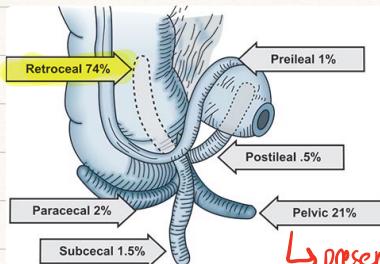
- Anatomical Positions (Image 027):
- Retrocecal (74%): Appendix behind cecum; peritonitis masked; mimics renal colic; positive Psoas sign.
- Pelvic (21%): Near right adnexa; causes dysuria, diarrhea, tenesmus; positive Obturator sign; tender on DRE/pelvic exam.
- Paracecal (2%), Subcecal (1.5%), Pre-ileal (1%), Post-ileal (0.5%): Post-ileal has highest rate of missed diagnosis and early peritonitis.
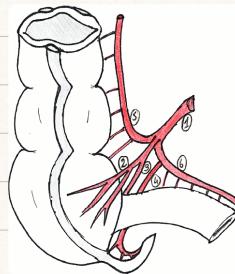
- Vascular Supply: Appendicular Artery, an anatomical end-artery from the inferior division of the ileocolic artery, runs in the free edge of the mesoappendix.
- 6 Branches of Ileocolic Artery (Image 028): 1) Ileocolic trunk, 2) Anterior cecal artery, 3) Posterior cecal artery, 4) Appendicular artery, 5) Ascending colic branch, 6) Descending ileal branch.
- Thrombosis causes rapid gangrene and perforation on the antimesenteric border near the tip.
B. Pathogenesis, Natural History & High-Risk Groups
- Etiology: Young patients: submucosal lymphoid hyperplasia (viral MALT, peak age 16–18y); older adults: fecalith / appendicolith; rare: carcinoid tumor, cecal carcinoma blocking orifice, pinworms (Enterobius).
- Microbiology: Rapid bacterial overgrowth dominated by Escherichia coli and Bacteroides fragilis.
- Visceral to Somatic Pain Progression:
- Visceral (T10): Intraluminal obstruction visceral stretch dull, poorly localized periumbilical / epigastric pain.
- Somatic (T11–L1): Transmural inflammation reaches parietal peritoneum pain shifts and sharpens to Right Iliac Fossa (RIF); worsened by movement and coughing.
- Cardinal Symptoms: Anorexia (“Hamburger Sign” — hungry patient makes appendicitis unlikely); nausea/vomiting starting AFTER pain; low-grade fever ().
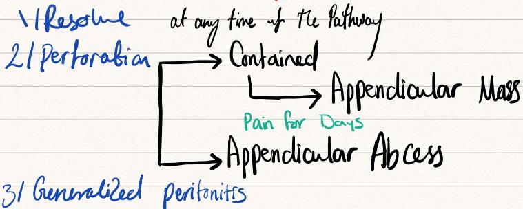
- The Natural History & Fate of Appendicitis (Image 032):
- Spontaneous Resolution: Very rare.
- Perforation:
- Most Common Site: Midpoint on the antimesenteric border (watershed area with poorest collateral supply).
- Timing: Typically occurs between 12 and 24 hours from onset.
- Contained Perforation: Omentum and bowel loops wall off defect Appendicular Mass (phlegmon) or Abscess (presents late with of pain and palpable RIF mass; subphrenic or periappendicular collection).
- Uncontained Perforation: Host defenses fail Generalized Diffuse Peritonitis and septic shock.
- High-Risk Populations:
- Pediatrics (< 5 years): Wide lumen, thin wall, and rudimentary, underdeveloped omentum unable to wall off perforation rapid generalized peritonitis and shock.
- Elderly (> 65 years): Atherosclerotic appendicular artery early ischemic gangrene; fibrosed appendix; blunted inflammatory response (minimal fever/pain/WBC) delayed presentation and high mortality.
- Pregnancy: Appendix displaced upward and laterally into RUQ by gravid uterus; perforation causes preterm labor and fetal mortality (up to 30%).
- Prior Laparotomy: Intra-abdominal adhesions anchor omentum, preventing migration to RIF immediate uncontained peritonitis.
C. Consultant Surgical Dilemmas
- Q: Finding a Macroscopically Normal Appendix at Laparoscopy?
- Perform Appendectomy: Eliminates future diagnostic confusion; rules out microscopic mucosal endoluminal appendicitis.
- Systematically Search Alternative Pathology:
- Run 100 cm (4 feet) of terminal ileum retrograde for Crohn’s ileitis and Meckel’s Diverticulum (rule of 2s: antimesenteric border, 2 feet / 60 cm from IC valve).
- Inspect female pelvis: PID, ruptured ectopic pregnancy, ovarian torsion, ruptured hemorrhagic follicular cyst (Mittelschmerz).
- Inspect mesenteric root: Mesenteric lymphadenitis (enlarged mesenteric lymph nodes; common in infants and children following acute sore throat / viral URI).
- Inspect cecum: Cecal diverticulitis, cecal adenocarcinoma.
- Inspect RUQ: Perforated duodenal ulcer fluid tracking down right paracolic gutter (Valentino’s Sign; erect CXR confirms pneumoperitoneum).
- Q: Finding an Unexpected Appendiceal Neoplasm (Carcinoid / Neuroendocrine)?
- Tumor at tip/body without mesoappendiceal invasion: Simple Appendectomy.
- Tumor , or involving base of appendix / cecum, or adenocarcinoma: Formal Right Hemicolectomy.
D. Physical Examination & Classic Signs
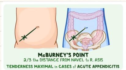
- McBurney’s Point: Junction of lateral 1/3 and medial 2/3 of line between ASIS and umbilicus; maximal tenderness and guarding.
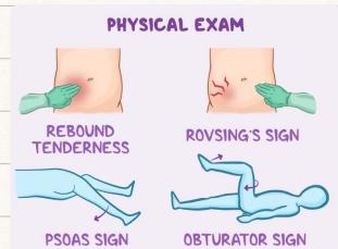
- Rovsing’s Sign: Deep palpation of LIF produces sharp pain in RIF (retrograde gas distension of cecum).
- Psoas Sign: Passive hyperextension of right hip in left lateral decubitus produces RIF pain (inflamed retrocecal appendix irritating psoas major).
- Obturator Sign: Passive internal rotation of flexed right hip produces pelvic/hypogastric pain (inflamed pelvic appendix irritating obturator internus).
- Dunphy’s Sign: Accentuation of sharp RIF pain on coughing.
- Blumberg’s Sign: Rebound tenderness indicating localized parietal peritonitis.
- Valentino’s Sign: Perforated peptic ulcer fluid tracking down right paracolic gutter to RIF, mimicking acute appendicitis; confirmed by pneumoperitoneum on erect CXR.
E. Alvarado (MANTRELS) Scoring System
| Component | Clinical Feature | Score |
|---|---|---|
| M | Migration of pain to RIF | 1 |
| A | Anorexia | 1 |
| N | Nausea or vomiting | 1 |
| T | Tenderness in RIF | 2 |
| R | Rebound tenderness in RIF | 1 |
| E | Elevated temperature () | 1 |
| L | Leukocytosis (WBC ) | 2 |
| S | Shift of neutrophils to left () | 1 |
| Total | 10 |
- Interpretation:
- 1–4: Unlikely appendicitis; search other causes or discharge with safety net.
- 5–6: Compatible; admit for serial physical exams and US/CT.
- 7–8: Probable; surgical consult and urgent diagnostic laparoscopy.
- 9–10: Definite; proceed directly to emergency appendectomy.
F. Diagnostic Workup & Management
- Imaging:
- Ultrasound (First-line in children & pregnancy): Non-compressible, blind-ending tubular structure outer diameter, target/bullseye sign, appendicolith, periappendiceal fluid.
- Contrast-Enhanced CT (Gold standard in adults / when in doubt): Sensitivity/specificity ; appendix , wall hyperenhancement, pericecal fat stranding, and obstructing fecalith / appendicolith. (MRI in pregnant patients if US inconclusive).
- Appendicular Mass Management (Ochsner-Sherren Regimen):
- For late presentation () with palpable RIF mass (phlegmon or abscess) and no diffuse peritonitis.
- Conservative therapy: Bed rest, NPO, IV fluids, IV broad-spectrum antibiotics, serial vital signs, mark mass margins daily on abdominal skin.
- If mass enlarges, spikes high swinging fever, or peritonitis develops CT scan and percutaneous catheter drainage of abscess or emergency laparotomy.
- On resolution Interval Appendectomy at 6 to 12 weeks.
- Mandatory Interval Colonoscopy: Required in all patients to rule out underlying cecal adenocarcinoma.
- Pylephlebitis (Portal Pyemia): Suppurative thrombophlebitis of portal vein system (high fever, rigors, jaundice, portal gas, liver abscesses).
Station 9: Groin & Abdominal Wall Hernias Examination & Surgical Repair
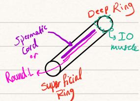
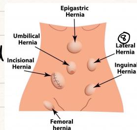

A. Surgical Anatomy of the Inguinal Canal & Spermatic Cord (“Rule of 3s”)
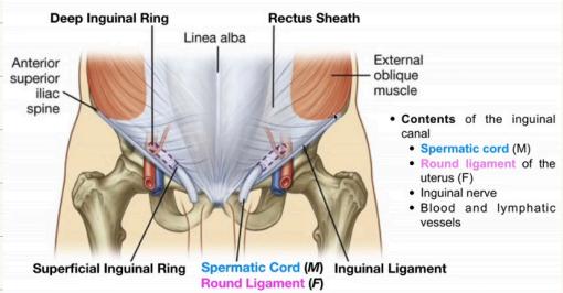
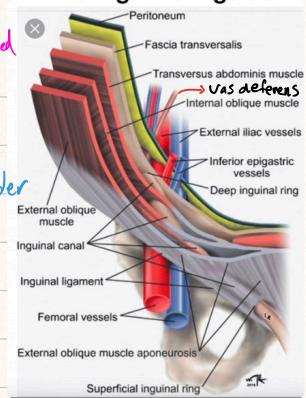
- Dimensions & Route: Oblique passage long, directed downwards and medially from deep inguinal ring to superficial inguinal ring, 2–4 cm above inguinal ligament.
- Canal Boundaries (Mnemonic “MALT”):
- Roof (Superior): Arching fibers of Internal Oblique and Transversus Abdominis.
- Anterior Wall: External Oblique Aponeurosis throughout, reinforced laterally by fleshy internal oblique fibers.
- Floor (Inferior): Upper surface of Inguinal Ligament (Poupart’s), reinforced medially by Lacunar Ligament (Gimbernat’s).
- Posterior Wall: Fascia Transversalis throughout, reinforced medially by Conjoint Tendon (internal oblique + transversus abdominis aponeurotic insertion into pubic crest/pectineal line).
- Rings & Spermatic Cord (“Complete Rule of 3s”):
- Deep Ring: Oval defect in fascia transversalis, located 1.5 cm above mid-inguinal point; inferior epigastric vessels lie immediately medial.
- Superficial Ring: Triangular defect in external oblique aponeurosis, superolateral to pubic tubercle.
- 3 Fascias / Coverings:
- External spermatic fascia: Derived from External Oblique aponeurosis.
- Cremasteric fascia & muscle: Derived from Internal Oblique muscle.
- Internal spermatic fascia: Derived from Fascia Transversalis.
- 3 Arteries:
- Testicular artery: Direct branch from the abdominal aorta ().
- Cremasteric artery: Branch of the Inferior Epigastric artery.
- Artery to vas deferens (Deferential artery): Branch of the Inferior Vesical artery (from internal iliac).
- 3 Veins:
- Testicular vein: Drains into pampiniform plexus (right testicular vein drains directly into IVC; left drains into left renal vein at ).
- Cremasteric vein.
- Deferential vein.
- 3 Nerves:
- Ilioinguinal nerve (L1): Runs on the anterior surface of the cord (outside the cord coverings); sensory to upper inner thigh and root of penis/anterior scrotum.
- Genital branch of genitofemoral nerve (L1/L2): Runs inside the cord; motor to cremasteric muscle and sensory to lateral scrotum / labium majus.
- Sympathetic nerve fibers: Postganglionic fibers (T10–T11) accompanying the testicular artery.
- 3 Other Important Structures:
- Vas deferens (in males) or Round ligament of the uterus (in females).
- Pampiniform venous plexus / Lymphatics (draining to para-aortic lymph nodes).
- Remnant of processus vaginalis / Hernia sac (if indirect hernia present).
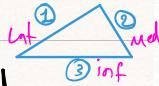
- Hesselbach’s Triangle (Direct Inguinal Hernia Site):
- Medial: Lateral border of Rectus Abdominis (linea semilunaris).
- Superolateral: Inferior Epigastric Vessels.
- Inferior: Inguinal Ligament.
- Floor: Fascia transversalis (site of acquired weakness).
B. Comprehensive Hernia Classification & Direct vs. Indirect Comparison
| Feature | Indirect Inguinal Hernia | Direct Inguinal Hernia |
|---|---|---|
| Pathophysiology | Congenital patent processus vaginalis; passes through deep ring | Acquired weakness in fascia transversalis of Hesselbach’s triangle |
| Relation to Epigastrics | Lateral to Inferior Epigastric vessels | Medial to Inferior Epigastric vessels |
| Descent to Scrotum | Frequently descends into scrotum | Rarely descends into scrotum (diffuse groin bulge) |
| Course through Canal | Traverses complete length of canal obliquely | Pushes directly forward through posterior wall |
| Trajectory of Reduction | Reduced upward, then laterally and backward | Reduced upward, then straight backward |
| Deep Ring Occlusion Test | Controlled by pressure over deep ring | Not controlled; escapes medially through Hesselbach’s |
| Palpation of Defect | Defect not palpable (behind external oblique aponeurosis) | Defect felt in abdominal wall above pubic tubercle |
| Typical Demographic | Children, adolescents, young adult males; association with undescended testis / hydrocele | Older males, chronic cough, constipation, prostatism, heavy lifting |
| Feature | Inguinal Hernia | Femoral Hernia |
|---|---|---|
| Relation to Pubic Tubercle | Emerges Above and Medial to pubic tubercle | Emerges Below and Lateral to pubic tubercle |
| Sex Predilection | Far more common in Males () | More common in Females (middle-aged multiparous) |
| Anatomical Route | Passes through inguinal canal | Passes through femoral canal (medial to femoral vein) |
| Strangulation Risk | Lower () | Extremely High () due to rigid lacunar ligament |
| Surgical Necessity | Can be observed if asymptomatic/frail | Mandatory prompt surgery due to strangulation risk |
- Other External, Internal & Diaphragmatic Hernias:
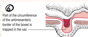
- Richter’s Hernia (Image 039): Part of antimesenteric bowel circumference trapped; strangulates without mechanical bowel obstruction.
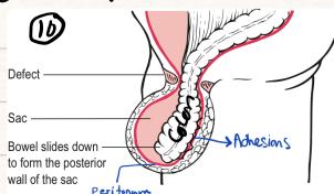
- Sliding Hernia (Image 041): Retroperitoneal organ forms posterior wall of sac (sigmoid colon on left, cecum/bladder on right).
- Obturator Hernia: Through obturator foramen; elderly emaciated females; SBO + Howship-Romberg Sign (pain down medial thigh on hip extension/abduction).
- Diaphragmatic Hernias:
- Bochdalek Hernia: Posterolateral diaphragmatic defect due to failure of pleuroperitoneal canal closure; commonest congenital diaphragmatic hernia (, predominantly left-sided).
- Morgagni Hernia: Retrosternal / parasternal anterior defect through the space of Larrey / foramen of Morgagni.
- Traumatic Diaphragmatic Hernia: Secondary to blunt thoracoabdominal trauma or penetrating stab/bullet wounds (usually left-sided due to protective hepatic mass on right).
C. Consultant Pearls & Critical Landmarks
- The 3 Lumps & Dynamic Actions ():
- Thyroid Lump: Command patient to Swallow (moves upward with pretracheal fascia).
- Breast Lump: Command patient to Press hands firmly on hips (contracts pectoralis major to test deep fixation).
- Hernia Lump: Command patient to Cough (elicits visible/palpable expansile cough impulse).
- The 12 Clinical Palpation Criteria for Any Lump / Swelling (Image 067 Flowchart):
-
- Site, 2) Size, 3) Shape, 4) Surface, 5) Consistency, 6) Borders/Edges, 7) Compressibility, 8) Reducibility, 9) Attachment to skin/muscle, 10) Pulsatility, 11) Fluctuation, 12) Transillumination.
-
- Midpoint of Inguinal Ligament vs. Midinguinal Point:
- Midpoint of Inguinal Ligament: Halfway between ASIS and Pubic Tubercle. Landmark for Deep Inguinal Ring (1.5 cm above; site for digital thumb occlusion).
- Midinguinal Point: Halfway between ASIS and Pubic Symphysis. Landmark for Femoral Artery Pulse.
D. Bedside Inguinal Hernia Examination Checklist
- Preparation: WIPES, chaperone, exposure from nipples to mid-thigh. Examine patient STANDING first (gravity and abdominal pressure maximize descent and size).
- Inspection (Standing):
- Note location relative to pubic tubercle: Inguinal hernia emerges above and medial; femoral hernia lies below and lateral.
- Instruct patient to turn head and cough: observe expansile cough impulse. Inspect overlying skin (erythema/edema indicate strangulation).
- Palpation (Standing & Supine):
- Scrotal Neck Test (“Getting Above the Mass”):
- Pinch fingers above the upper pole at scrotal neck.
- Can get above mass (empty cord felt): Purely scrotal swelling (hydrocele, cyst, varicocele, tumor).
- CANNOT get above mass: Mass originates from groin/abdomen = Inguinoscrotal Hernia.
- Reducibility:
- Ask patient to reduce mass supine, or gently guide upwards and laterally.
- Feel: Smooth, slippery, resonant with a gurgle = Small bowel (enterocele); doughy, granular, non-gurgling, hard to reduce = Omentum (omentocele).
- Incarceration: Irreducible hernia due to adhesions between sac and contents (without acute ischemia).
- Deep Ring Occlusion Test:
- Reduce hernia fully with patient supine.
- Firmly occlude deep ring with thumb: 1.5 cm above midpoint of inguinal ligament (halfway between ASIS and pubic tubercle).
- Instruct patient to stand and cough:
- Hernia is CONTROLLED: Indirect Inguinal Hernia.
- Hernia ESCAPES medial to thumb: Direct Inguinal Hernia.
- Zieman / Invagination Test: Invaginate scrotal skin with pinky through external ring; cough impulse on fingertip = Indirect; impulse on pulp/side = Direct.
- Scrotal Neck Test (“Getting Above the Mass”):
- Percussion & Auscultation: Tympanitic (bowel) vs. dull (omentum). Auscultate peristaltic bowel sounds over scrotum.
E. Complications & Surgical Repair
- Complication Spectrum: Reducible Incarcerated (irreducible without vascular compromise) Obstructed (colicky pain, distension, obstipation) Strangulated (Surgical Emergency):
- Constriction at neck venous thrombosis arterial ischemia gangrene and perforation.
- Signs: Exquisitely tender, tense, red, warm, irreducible lump with loss of cough impulse and systemic toxicity.
- Rule: Never attempt forceful manual reduction of a strangulated hernia (risk of reduction en masse or fatal intraperitoneal rupture).
- Surgical Principles:
- Pediatric: Herniotomy alone (high ligation and division of patent processus vaginalis at deep ring; no wall repair).
- Adult Open Gold Standard: Lichtenstein Tension-Free Hernioplasty (polypropylene mesh sutured to pubic tubercle, inguinal ligament, and conjoint tendon). Indicated in Class I Clean wounds.
- Laparoscopic Repairs: TAPP (transabdominal preperitoneal) or TEP (totally extraperitoneal). Indicated for bilateral hernias, recurrent hernias, and active working individuals.
- Mesh Contraindication: Strangulated hernia with bowel gangrene, perforation, or purulent peritonitis (Class III/IV wound; mesh causes severe sepsis; perform tissue suture repair: Shouldice or Bassini technique).
Station 10: Breast Lump Clinical Scenario, Physical Examination & Triple Assessment
A. Clinical Objectives & Prof. Alam’s Scenario
- Solitary painless breast lump is the #1 presentation of breast carcinoma.
- Systematic execution of Triple Assessment protocol (accuracy ).
- Step-by-step bedside clinical examination of breasts and regional lymph nodes.
B. High-Yield Consultant Teaching (Prof. Alam)
- Risk Factors for Breast Cancer:
- Early menarche (), late menopause (), nulliparity, first child after age 30, prolonged HRT, family history (BRCA1/BRCA2).
- Protective Factors: Prolonged breastfeeding, multiparity, early childbearing, regular exercise.
- Nipple Eczema vs. Paget’s Disease:
- Eczema: Typically bilateral, begins on areola and spreads to nipple, itchy, responds to topical corticosteroids.
- Paget’s Disease: Typically unilateral, begins on nipple and spreads outward to areola, does not respond to steroids; represents epidermotropic intraepithelial migration of underlying DCIS or invasive ductal carcinoma.
- Palpation Technique: Palpate breast tissue using the palmar pads of the fingers flat against the chest wall in circular compressing motions. Use fingertips secondarily to delineate lump margins and attachment.
- Axillary Lymph Node Levels (in relation to Pectoralis Minor):
- Level I: Lateral to lateral border of pectoralis minor (pectoral, subscapular, humeral groups).
- Level II: Deep/posterior to pectoralis minor (central group).
- Level III: Medial to medial border of pectoralis minor (apical group).
- Histological Grading (Bloom-Richardson System):
- Grade 1: Well-differentiated (tubule formation , low mitotic rate).
- Grade 2: Moderately differentiated.
- Grade 3: Poorly differentiated / anaplastic (marked pleomorphism, high mitotic index).
- Receptor Status & Adjuvant Therapy:
- Core biopsy confirms grade and receptors: ER/PR +ve respond to endocrine blockade (Tamoxifen in pre/postmenopausal; Aromatase Inhibitors [Anastrozole, Letrozole] in postmenopausal). HER2 +ve tumors receive targeted monoclonal antibody therapy (Trastuzumab / Herceptin).
- Breast Conserving Therapy (BCT) Contraindication:
- Lumpectomy with clear margins + whole breast radiotherapy.
- Absolute Contraindication: Systemic Lupus Erythematosus (SLE) or active scleroderma (cannot tolerate radiotherapy; causes severe vasculitic soft tissue necrosis).
- Nipple Discharge Dilemmas:
- #1 Cause of bloody nipple discharge: Intraductal Papilloma (IDP) (benign fibrovascular polyp in lactiferous duct).
- Pathologic / Suspicious Discharge: 1) Bloody (sanguineous), 2) Unilateral, and 3) Spontaneous from a single duct.
C. The Triple Assessment Protocol
                       [ TRIPLE ASSESSMENT PROTOCOL ]
                                      |
         +---------------------------+---------------------------+
         |                                                       |
[ 1. Clinical Assessment ]                              [ 2. Radiological Imaging ]
  - Complete History & Risk Factors                       - Age < 35: Targeted Breast Ultrasound
  - Stepwise Physical Exam                                - Age >= 35: Bilateral Digital Mammography
                                                                      + Targeted Ultrasound
                                                                      + Breast MRI (selected)
                                      |
                                      v
                         [ 3. Pathological Tissue Diagnosis ]
                           - Core Needle Biopsy (CNB) (Gold Standard - 14G)
                           - Fine Needle Aspiration Cytology (FNAC)
- Each component scored 1 to 5 (1 Normal, 2 Benign, 3 Uncertain, 4 Suspicious, 5 Malignant). Diagnostic concordant accuracy .
- Imaging Strategy:
- Women : Targeted Ultrasound (dense fibroglandular parenchyma obscures mammography; US accurately differentiates cyst vs. solid; preferred in fibroadenoma).
- Women : Bilateral Digital Mammography (CC and MLO views) PLUS targeted Ultrasound. Malignant signs: Spiculated high-density mass, clustered pleomorphic microcalcifications, architectural distortion.
- Breast MRI: Indicated for occult primary (positive axillary node with negative mammogram/US), lobular cancer, BRCA carriers, silicone implant rupture, and neoadjuvant response.
- Pathology Strategy:
- Core Needle Biopsy (CNB) (14-gauge, Gold Standard): Obtains intact histological architecture; differentiates in situ (DCIS) from invasive carcinoma (evaluates basement membrane); tests ER, PR, HER2-neu, and Ki-67.
- FNAC: Cytological aspirate only; cannot differentiate in situ from invasive cancer; high insufficient sample rate.
D. Step-by-Step Breast Physical Examination Checklist (Leenah Turjoman)
- Introduction & Setup: WIPES, informed consent, ensure absolute privacy. Female chaperone is MANDATORY.
- Positioning & Exposure:
- Position: Patient semi-recumbent at 45° on couch.
- Rationale: Upright position makes breasts drop downwards; lying flat makes breast tissue slide laterally into axillae; 45° provides optimal, symmetric tissue spread over pectoralis major.
- Special Rule: If the patient states that the lump is felt only in a specific position, examine in the standard 45° semi-sitting position first, then re-examine in the specific position stated.
- Exposure: Fully undressed from waist upwards.
- Position: Patient semi-recumbent at 45° on couch.
- Inspection (Directly facing patient):
- At Rest (Hands on thighs):
- Symmetry & Contour: Asymmetry, visible bulge, dimpling, flattening.
- Skin: Erythema, puckering, visible veins, Peau d’orange (dermal lymphatic edema with pitted hair follicles from tumor emboli).
- Nipple-Areola Complex: Inversion/retraction (recent vs. congenital), deviation (points toward quadrant of tumor), discharge, Paget’s ulceration.
- Dynamic Maneuvers (The 4 Classic Positions):
- Hands relaxed on thighs / by side: Baseline appearance.
- Arms raised above head: Stretches Cooper’s suspensory ligaments, elevates breasts, exposes inframammary fold and lower quadrants.
- Hands pressed firmly on hips/waist pushing inward: Contracts Pectoralis Major reveals skin dimpling or tumor tethering to pectoral fascia.
- Leaning forward: Breasts hang freely, accentuating deep chest wall tethering and asymmetry in pendulous breasts.
- At Rest (Hands on thighs):
- Palpation (Stand at patient’s right side; examine UNAFFECTED breast first):
- Patient semi-recumbent with ipsilateral hand placed behind head (flattens breast over ribs).
- Use pads of middle 3 fingers flat against chest wall with gentle circular compressing movements.
- Examine in a systematic radial clock-face pattern (examining at each ‘hour’ from periphery towards nipple) or concentric circles covering all 4 quadrants (UOQ harbor of cancers).
- Palpate the Axillary Tail of Spence (extends through deep fascia foramen of Langer into axilla).
- Palpate subareolar tissue and nipple; if discharge was reported, ask the patient to gently express/reproduce it herself; if not successful, gently massage breast tissue and compress areolar base.
- Lump Characterization:
- Site: Clock-face position and distance in centimeters from nipple center.
- Size: Measured in centimeters with a calibrated ruler (e.g., ).
- Consistency: Soft (lipoma), cystic/fluctuant (cyst), firm/rubbery (fibroadenoma), stony hard (carcinoma).
- Margins: Smooth/well-defined (benign) vs. irregular/infiltrative (malignant).
- Mobility & Fixation Tests:
- Skin Tethering: Pinch overlying skin; inability to pinch indicates dermal invasion of Cooper’s ligaments.
- Pectoralis Major Fixation: Test lump mobility parallel/perpendicular to muscle fibers with muscle relaxed, then repeat with patient pressing hands into hips (contracted): mobility restricted when contracted = Fixed to pectoral fascia.
- Chest Wall Fixation: Lump completely immobile whether muscle is relaxed or contracted = Fixed to rib/intercostal muscle/serratus anterior.
- Axillary & Regional Lymph Node Palpation:
- Patient seated upright.
- Golden Technique: Fully support the weight of the patient’s arm at the elbow/forearm with your ipsilateral arm so the pectoral and shoulder girdle muscles are completely relaxed.
- Examining Left axilla: Support patient’s left arm with your left hand; palpate with your Right hand (and vice versa).
- Systematically palpate all 5 walls/groups: 1) Anterior / Pectoral (along lower border of pectoralis major), 2) Posterior / Subscapular (along anterior border of latissimus dorsi), 3) Lateral / Humeral (along upper humerus/axillary vein), 4) Central / Medial (deep against chest wall/serratus anterior), 5) Apical (at the apex of the axilla behind the clavicle).
- Palpate Supraclavicular and Infraclavicular fossae bilaterally (ask patient to slightly shrug shoulders to relax clavicular spaces).
- Metastatic Screen: Palpate abdomen for nodular hepatomegaly and ascites; auscultate/percuss lung bases for pleural effusion; palpate thoracic/lumbar spine for vertebral bony tenderness.
Station 11: Thyroid Gland & Solitary Neck Mass Physical Examination & Assessment

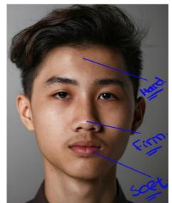
A. Surgical Anatomy & Neurovascular Relations
- Anatomy & Fascial Compartments:
- Located in the visceral compartment of anterior neck, deep to sternohyoid and sternothyroid strap muscles.
- Two lateral lobes (extending from thyroid cartilage down to 5th/6th tracheal rings), joined across midline by isthmus overlying the 2nd, 3rd, and 4th tracheal rings.
- Pyramidal Lobe: Present in up to of individuals (remnant of embryonic thyroglossal duct), ascending superiorly from isthmus toward hyoid bone.
- Gland enveloped by visceral layer of pretracheal fascia, anchored firmly to cricoid cartilage and upper tracheal rings via the suspensory ligament of Berry (causes thyroid to move upward with deglutition).
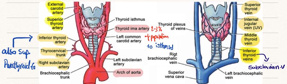
- Arterial Blood Supply:
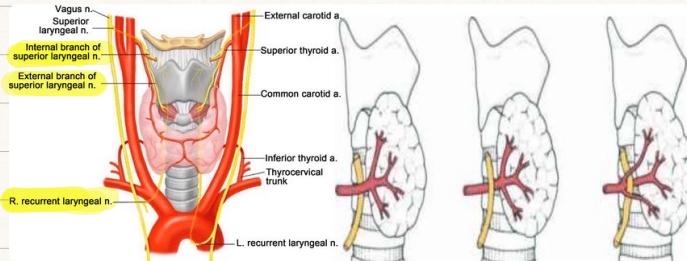
- Superior Thyroid Artery (STA): 1st anterior branch of External Carotid Artery; intimately related to External Branch of Superior Laryngeal Nerve (EBSLN) (injury during high vascular ligation paralyzes cricothyroid muscle loss of high-pitch phonation, decreased vocal projection, and vocal fatigue).
- Inferior Thyroid Artery (ITA): From Thyrocervical Trunk (subclavian); crossed intimately by Recurrent Laryngeal Nerve (RLN).
- Thyroidea Ima Artery: Present in from brachiocephalic trunk or aortic arch (major bleeding hazard during emergency tracheostomy).
- Venous Drainage: Superior and Middle Thyroid Veins drain directly into Internal Jugular Vein (IJV); Inferior Thyroid Veins form a plexus draining into Left Brachiocephalic Vein.
- Recurrent Laryngeal Nerve (RLN):
- Right RLN loops around right subclavian artery; Left RLN loops around aortic arch.
- Ascend in tracheoesophageal groove to enter larynx behind cricothyroid joint; motor to all intrinsic laryngeal muscles except cricothyroid.
- Unilateral Injury: Paramedian cord palsy hoarseness, bovine cough, weak phonation.
- Bilateral Injury: Both cords paralyzed in paramedian position acute airway obstruction, inspiratory stridor, emergency intubation/tracheostomy.
B. Thyroid Hormone Kinetics & Clinical Physiology
| Hormone | Secretion Proportion | Bound State & Potency | Serum Half-Life | Clinical Pearl |
|---|---|---|---|---|
| Thyroxine () | Prohormone; tightly bound to TBG | 5 to 7 days | Medical antithyroid therapy (Carbimazole, PTU) requires 3 to 6 weeks to deplete circulating and achieve euthyroid state | |
| Triiodothyronine () | (90% from peripheral deiodination) | Biologically active form; more potent | 1 to 3 days () | Rapid biological effect, short-acting |
C. Clinical History & Functional Assessment
- History Red Flags: Solitary hard mass growing rapidly over is highly suspicious for aggressive malignancy; compression symptoms (dysphagia, dyspnea/stridor); infiltration symptoms (dysphonia/hoarseness RLN invasion).
- Systemic Symptoms (Handwritten Notes Comparison):
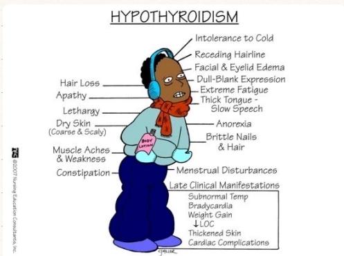
- Hypothyroidism (Image 044): Cold intolerance, facial and periorbital edema, dull-blank apathetic expression, diffuse hair loss, coarse dry scaly skin, thick tongue, slow slurred speech, lethargy, muscle aches, constipation, brittle nails, menorrhagia, weight gain, hypothermia, sinus bradycardia. (Treatment: Thyroxine replacement).
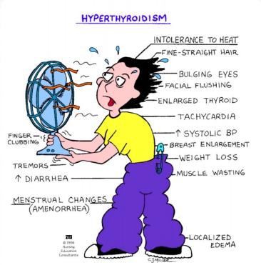
- Hyperthyroidism / Thyrotoxicosis (Image 045): Heat intolerance, diaphoresis, warm moist palms, fine straight hair, bulging eyes (proptosis/exophthalmos), facial flushing, palpitations, resting sinus tachycardia, systolic hypertension (wide pulse pressure), high-output collapsing water-hammer pulse, fine postural tremor, weight loss despite hyperphagia, diarrhea/hyperdefecation, oligomenorrhea, proximal muscle wasting (hypotrophic muscles), finger clubbing. (Treatment: Antithyroid medications [Carbimazole/PTU], radioactive iodine ablation, surgical subtotal/total thyroidectomy).
- Compressive Symptoms: Dyspnea/stridor (tracheal compression), dysphagia (esophageal compression).
- Pemberton’s Sign: Facial plethora, cyanosis, distended neck veins, and inspiratory stridor induced by elevating both arms above head for 1 minute (thoracic inlet obstruction by retrosternal goiter).
- Malignancy Risk Factors: Childhood head/neck ionizing radiation, family history of MEN 2A/2B (RET proto-oncogene) or medullary thyroid cancer, rapidly growing hard solitary nodule in young male or elderly ().
D. Step-by-Step Bedside Physical Examination (‘s Protocol)
- Preparation & Exposure: WIPES, seated on backless chair. Exposure: Chin to Nipple Line (2nd ICS) (mandatory to detect dilated chest wall collateral veins and retrosternal extensions).
- Inspection (Directly in Front):
- The 3 S’s:
- 1. Scars: Low horizontal collar scar (Kocher incision) in skin crease.
- 2. Swellings: Symmetry, location, lobar contour, butterfly goiter.
- 3. Skin changes / Superficial veins: Erythema, dilated collateral veins over manubrium.
- Dynamic Swallowing Maneuver: Patient sips water, holds, swallows on command while watching the neck:
- Observation: Thyroid gland and thyroglossal cysts move upward with deglutition (enclosed in pretracheal fascia anchored to larynx). Cervical lymph nodes do not move.
- Dynamic Tongue Protrusion Maneuver (Indications):
- Mandatory if patient is young with a small () midline neck mass.
- Observation: Thyroglossal Duct Cyst moves UPWARD with tongue protrusion (attached to hyoid bone and foramen cecum via thyroglossal tract). Thyroid goiters/nodules do NOT move with tongue protrusion.
- The 3 S’s:
- Tracheal Position:
- Evaluated while standing directly in front of patient: index and ring fingers placed on the sternoclavicular heads, middle finger palpates tracheal rings in the suprasternal notch to detect lateral tracheal deviation.
- Palpation (Examiner Standing BEHIND Patient):
- Position: Patient flexes neck slightly forward to relax sternocleidomastoid (SCM) and strap muscles. (Never extend neck; tell patient to face forward).
- Thumbs point upwards resting on the nape/back of neck to support the head; examining fingers curve around the lateral lobes.
- Golden Rule: NEVER MOVE BOTH EXAMINING HANDS SIMULTANEOUSLY (compresses trachea severe patient choking). Gently apply pressure to one side of the larynx to displace the trachea and make the contralateral thyroid lobe prominent for palpation, then switch.
- Palpate each lobe systematically while patient swallows a small sip of water.
- Retrosternal Assessment (High-Yield Verbal Statement): State to examiner: “I am palpating to determine if I can get below the lower border of both thyroid lobes.” Inability to reach lower border confirms Retrosternal Extension / Goiter.
- The 4 S’s and C on Palpation:
- Site, Size, Shape, Surface (Smooth in Graves’/diffuse goiter vs. Multinodular).
- Consistency: Firm in 99% of exam cases ( note); stony-hard in anaplastic/carcinoma; soft in colloid goiter; rubbery in Hashimoto’s.
- Facial Tactile Landmarks (Image 068):
- Forehead: Hard (bone / invasive carcinoma).
- Tip of Nose: Firm / Rubbery (normal thyroid tissue / benign multinodular goiter).
- Lower Lip / Chin: Soft (colloid nodule / cystic degeneration).
- Carotid Pulse & Berry’s Sign:
- Palpate carotids against C6 transverse tubercle (Chassaignac’s tubercle) bilaterally.
- Positive Berry’s Sign: Carotid pulse obliterated/encased by invasive thyroid carcinoma. (In benign goiter, pulse remains palpable, displaced posterolaterally; superficial temporal pulse remains easily palpable).
- Cervical Lymph Nodes:
- Palpate systematically by anatomical names (submental, submandibular, upper deep cervical / jugulodigastric, middle deep cervical, lower deep cervical / supraclavicular, posterior triangle, preauricular, postauricular, occipital). Names are more professional than numeric levels in viva.
- Ask patient to slightly shrug shoulders during supraclavicular fossa palpation.
- Percussion: Directly percuss over manubrium sterni; dullness confirms retrosternal goiter.
- Auscultation: Auscultate superior thyroid poles with stethoscope bell for systolic bruit (machinery vascular flow in vascular Graves’). Differentiate from transmitted carotid bruit / aortic stenosis.
- Eye Signs & Systemic Examination:
- Graves’ Eye Signs: Proptosis/exophthalmos, Lid retraction (Dalrymple’s sign) (sclera visible above superior limbus), Lid lag (von Graefe’s sign) (upper lid lags behind downward gaze), infrequent blinking (Stellwag’s), poor convergence (Mobius’), ophthalmoplegia.
- Hands & Pulse: Fine postural tremor (paper test), warm sweaty palms, palmar erythema, Plummer’s nails (onycholysis). Pulse: Resting sinus tachycardia, AF, or high-output collapsing water-hammer pulse; bradycardia in hypothyroidism.
- Facies: Coarse hair, loss of outer 1/3 of eyebrows (Queen Anne’s sign / Hertoghe’s sign) in hypothyroidism.
- Lower Limbs: Pretibial myxedema (peau d’orange violaceous non-pitting plaques over shins in Graves’), brisk reflexes (or delayed Achilles relaxation in hypothyroidism).
E. Histopathological Types of Thyroid Carcinoma
| Tumor Type | Frequency | Origin & Pathology | Spread | Prognosis & Surgical Treatment |
|---|---|---|---|---|
| Papillary (PTC) | 80–85% | Follicular cells; Orphan Annie nuclei, Psammoma bodies | Lymphatic (cervical nodes) | Excellent ( 10y survival). Total thyroidectomy / hemithyroidectomy central neck dissection + ablation () |
| Follicular (FTC) | 10–15% | Follicular cells; capsular / vascular invasion | Hematogenous (bone, lungs) | Good ( 10y survival). Diagnostic hemithyroidectomy completion thyroidectomy + ablation |
| Medullary (MTC) | 5% | Parafollicular C-cells; secretes Calcitonin; MEN 2A/2B (RET) | Lymphatic & hematogenous | Intermediate. Total thyroidectomy + central neck dissection (radioiodine ineffective) |
| Anaplastic (ATC) | 1–2% | Undifferentiated; elderly (); rapid stony-hard mass | Rapid local invasion | Extremely poor (); airway compromise; palliative tracheostomy/radiation |
| Thyroid Lymphoma | 1–2% | Non-Hodgkin B-cell; arises on Hashimoto’s | Rapid diffuse growth | Responsive to Systemic Chemotherapy + Radiotherapy (surgery for biopsy only) |
- Surgical Definitions:
- Total Thyroidectomy: Removal of both lobes, isthmus, and pyramidal lobe (Graves’, bilateral goiter, cancer ).
- Hemithyroidectomy (Lobectomy): Removal of one lobe + isthmus (indeterminate solitary nodule / Bethesda IV).
- Completion Thyroidectomy: Excision of residual lobe after histology reveals invasive cancer.
- Radioiodine Ablation (): Destroys microscopic thyroid remnants in well-differentiated cancers (PTC/FTC ).
F. Diagnostic Workup & Bethesda Cytopathology
- TSH: First-line screening. Suppressed in hyperthyroidism, elevated in hypothyroidism.
- Subclinical Thyroid Dysfunction (): Abnormal TSH with completely normal free and free .
- Calcitonin: Mandatory if suspecting MTC or MEN 2.
- Thyroid Scintigraphy (Radionuclide Scan): Indicated in hyperthyroidism to differentiate diffuse uptake (Graves’) from focal hot nodule (toxic adenoma) or patchy uptake (toxic MNG). Hot nodules are rarely malignant (); cold nodules carry a malignancy risk.
- Ultrasound (TI-RADS): Microcalcifications, hypoechoic, irregular margins, “taller-than-wide”, cervical lymphadenopathy.
- Fine Needle Aspiration Cytology (Bethesda Reporting):
- Bethesda I: Non-diagnostic (repeat FNA).
- Bethesda II: Benign ( malignancy; observe).
- Bethesda III: AUS / FLUS (; repeat FNA or molecular testing).
- Bethesda IV: Follicular Neoplasm (; cytology cannot distinguish adenoma from carcinoma due to lack of capsular evaluation; Diagnostic Hemithyroidectomy).
- Bethesda V: Suspicious for malignancy (; surgery).
- Bethesda VI: Malignant (; Total Thyroidectomy).
G. The 3 Main Indications for Thyroid Surgery ()
- FNA Confirmed Malignancy (Bethesda VI: Papillary, Medullary, Anaplastic Carcinoma).
- FNA Suspicion of Malignancy (Bethesda IV: Follicular Neoplasm; Bethesda V: Suspicious).
- Benign Goiter with Compressive Pressure Symptoms:
- Dysphagia (esophageal compression).
- Dyspnea / Stridor (tracheal compression).
- Severe cosmetic deformity.
- (Also: Large nodule , or refractory thyrotoxicosis failing medical/radioiodine therapy).
H. Post-Thyroidectomy Complications
- 1. Reactionary Hemorrhage / Neck Hematoma (First 6 Hours):
- Arterial bleeding under tension compresses trachea acute airway obstruction, stridor, cyanosis.
- Emergency Bedside Protocol: Immediately cut/remove skin clips and sutures at bedside, open deep cervical fascia, evacuate hematoma with sterile gloved fingers to relieve tracheal compression, then transfer to OR for formal exploration.
- 2. Recurrent Laryngeal Nerve Injury: Unilateral: hoarseness, bovine cough; Bilateral: acute stridor, emergency tracheostomy.
- 3. Superior Laryngeal Nerve (External Branch) Injury: Cricothyroid muscle paralysis loss of high-pitch sound/voice and early vocal fatigue.
- 4. Postoperative Hypocalcemia (Hypoparathyroidism):
- Inadvertent devascularization/excision of parathyroid glands; onset at 24 to 72 hours.
- Paresthesias of lips/fingers, carpopedal spasm. Chvostek’s sign (facial twitching on tapping facial nerve); Trousseau’s sign (carpopedal spasm on BP cuff inflation for 3 minutes).
- Treatment: Mild: Oral calcium + calcitriol; Severe: 10 mL of 10% IV Calcium Gluconate infused over 10 minutes under ECG monitoring.
- 5. Thyroid Storm: Fever , tachycardia , delirium; treat with beta-blockers, PTU, Lugol’s iodine, IV hydrocortisone.
Station 12: Lower Limb Peripheral Vascular Physical Examination & Arterial Disease
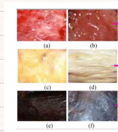
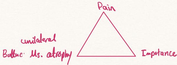
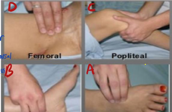
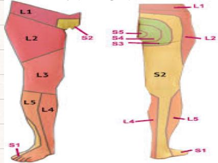
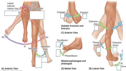
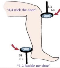
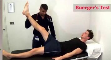
A. Clinical Spectrum of Peripheral Arterial Disease (PAD)
- Clinical History Taking Questions (Handwritten Notes):
- Chief Complaint: Severe unrelenting lower extremity ischemic pain lasting weeks.
- Intermittent Claudication: Screening questions (“Do you get cramping muscle fatigue when you walk that eases when you stand still? What exact distance can you walk before the pain stops you?”).
- Ischemic Rest Pain: Dependency relief (“Patients characteristically hang their leg down off the edge of the bed to the floor so gravity restores perfusion and relieves the pain”).
- Medications: Treatment modality (“Are you taking subcutaneous insulin injections or oral hypoglycemic tablets?”).
- Fontaine Classification & Claudication Distance Progression:
- Stage I: Asymptomatic.
- Stage II: Intermittent Claudication: Cramping calf/thigh/buttock pain provoked by walking, relieved within 5–10 minutes of standing still.
- Historical distance progression: 1 km 500 m 100 m Rest Pain Gangrene.
- Anatomical levels: Calf = Superficial Femoral / Popliteal; Thigh = Common Femoral / External Iliac; Buttock/Hip = Common Iliac / Distal Aorta.
- Stage III: Ischemic Rest Pain: Severe, burning forefoot/toe pain; worse at night when supine in bed (loss of gravity perfusion); relieved by hanging foot over bed or sleeping in chair.
- Stage IV: Ischemic Ulceration & Gangrene (Tissue Loss).
- Critical Limb Ischemia (CLI): Rest pain or ulcer/gangrene, plus ankle systolic pressure or toe pressure .
- Leriche Syndrome Triad (Aortoiliac Occlusion):
- Bilateral buttock and thigh claudication.
- Absent or markedly diminished bilateral femoral pulses.
- Erectile dysfunction / impotence in males (internal iliac / pudendal ischemia).
B. Step-by-Step Bedside Vascular Examination Checklist (Leenah Turjoman)
- Preparation: WIPES, supine, exposure from groin to toes (both legs exposed to compare).
- Inspection (Compare Bilaterally with Normal Side):
- Trophic Changes: Shiny, thin, atrophic skin; hair loss over shins/dorsum of feet; thickened, brittle, slow-growing dystrophic toenails.
- Color: Pallor on elevation, fixed rubor, cyanosis, mottling.
- Muscular Atrophy: Calf/thigh wasting.
- Arterial Ulcers: Distal extremities (toe tips, interdigital spaces, pressure points, heel, lateral malleolus); “punched-out” circular margins, pale slough base, minimal bleeding, excruciating pain.
- Gangrene: Dry (mummified, black, sharp demarcation) vs. Wet (edematous, purulent, foul-smelling, spreading, crepitus — surgical emergency).
- Scars: Groin incisions, medial thigh fem-pop bypass scars, saphenous vein harvest scars.
- Golden Rule: DON’T FORGET THE BACK OF THE LEG AND HEEL (elevate limb to inspect Achilles tendon and plantar heel for occult decubitus ulcers).
- Palpation:
- Temperature Gradient: Use dorsum of hand from thighs down to toes bilaterally; sudden cold cutoff demarcates level of arterial occlusion.
- Pitting Edema Check: Apply firm pressure over the medial aspect of the tibial shaft for 10 seconds to assess for pitting versus non-pitting edema.
- Capillary Refill Time (CRT): Compress great toe pulp 5s; normal refill ; delayed indicates poor perfusion. (Cannot be reliably assessed in patients with poor hygiene/subungual hyperkeratosis).
- Systematic Pulse Palpation (Bilaterally):
- Abdominal Aorta: Epigastrium slightly left of midline with flat hands ( expansile pulsation suggests AAA).
- Femoral Artery: At midinguinal point (halfway between ASIS and pubic symphysis; palpate with 3 fingers).
- Popliteal Artery: Deepest pulse; knee flexed 90° to 120°; two hands wrap around knee with fingers pressing deeply into popliteal fossa against tibial plateau.
- Posterior Tibial Artery: inferior and posterior to medial malleolus (apply deep relaxed 2-finger pressure).
- Dorsalis Pedis Artery: Dorsum of foot, immediately lateral to extensor hallucis longus tendon over navicular bone.
- Auscultation: Auscultate abdominal aorta, iliac, and femoral arteries for systolic bruits (indicates stenosis).
- Special Provocative Test — Buerger’s Test:
- Explain to patient: “I am going to raise your leg to test your circulation.”
- Phase 1 (Elevation): Elevate legs for 1–2 minutes. (Healthy limb tolerates up to without pallor).
- Elevation Pallor: Ischemic limb blanches.
- Buerger’s Angle of Circulatory Suffering: Angle at which pallor appears; angle indicates critical limb ischemia. Note venous guttering.
- Phase 2 (Dependency): Patient sits upright with legs hanging over couch edge.
- Dependency Rubor (“Sunset Foot”): Delayed deep dusky red / violaceous hyperemic flush (reactive capillary dilation from severe hypoxia).
- Venous refill time: Normal ; prolonged indicates severe arterial disease.
- Neurological Screen: Sensory across L1–S1; vibration (128 Hz tuning fork) tested at 3 sites: 1) Great toe tip, 2) Medial malleolus, 3) Tibial tuberosity; proprioception; knee jerk (L3/L4) and ankle jerk (S1/S2); motor power and tone.
- “What Else Do You Want to Examine?” (High-Yield Viva Question):
-
- Fundoscopy: Diabetic and hypertensive retinopathy (use ophthalmoscope).
-
- Carotid Examination: Auscultate for carotid bruits ( stenosis stroke risk).
-
- Full Cardiovascular Exam: Silent ischemic heart disease, murmurs, atrial fibrillation.
-
- Urine Dipstick: Microalbuminuria / proteinuria (early diabetic nephropathy).
-
C. Vascular Pathology Pearls
- Eggshell Calcification of AAA: Plain AXR may show curvilinear calcification outlining dilated aortic wall. Detached mural thrombus showers microemboli to lower extremities acute “trash foot” (blue toe syndrome) or stroke.
- Buerger’s Disease (Thromboangiitis Obliterans — TAO):
- Non-atherosclerotic segmental inflammatory occlusive vasculitis of small/medium vessels.
- Young male heavy smokers ().
- Triad: Distal claudication (foot instep arch pain), superficial migratory thrombophlebitis, Raynaud’s phenomenon.
- Palpable femoral/popliteal pulses with absent pedal pulses; corkscrew collaterals on angiogram.
- Management: Absolute permanent smoking cessation is the ONLY effective treatment; failure leads inevitably to digital gangrene and amputation.
D. Diagnostic Investigations
- Ankle-Brachial Pressure Index (ABPI):
- : Incompressible calcified vessels (Monckeberg’s sclerosis; common in diabetes/ESRD; requires Toe-Brachial Index [TBI]).
- : Normal perfusion. (If ABI normal no further vascular imaging required).
- : Mild/moderate PAD (intermittent claudication).
- : Severe ischemia / Critical Limb Ischemia (rest pain, tissue necrosis).
- Arterial Duplex Ultrasound: First-line non-invasive imaging.
- Digital Subtraction Angiography (DSA) / CTA / MRA: Road-mapping to distinguish focal, reconstructible/stentable lesions from diffuse inoperable occlusion.
Station 13: Diabetic Foot Ulcer Examination & Surgical Management (Amputations)

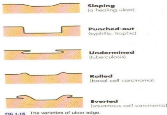
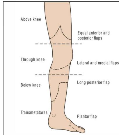
A. Pathophysiology: The Diabetic Triad
- Neuropathy:
- Hyperglycemia polyol pathway (aldose reductase converts glucose to sorbitol) osmotic swelling, axonal and Schwann cell degeneration.
- Sensory: Glove-and-stocking loss of protective sensation (“Loss of protective pain sensation patient steps on nails, pins, or glass without feeling anything, noticing only upon bleeding or odor”).
- Motor: Lumbrical/interosseous atrophy flexor/extensor imbalance claw toes, hammer toes, prominent metatarsal heads with focal plantar pressure points.
- Autonomic: Anhidrosis (dry cracked skin) + cutaneous Arteriovenous (AV) shunting (warm pink foot with nutrient capillary starvation).
- Macro/Microvascular Disease: Accelerated tibial/peroneal occlusive disease.
- Immunopathy: Impaired PMN leukocyte chemotaxis, adhesion, and intracellular phagocytosis predisposition to virulent and opportunistic infections (Candida).
B. Autonomic Mechanisms & Patient Education
- Mechanisms:
- Sweat gland denervation anhidrosis skin fissures and portals of entry.
- Cutaneous smooth muscle denervation Arteriovenous (AV) shunting deceptively warm skin with microvascular ischemia.
- Diabetic Foot Patient Education Checklist:
- Daily foot inspection using a hand-held mirror (or family assistance).
- Never walk barefoot (even indoors; prevent silent pin/glass puncture wounds).
- Feel inside shoes for pebbles or rough linings before wearing.
- Avoid heat sources: Thermal burn warning (“Never warm feet near electric heaters, radiators, or hot water bottles severe painless full-thickness contact burns”).
- Wear custom diabetic shoes with wide toe-box; break in gradually ().
- Cut toenails straight across; never round edges; never self-shave calluses with razor blades.
C. Step-by-Step Bedside Diabetic Foot Examination (‘s Protocol)
- The Golden Rule (): ALWAYS KEEP THE ULCER FOR THE VERY LAST STEP. Examine limb, alignment, skin, pulses, and nerves before touching the ulcer.
- The 3 Hidden Sites to Touch on Inspection (): 3. In between the toes and behind the heels. 4. Underside of the breast (inframammary fold). 5. Abdominal pannus / panniculus fold.
- General Foot Inspection:
- Skeletal deformities: Claw toes, hammer toes, hallux valgus.
- Charcot Foot Triad: 1) Warm erythematous swollen foot/ankle (mimics gout/cellulitis), 2) Loss of medial longitudinal arch (rocker-bottom flat foot), 3) Bony subluxation, destruction, and fractures on X-ray.
- Skin: Anhidrosis, hyperkeratotic callous over 1st/5th metatarsal heads, tinea pedis.
- Pitting Edema Assessment: Compress over medial tibial shaft for 10 seconds to differentiate pitting from non-pitting edema.
- The 6 Ulcer Characterization Criteria (Image 053):
- 1. Site: Exact anatomical location (plantar 1st metatarsal head, great toe tip, heel).
- 2. Size: Measured in centimeters (Length Width, e.g. ).
- 3. Depth: Superficial vs. full-thickness vs. deep (exposing tendon, joint capsule, bone).
- 4. Base / Floor: Granulation (healthy red) vs. Slough (yellow cotton-like dying tissue) vs. Gangrenous black eschar. (Pearl: Floor you can see; Base you can touch/feel).
- 5. Edge Morphology:
- Punched-out / Perpendicular: Neuropathic and ischemic ulcers.
- Sloping: Venous ulcers (and healing diabetic ulcers).
- Undermined: Tuberculosis, syphilis, decubitus bed sores.
- Rolled / Raised: Basal cell carcinoma (rodent ulcer; sun-exposed face/nose; never metastasizes).
- Everted: Squamous cell carcinoma (Marjolin’s ulcer in chronic burn scar/sinus).
- 6. Margin & Surrounding Skin: Erythema ( = severe cellulitis), warmth, callous halo, whitish maceration, black necrotic discoloration.
- Special Ulcer Palpation Techniques ():
- Technique 1: Squeeze surrounding skin of the ulcer (milking from distal to proximal) to express hidden purulent discharge (pus release confirms deep space infection/abscess).
- Technique 2: Feel the base of the ulcer with a gloved thumb: Normal cortical bone feels smooth and hard. A diseased bone crumbles under constant pressure (feels like crushing a dry biscuit), pathognomonic for Osteomyelitis (NOT osteoporosis). Indicates long-term IV antibiotics, surgical debridement, or amputation.
- Neurological & Vascular Testing:
- 10g Semmes-Weinstein Monofilament: 10 plantar sites; missed sites confirms loss of protective sensation.
- 128 Hz tuning fork over hallux; Achilles reflex. Palpate pedal pulses, CRT, Buerger’s test.
D. Wagner-Meggitt Classification
| Grade | Clinical Feature | Management |
|---|---|---|
| 0 | Intact skin; bony deformities, hyperkeratotic calluses | Preventive offloading, custom shoes, education |
| 1 | Superficial ulcer; no tendon/capsule penetration | Debridement, dressings, offloading |
| 2 | Deep ulcer penetrating to tendon, capsule, or bone; NO abscess/osteomyelitis | Surgical debridement, antibiotics, offloading |
| 3 | Deep ulcer with Abscess, Osteomyelitis, or Septic Arthritis | Urgent surgical drainage, bone debridement, IV antibiotics |
| 4 | Gangrene localized to Forefoot or Heel | Urgent vascular evaluation, localized amputation (Ray/TMA) |
| 5 | Extensive Gangrene involving entire foot | Major limb amputation (BKA or AKA) |
E. Microbiology, Laboratory Workup & Medical Optimization
- Microbiology: Most common single isolate: Staphylococcus aureus. Established ulcers are POLYMICROBIAL (Gram+ cocci: Staph, Strep; Gram- bacilli: E. coli, Klebsiella, Proteus, Pseudomonas; Anaerobes: Bacteroides fragilis).
- Sampling Protocol (Mandatory OSCE Statement): “I will obtain a deep tissue biopsy or curettage from the debrided ulcer base (or bone biopsy) for Gram stain and C&S BEFORE initiating antibiotics. Superficial swabs are avoided as they merely culture non-pathogenic skin colonizers.”
- Empirical Antibiotics (Bonus Exam Question):
- Tazocin (Piperacillin/Tazobactam 4.5g IV q8h) Metronidazole.
- In severe sepsis or penicillin allergy: Clindamycin or Vancomycin PLUS Ciprofloxacin/Carbapenem (Imipenem/Meropenem).
- Anemia Correction: Target to optimize tissue oxygen delivery for collagen synthesis and granulation. Normocytic anemia common in diabetes due to erythropoietin (EPO) deficiency from diabetic nephropathy (CRF).
- Hyperglycemic Dilutional Pseudohyponatremia (Elevated Glucose = Reduced Serum Sodium): Elevated glucose draws intracellular water into vascular space.
- Correction Formula: Add to measured serum sodium for every glucose elevation above normal ().
- Target Glycemic Control: HbA1c normal range .
F. High-Yield Consultant Exam Pearls ()
- Multi-Disciplinary Team (MDT) Approach (The #1 Exam Answer):
- In the exam, when asked how to manage a diabetic foot patient, immediately state: “This patient requires a Multi-Disciplinary Team (MDT) approach.”
- Team members: General Surgeon, Diabetologist / Endocrinologist, Nephrologist, Radiologist, Vascular Surgeon, Infectious Disease Specialist, Podiatrist / Wound Care Specialist, Orthotist, and Physiotherapist.
- The 2 Types of Gangrene in Diabetic Foot:
- Dry Gangrene: Arterial occlusion with NO underlying infection; dry, shriveled, mummified black tissue with clear demarcation line; stable outpatients can be managed conservatively at home.
- Wet Gangrene: Underlying virulent bacterial infection with liquefactive necrosis; swollen, boggy, purulent, foul-smelling; surgical emergency requiring immediate hospitalization, IV antibiotics, and urgent surgical debridement/amputation.
- Foot Radiographs & MRI: Plain X-ray evaluates soft tissue gas, radiopaque foreign bodies, cortical disruption, and periosteal reaction (Osteomyelitis). MRI is the gold standard imaging modality for osteomyelitis. Probe-to-Bone test has positive predictive value for osteomyelitis if metallic probe hits hard bone.
G. Lower Limb Amputations: Indications & Levels
- The “3 D’s” for Amputation:
- Dead: Irreversible tissue gangrene beyond revascularization.
- Deadly: Spreading life-threatening sepsis (wet gangrene, ascending necrotizing fasciitis).
- Dead Loss (Severe Disability): Intractable rest pain, non-healing ulceration, or severe flexion contracture rendering limb non-functional.
- Surgical Rule: Amputation should only be considered when arterial reconstruction is deemed inappropriate or impossible by a vascular surgeon, or when immediate amputation is life-saving in profound sepsis.
- Levels of Lower Limb Amputation:
- Toe Amputation: Disarticulation at MTP joint (removal of one or more toes; affects balance and push-off).
- Ray Amputation: Excision of toe + corresponding metatarsal head.
- Transmetatarsal (TMA): Transection across metatarsal shafts with plantar flap; preserves heel and ankle.
- Ankle Disarticulation (Syme’s Amputation): Amputation of foot at ankle joint; retains heel pad allowing patient to bear weight and ambulate short distances without a prosthesis.
- Below-Knee Amputation (BKA / Transtibial): Preferred major level; preserves knee joint (vastly superior prosthetic rehabilitation and lower cardiac workload than AKA). Ideal stump length: 12 to 15 cm below tibial plateau (or 8–12 cm). Uses Burgess Posterior Myocutaneous Flap (gastrocnemius-soleus). Dressed with crepe bandage.
- Through-Knee (Disarticulation): End-weight-bearing stump retaining the entire femur with lateral and medial flaps.
- Above-Knee Amputation (AKA / Transfemoral): Severe knee contracture, high gangrene, or failed BKA. Equal anterior and posterior flaps; ideal length: 25 cm below greater trochanter (10–12 cm above knee for prosthetic joint).
- Hip Disarticulation: Amputation through hip joint; radical worst-case scenario for ascending necrotizing infection or high sarcoma.
Station 14: Digital Rectal Examination (DRE / PR Exam) & Proctoscopy
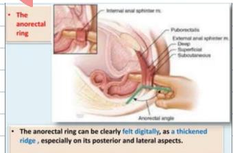
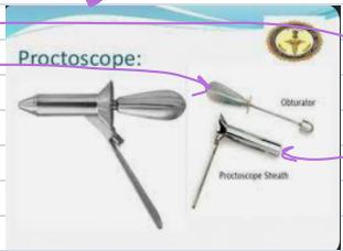
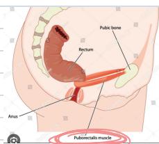
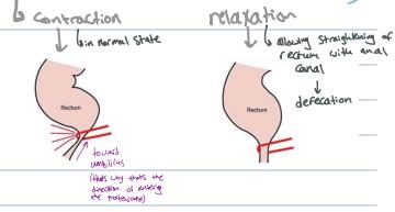
A. Indications & Contraindications
- Indications: Altered bowel habits (diarrhea, progressive constipation, tenesmus), rectal bleeding/melena, pelvic/rectal mass, prostate assessment (BPH vs. cancer), sphincter tone (incontinence/impaction), acute abdomen (pelvic appendicitis/abscess tenderness in pouch of Douglas), trauma (sphincter tone, bony pelvic fragments).
- Contraindications:
- Absolute: Imperforate anus, impassable anal stricture, extreme agony without anesthesia.
- Relative: Acute anal fissure (extreme pain), thrombosed external hemorrhoids, recent acute myocardial infarction, severe neutropenia/immunocompromise (risk of fatal bacteremia/perianal sepsis), unwilling patient.
B. Step-by-Step Bedside DRE Examination Checklist
- Communication & Consent: WIPES, informed consent, ensure privacy. Chaperone is MANDATORY. Reassure the patient continuously that relaxing avoids pain.
- Positioning: Left Lateral (Sims) Position (buttocks at couch edge, hips and knees flexed towards chest). Drape genitalia appropriately.
- Perianal Inspection (Separate buttocks with both hands):
- Excoriations, pruritus ani, fecal soiling.
- External hemorrhoids (thrombosed/prolapsed, 3rd degree piles visible externally).
- Anal Fissure: Longitudinal tear in anoderm (90% posterior midline; 10% anterior; lateral suggests Crohn’s, TB, HIV, syphilis); look for distal sentinel skin tag.
- Fistula-in-ano External Opening: Pus/blood discharge. Goodsall’s Rule: Openings anterior to transverse anal line track radially straight into canal; openings posterior track along a curved path to posterior midline.
- Perianal abscess, condylomata acuminata (warts), anal canal ulcer/mass, hyperemia.
- Digital Palpation:
- Generously lubricate gloved index finger (apply lubricant to the pulp/bulb of finger).
- Place finger bulb flat against anal verge; perform gentle circular massage to relax the sphincter opening while instructing patient to breathe deeply and relax.
- Insert finger smoothly through canal aiming toward umbilicus.
- Sphincter Tone: Assess resting tone (internal sphincter; normally strong; weak tone warns of post-op fecal incontinence), then active voluntary squeeze tone (external sphincter and puborectalis).
- Anorectal Ring: Palpate muscular sling of puborectalis posteriorly.
- Systematic 360° Sweep: Advance into rectal ampulla, sweep anterior, lateral, posterior walls. Palpate against sacrum and coccyx.
- Anterior Palpation:
- Prostate (Males):
- Normal: Smooth, firm (like tip of nose), elastic, non-tender, heart-shaped with distinct central median sulcus.
- BPH: Symmetrically enlarged, smooth, rubbery/firm, non-tender, preserved median sulcus.
- Prostate Cancer: Hard, stony, nodular, asymmetrical, loss/obliteration of median sulcus, pelvic fixation.
- Females: Palpate cervix and pouch of Douglas (tenderness, drop metastases / Blumer’s shelf).
- Prostate (Males):
- Withdrawal & Glove Inspection:
- Withdraw finger; immediately inspect glove tip for: Fresh red blood (rectal bleed/tumor/piles), melena, pus/mucus (proctitis/IBD), pale/acholic stool (obstructive jaundice). Clean patient, dispose gloves, explain findings.
C. Proctoscopy (Rigid Proctoscope)
- Instrument: Rigid hollow cylinder with rounded obturator and illumination (fenestrated or non-fenestrated).
- Technique: Generously lubricate, insert aiming toward umbilicus, redirect into rectal ampulla, withdraw obturator, advance light, inspect mucosa during gradual circular withdrawal.
- Normal Landmark: Can clearly visualize the dentate line (pectinate line).
- Internal Hemorrhoids (Piles): Normal vascular cushions; CANNOT be felt digitally on DRE (compressible veins) unless thrombosed or 4th degree. Proctoscopy visualizes them prolapsing into lumen at classic 3, 7, and 11 o’clock positions (left lateral, right anterior, right posterior). Also visualizes polyps, proctitis, ulcers.
Station 15: Nasogastric Tube (NGT) Insertion Procedural Station
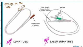
A. Indications & Contraindications
- Indications:
- Gastric Decompression: Mechanical SBO, paralytic ileus, gastric dilatation/volvulus, nasal obstruction decompression, aspiration prevention in unconscious trauma resuscitation, post-laparotomy decompression.
- Enteral Feeding: Dysphagia, CVA/stroke, head injury (convert to PEG/PEJ if feeding ; change tube every 2 weeks).
- Diagnostic/Therapeutic: Gastric aspirate analysis, upper GI bleed assessment, contrast/activated charcoal administration.
- Contraindications:
- Absolute: Severe midface trauma, suspected base of skull fracture (Battle’s sign, raccoon eyes, CSF rhinorrhea/otorrhea, hemotympanum fatal intracranial passage through cribriform plate into brain).
- Relative: Severe coagulopathy (INR ; patient on warfarin; catastrophic epistaxis risk), nasopharyngeal obstruction (large tumor, carcinoma of nasopharynx), esophageal strictures/burns, varices (pass with gentleness), recent gastric/bariatric surgery.
B. Tube Types, Sizing & Equipment
- Tube Types:
- Levin Tube: Single-lumen; indicated for enteral feeding and diagnostic sampling.
- Salem Sump Tube: Double-lumen; gold standard for suction and decompression (large lumen for suction; blue vent “pigtail” open to atmosphere prevents vacuum mucosal adherence/ulceration).
- Sizing & Material:
- 12 Fr: Narrow bore, soft, flexible, more comfortable for long-term enteral feeding, but harder to insert as it may bend in the pharynx.
- 14–16 Fr: Standard adult, stiffer, easier to insert without bending; essential for active gastric suction and decompression (uncomfortable for patient).
- Silicon-based or polyurethane materials.
- Assembly: NGT, 2% lidocaine water-soluble lubricating gel (up to 10–15 mL / cc on tip), 50 mL catheter-tip syringe, CE-marked pH test strips (0–6), cup of water with straw, vomit bowl, split hypoallergenic tape, towel over chest, gloves.
C. Step-by-Step Procedural Checklist
- Preparation: WIPES (wash hands, introduce, permission, explain procedure, privacy), confirm patient identity (name and MRN), verify INR , chaperone. Agreed Distress Signal: Establish stop hand-raise signal. If patient is uncooperative, have assistant/nurse support.
- Positioning & Patency: High Fowler’s position (semi-sitting 45°–90° upright, flexed hips and knees). Inspect nostrils for polyps or severe septal deviation; test patency (occlude one side and sniff; select more patent nostril).
- NEX Measurement (Calibration): Measure distance from Nose tip (N) Earlobe (E) Xiphoid process (X). Note pre-printed centimeter mark (typically 50 to 55 cm in adults). Note radiopaque blue line for X-ray visibility.
- Initial Insertion: Lubricate distal 10–15 cm with water-soluble gel. Insert tip into nostril, advancing HORIZONTALLY along floor of nasal cavity (parallel to hard palate, aiming toward ear/occiput, NEVER vertically upwards).
- Advancement with Swallowing:
- At posterior pharynx (12–15 cm; gagging felt): instruct patient to flex head forward (chin to chest) (closes epiglottis, opens esophagus).
- Instruct patient to take continuous small sips of water through straw and swallow continuously.
- Advance tube smoothly 5 to 10 cm per swallow until NEX mark reaches nares.
- EMERGENCY RULE: If patient coughs violently, chokes, becomes cyanotic, lacrimates, or dyspneic IMMEDIATELY STOP AND WITHDRAW TUBE INTO PHARYNX (accidental tracheal entry).
- Confirmation of Placement:
- Method 1: Gastric Aspirate pH (Bedside Standard): Aspirate 2–5 mL fluid; inspect character (clear, yellow, green, brown bilious; note: empty stomach if patient fasting); test on pH strip:
- pH : Confirms Acidic Gastric Placement (gastric acidity is 2–3; safe to use).
- pH : Inconclusive (respiratory/intestinal fluid); DO NOT use; obtain X-ray.
- Method 2: Chest/Abdominal X-Ray (Gold Standard): Mandatory if pH or no aspirate. 4 criteria: 1) Down midline crossing carina, 2) Bisects left diaphragm, 3) Tip and side-ports below GE junction in stomach, 4) In gastric bubble.
- The “Whoosh” Test (Air Auscultation): Push 30 mL (cc) of air with syringe while auscultating epigastrium; CONDEMNED AND UNRELIABLE (sounds transmit from respiratory tract).
- Method 3: Capnography / : Colorimetric/infrared detector on proximal port; high detection confirms tracheal/lung cannulation withdraw immediately.
- Method 1: Gastric Aspirate pH (Bedside Standard): Aspirate 2–5 mL fluid; inspect character (clear, yellow, green, brown bilious; note: empty stomach if patient fasting); test on pH strip:
- Securing, Complications & Documentation:
- Secure to nose bridge with split tape; pin to gown with slack.
- Complication: Tube splints open lower esophageal sphincter (LES), predisposing to exaggerated GERD symptoms and chemical esophagitis.
- Document tube type, size, nostril, depth mark, aspirate pH, date/time.