collected from Faisal Alkharji, Faisal Alturki, Haneen H. Alsultan, Khalid Alanazi, Lina Serhan, Moath Bin Abdullah, Sultan AlDajani, Talal M. Ak.
اللهم يا معلّم موسى علّمني، ويا مفهم سليمان فهّمني، ويا مؤتي لقمان الحكمة وفصل الخطاب آتني الحكمة وفصل الخطاب اللهم اجعل ألستنا عامرة بذكرك، وقلوبنا بخشيتك، وأسرارنا بطاعتك، إنك على كل شيء قدير، حسبنا الله ونعم الوكيل
CS
- [[#CS#Main cases|Main cases]]
- [[#CS#General Principles & Perioperative Care|General Principles & Perioperative Care]]
- [[#CS#Trauma & Emergency Surgery|Trauma & Emergency Surgery]]
- [[#CS#Neurosurgery|Neurosurgery]]
- [[#CS#Breast|Breast]]
- [[#CS#Upper GI & Abdominal Surgery|Upper GI & Abdominal Surgery]]
- [[#CS#Urology|Urology]]
- [[#CS#Vascular|Vascular]]
- [[#CS#Skin & Soft Tissue|Skin & Soft Tissue]]
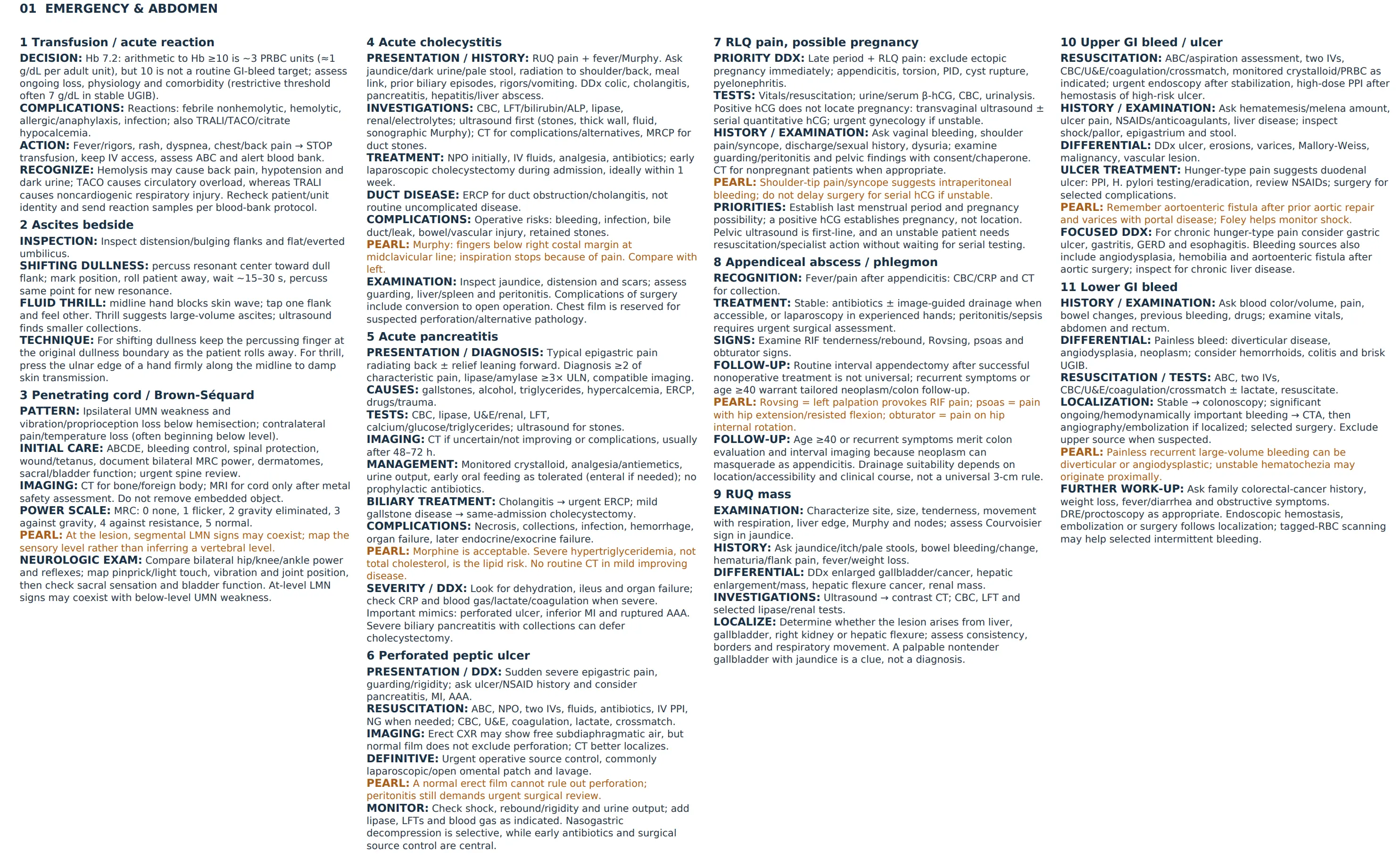
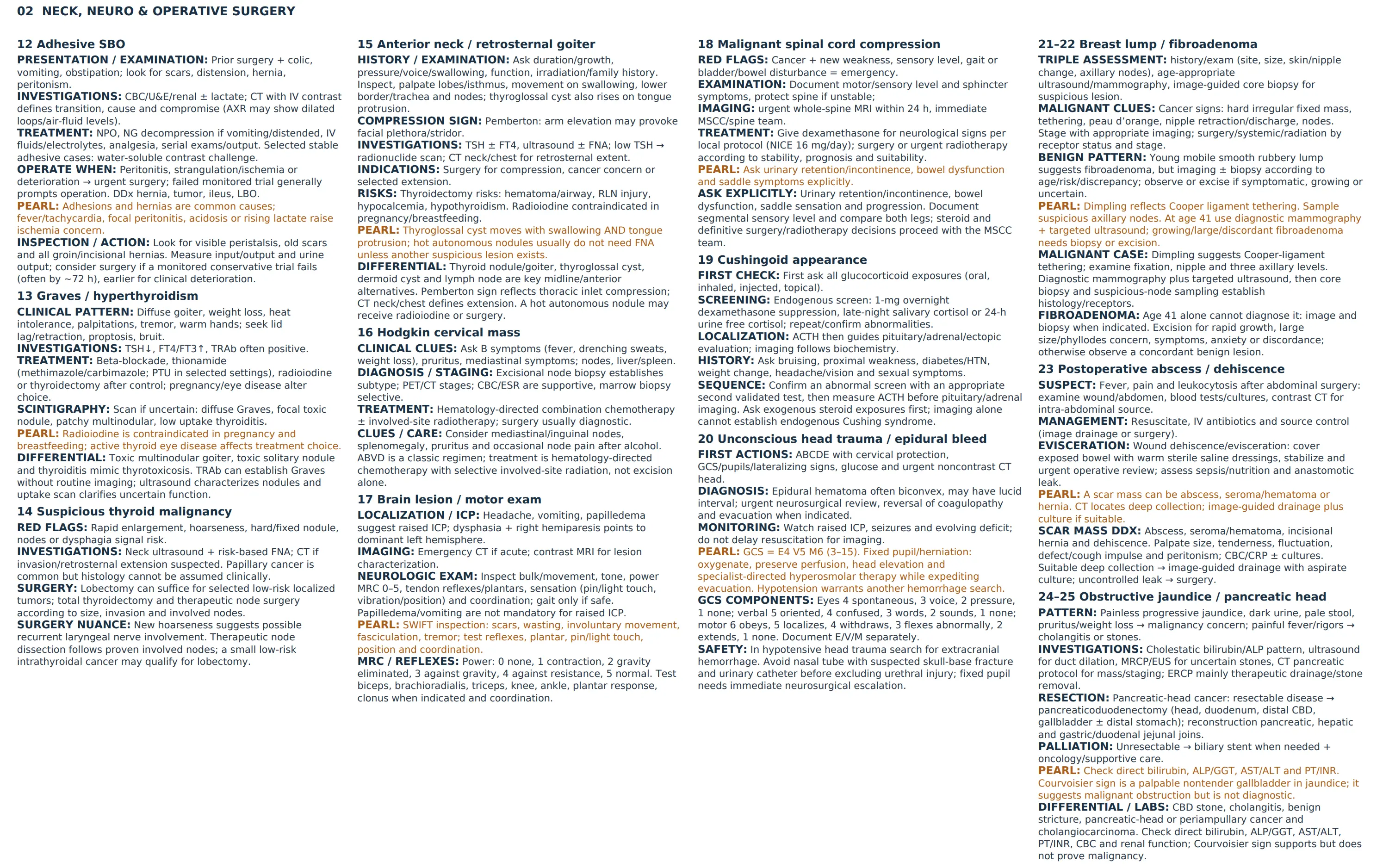
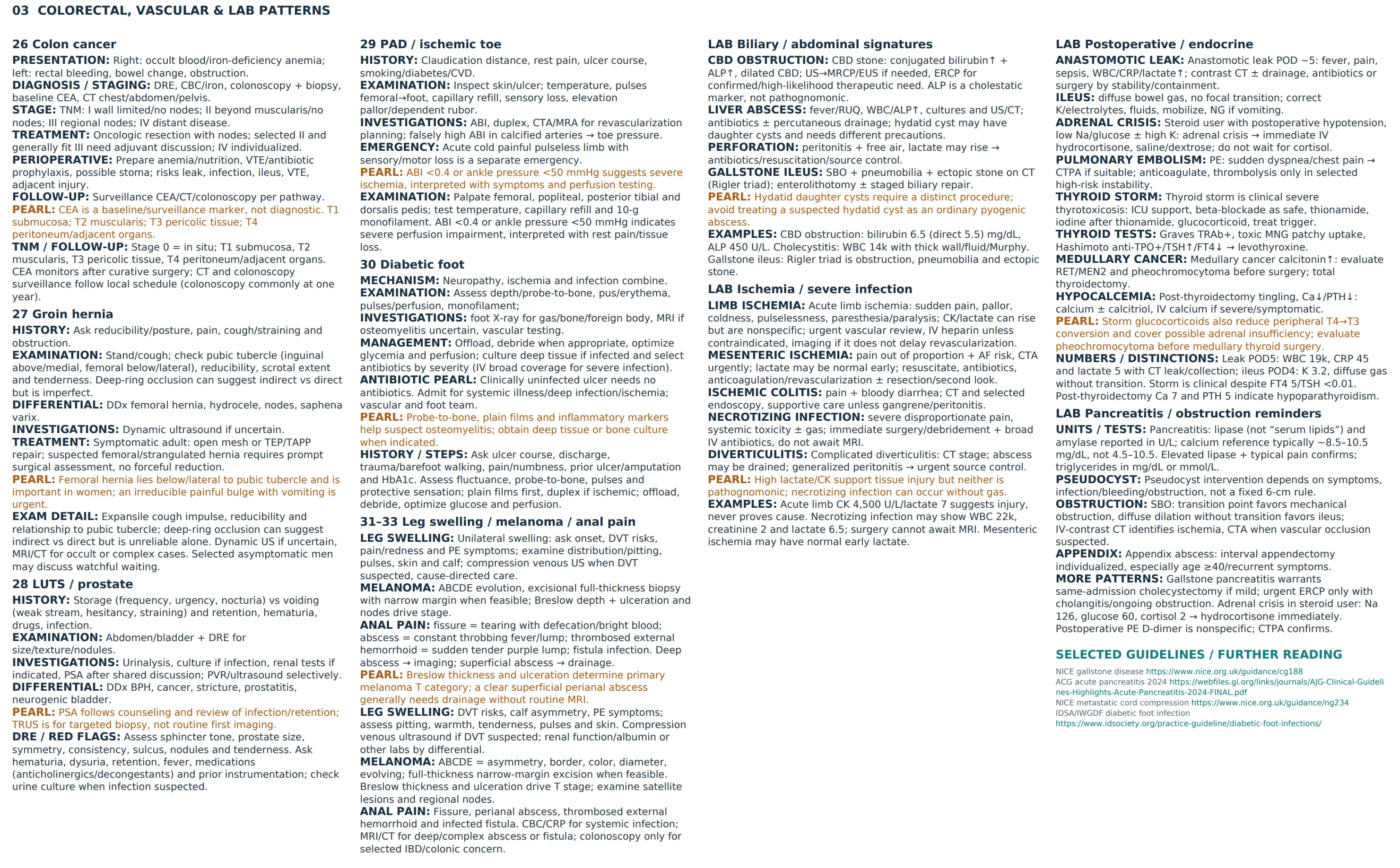
Main cases
Case 1: Graves’ Disease (Toxic Diffuse Goiter)
Case: A 46-year-old female has neck swelling, weight loss, palpitations, warm wet palms, tachycardia/arrhythmia and an enlarged thyroid; another stem describes diffuse anterior neck enlargement with the same thyrotoxic features.
Q1: What is the most likely clinical diagnosis?
- Answer: Graves’ Disease (toxic diffuse goiter / hyperthyroidism).
Q2: Which TWO further signs (or FIVE additional signs) would you seek?
- Answer: For a two-sign answer, choose lid lag and lid retraction. For five additional signs, seek lid lag, lid retraction, proptosis, fine tremor and thyroid bruit. Warm wet palms and tachycardia are already supplied by the stem; ask about heat intolerance rather than listing it as a physical sign.
Q3: Which laboratory investigations are relevant?
- Answer: Core assessment is TSH with free T4/free T3 and TRAb. Anti-TPO is supplementary and may support autoimmune thyroid disease when indicated.
Q4: Mention THREE treatment modalities available for Graves’ disease.
- Answer:
- Antithyroid medications: Thionamides (Methimazole / Carbimazole, or Propylthiouracil).
- Radioactive Iodine Ablation ().
- Surgery: Total or near-total thyroidectomy (after achieving euthyroid state with medications). Beta-blockade controls tremor, palpitations and tachycardia while definitive treatment is planned; choice considers pregnancy, age, goiter/compression and thyroid eye disease.
Q5: Which TWO other differential diagnoses and TWO imaging investigations are useful?
- Answer: Toxic multinodular goiter and a toxic solitary nodule; thyroiditis is another cause of thyrotoxicosis. Ultrasound characterizes structural nodules; a radionuclide uptake/scan helps distinguish diffuse Graves uptake, focal autonomous uptake, and low-uptake thyroiditis when the cause is uncertain. Imaging is not obligatory when TRAb/clinical findings establish Graves disease.
Case 2: Thyroid Malignancy (Suspected Papillary Thyroid Carcinoma)
Case: A female has had a neck swelling for 2 years, with rapid progression and new hoarseness over 2 months.
Q1: What is the most likely diagnosis?
- Answer: Thyroid Carcinoma (most commonly Papillary Thyroid Carcinoma).
Q2: What is the primary surgical management?
- Answer: Thyroid surgery is tailored to size, invasion, nodal disease, and risk. This suspected invasive case may require total thyroidectomy and therapeutic dissection of involved nodes; low-risk localized disease can be treated with lobectomy. Central/lateral neck dissection is not automatic for every papillary carcinoma.
Q3: What FIVE clinical features would support malignancy, and which TWO investigations are initially requested?
- Answer: Supporting features of malignancy include rapid growth and hoarseness (as in this stem), a hard/fixed nodule, cervical nodes, and dysphagia/compressive symptoms. Initial investigations are neck ultrasound and ultrasound-guided FNA; CT assesses suspected invasion/retrosternal disease. Note that “papillary adenoma” is an incorrect label for thyroid carcinoma.
Case 3: Approach to Anterior Neck Swellings and Retrosternal Goiter
Case: A 50-year-old male has painless neck swelling for more than 2 years, moving on swallowing; a 53-year-old female has neck swelling with a question about retrosternal extension.
Q1: What clinical examination sign evaluates retrosternal extension?
- Answer: Pemberton’s sign (raising both arms above the head for 1 minute produces facial congestion, cyanosis, and respiratory stridor from thoracic inlet compression).
Q2: Mention TWO indications for surgical intervention.
- Answer:
- Compressive symptoms (dyspnea, dysphagia, stridor).
- Suspected or confirmed malignancy (or retrosternal extension, cosmetic deformity).
Q3: What FIVE history questions are relevant?
- Answer: Ask when/how the swelling was noticed, growth or fluctuation/pain, dysphagia/dyspnea/voice change, skin change or discharge/other lumps, and hyper-/hypothyroid symptoms. Also ask about neck irradiation, smoking, medication, past operations, and family thyroid disease/cancer. Palpitations, heat/cold intolerance, bowel habit, weight, menstrual changes, and sleep/mood symptoms help characterize function.
Q4: What FIVE examination steps should be performed on palpation?
- Answer: Assess site/size/consistency/tenderness and nodularity; confirm movement on swallowing; examine each thyroid lobe and isthmus from behind; assess the lower border/tracheal position; palpate cervical nodes. Inspect before palpating and auscultate for bruit when appropriate.
Q5: Give FOUR differential diagnoses of anterior/midline neck swellings.
- Answer: Thyroid enlargement/nodule, thyroglossal duct cyst, dermoid cyst, and lymphadenopathy. A plunging ranula may present below the floor of the mouth; a submental node is another location-specific possibility. Thyroglossal cysts characteristically move on tongue protrusion as well as swallowing.
Q6: Which TWO laboratory and TWO non-laboratory investigations are appropriate?
- Answer: TSH with free thyroid hormones as indicated, plus CBC if systemic illness/preoperative assessment warrants it. Ultrasound is the initial structural test; FNA depends on sonographic risk/size. CT neck/chest evaluates retrosternal extension. CXR and sternal percussion are historical adjuncts and cannot reliably exclude it; Pemberton’s sign indicates thoracic inlet compression rather than proving the full extent. Baseline CBC, U&E and ECG are reasonable before intervention.
Q7: How does thyroid function guide nodule evaluation, and how is a toxic solitary nodule treated?
- Answer: Low TSH prompts functional assessment with a radionuclide scan when appropriate; ultrasound/FNA evaluates nonfunctioning suspicious nodules. A hot nodule is usually benign, but history alone does not exclude cancer. Toxic solitary nodules may be treated with radioactive iodine or surgery, with medical symptom control while planning treatment.
Q8: What are the complications of thyroidectomy and contraindications to radioactive iodine?
- Answer: Bleeding/neck hematoma, recurrent laryngeal nerve injury, infection, hypothyroidism, and hypoparathyroidism/hypocalcemia. Radioiodine is contraindicated in pregnancy and breastfeeding; planning also considers conception timing and active thyroid eye disease. Children and prior external radiation are not universal absolute contraindications.
Case 4: Cervical Neck Mass in a Young Adult (Hodgkin Lymphoma)
Case: A photograph shows a neck mass in a 30-year-old male; the working diagnosis is Hodgkin lymphoma. B symptoms are not established in this patient.
Q1: What diagnostic investigation establishes lymphoma, and which additional tests may be requested?
- Answer: Excisional lymph-node biopsy with histopathology establishes the diagnosis/subtype. Bone-marrow assessment is selective; CXR can show mediastinal disease; ESR is supportive, not diagnostic. Tissue-access options also include core biopsy, mediastinoscopy and (rarely) thoracotomy; specialist choice depends on the site, and thoracotomy is not routine for an accessible neck node.
Q2: Which TWO staging tests are used?
- Answer: PET and CT. FDG-PET/CT commonly combines functional and anatomic staging; additional contrast CT is selected when needed rather than routinely ordering two redundant examinations.
Q3: What FIVE additional presenting features and baseline investigations should be considered?
- Answer: Cervical/mediastinal/inguinal lymphadenopathy, pruritus, alcohol-associated node pain, chest symptoms from mediastinal disease, and hepatosplenomegaly; B symptoms are unexplained fever, drenching night sweats and weight loss. CBC and ESR/other baseline bloods support assessment but do not establish histology. Excisional node biopsy is preferred; bone marrow assessment is selective rather than the routine first diagnostic test.
Q4: What are TWO principal treatment modalities?
- Answer: Hematology-directed combination chemotherapy with or without involved-site radiotherapy according to subtype/stage. ABVD is a classic regimen; newer regimens may be selected. Surgery is chiefly diagnostic, not routine definitive treatment; rituximab/methotrexate is not a classical-Hodgkin regimen.
Case 5: Lower GI Bleeding: Assessment and Initial Management
Case: A 45-year-old male has rectal bleeding; a 60-year-old male has two painless episodes within 4 hours (first bleeding episode, no medication, pulse 98/min, BP 108/78 mmHg, unremarkable abdomen); another 60-year-old has pulse 102/min, low BP, a normal abdomen and no palpable rectal mass.
Q1: What FIVE relevant questions will you ask?
- Answer:
- Color and quantity of blood (bright red vs dark maroon).
- Presence of abdominal pain or cramping.
- History of constipation, straining, or altered bowel habits.
- Previous episodes of GI bleeding or known diverticulosis/polyps.
- Medication history (anticoagulants, antiplatelets, NSAIDs).
Q2: Mention THREE most likely differential diagnoses.
- Answer:
- Diverticular bleeding (diverticulosis).
- Angiodysplasia (arteriovenous malformation).
- Colorectal cancer / polyps (or brisk upper GI bleed).
Q3: What FIVE initial laboratory tests and THREE special investigations are useful?
- Answer: CBC, renal function/electrolytes, coagulation profile, blood group/crossmatch, and lactate if significant hypoperfusion is suspected; add LFTs as indicated. Colonoscopy evaluates a stable lower GI source. CTA is prioritized in hemodynamic instability with suspected ongoing bleeding; a bedside estimate of a fixed mL/min bleeding rate is not needed. Upper endoscopy is indicated when a brisk upper source is suspected.
Q4: Outline initial management steps.
- Answer: ABC resuscitation, 2 large-bore IV lines, IV crystalloid fluids, blood transfusion if indicated, Foley catheter, and monitored unit admission.
Q5: Which further history questions and examination complete the assessment?
- Answer: Ask about family history of colorectal cancer, bleeding elsewhere, nausea/vomiting, fever/diarrhea, weight loss/anorexia, obstructive symptoms and metastatic symptoms. Examine the abdomen and perform DRE/proctoscopy when appropriate; assess anemia, shock, and masses. Additional DDx: hemorrhoids/fissure, IBD, ischemic or infectious colitis, rectal varices, and brisk upper GI bleeding.
Q6: How is source-directed treatment chosen, and what if colonoscopy cannot localize ongoing bleeding?
- Answer: Treat the identified cause: endoscopic hemostasis for amenable lesions, CTA localization followed by catheter angiography/embolization for appropriate active bleeding, and selected surgery when other measures fail. Tagged-red-cell scintigraphy is an option for selected intermittent bleeding where available. Gastrin testing, cystoscopy/cytology, routine NG insertion and PPI prophylaxis are not routine lower-GI-bleed investigations.
Case 6: Clinical Evaluation of Extrahepatic Biliary Obstruction
Case: A 55-year-old male has yellow discoloration of the eyes and skin; other stems have yellow eyes with tea-colored urine for two weeks, or jaundice with abdominal pain. Pain, progression, pale stool and pruritus are history questions rather than established findings.
Q1: Mention FIVE relevant questions regarding the history of present illness.
- Answer:
- Any abdominal pain (biliary colic vs painless jaundice).
- Change in urine color (tea-colored/dark) or stool color (pale/acholic/clay-colored).
- Generalized itching / pruritus.
- Constitutional symptoms (anorexia, early satiety, unintentional weight loss).
- History of gallstones, alcohol intake, viral hepatitis exposure, or blood transfusions.
Q2: Mention FOUR differential diagnoses.
- Answer:
- Choledocholithiasis (common bile duct stones).
- Carcinoma of the head of the pancreas.
- Cholangiocarcinoma (bile duct cancer / Klatskin tumor).
- Periampullary carcinoma (or benign biliary stricture).
Ascending cholangitis and biliary (gallstone) pancreatitis are additional causes of jaundice to consider.
Q3: What FIVE initial laboratory investigations should be requested?
- Answer:
- Total and Direct (conjugated) Bilirubin (predominantly direct hyperbilirubinemia).
- Alkaline Phosphatase (ALP) and Gamma-Glutamyl Transferase (GGT) (markedly elevated).
- AST and ALT (mild-to-moderate elevation).
- Coagulation profile: PT/INR and aPTT (prolonged PT may improve with vitamin K if deficiency is contributing).
- CBC and Renal Function Tests.
Q4: Mention THREE imaging/special investigations and their roles.
- Answer: Ultrasound initially; MRCP or EUS for duct evaluation; contrast CT when a mass/complication is suspected. ERCP is principally therapeutic for duct clearance/drainage, rather than a routine first diagnostic test in all jaundice.
Q5: Which additional history questions, examination steps, and laboratory tests are appropriate?
- Answer: Ask who noticed it/onset, meal association, nausea/vomiting, night sweats, travel/hepatitis/transfusion history, and previous biliary surgery. Examine for jaundice/excoriation, tenderness/Murphy sign, Courvoisier sign, hepatosplenomegaly and abdominal masses; palpate and percuss liver/spleen size. Add albumin, renal function, glucose, lipase for suspected pancreatitis, and viral serology when appropriate. Urine bilirubin/urobilinogen may assist context but does not replace blood tests.
Q6: What further differential causes and clinical distinction should be considered?
- Answer: CBD stones, cholangitis, benign biliary stricture, pancreatic head/periampullary cancer, cholangiocarcinoma, gallbladder cancer and primary sclerosing cholangitis. Hepatitis/liver abscess may cause jaundice but are not all extrahepatic obstruction. Sudden painful jaundice favors stones over a typical painless progressive malignant presentation, but pain alone does not exclude cancer.
Case 7: Acute Cholecystitis
Case: (1) A 48-year-old male has RUQ pain and fever for 3 days; temperature 38.5°C, pulse 100/min, no jaundice, with RUQ tenderness and guarding. (2) A 58-year-old male has right upper abdominal pain. (3) A patient presents with RUQ pain, nausea and vomiting.
Q1: What FIVE further relevant questions will you ask regarding the presenting complaint?
- Answer:
- History of jaundice, dark urine, or pale stools.
- Radiation of pain to the right shoulder or back (scapula).
- Association of pain with fatty or greasy meals.
- Previous similar episodes of biliary pain or known gallstones.
- Associated chills, rigors, or vomiting.
Q2: What is the most likely diagnosis and THREE relevant differential diagnoses?
- Answer: Acute calculous cholecystitis is most likely in the febrile RUQ case. Alternatives include biliary colic, ascending cholangitis, and acute pancreatitis. For a broader RUQ-pain prompt also consider pyogenic liver abscess and hepatitis.
Q3: What FIVE laboratory investigations will you request at admission?
- Answer:
- CBC (leukocytosis with left shift).
- Liver function tests: ALT, AST.
- Alkaline Phosphatase (ALP) and Bilirubin.
- Serum Amylase / Lipase (to rule out secondary gallstone pancreatitis).
- Serum Electrolytes / Renal function tests.
CRP and ESR are supportive inflammatory markers.
Q4: What TWO imaging studies are initially useful, and what THREE further imaging/special investigations may be requested?
- Answer: Ultrasound first: gallstones, gallbladder wall thickening, pericholecystic fluid, and sonographic Murphy sign. CT is useful for complications or alternative diagnoses; MRCP evaluates suspected duct stones. ERCP is generally reserved for therapeutic duct clearance/cholangitis, not routine uncomplicated cholecystitis. A plain/erect chest radiograph is appropriate if perforation is suspected.
Q5: Mention FIVE initial management steps after admission.
- Answer:
- NPO (bowel rest).
- IV fluid resuscitation.
- IV analgesics (NSAIDs or opioids).
- IV broad-spectrum antibiotics.
- Early laparoscopic cholecystectomy, ideally during the same admission; NICE recommends within one week of diagnosis. The old “72 hours, otherwise wait 2–3 months” rule is outdated.
Q6: Demonstrate Murphy’s sign and outline the relevant abdominal examination.
- Answer: Inspect for jaundice/distension/scars; palpate gently for RUQ tenderness and guarding. Place fingers below the right costal margin in the midclavicular line and ask the patient to inspire: inspiratory arrest due to pain is Murphy’s sign. Compare with the left, assess liver and spleen size, and check for peritonitis.
Q7: Mention recognized complications of cholecystectomy.
- Answer: Bleeding, wound/intra-abdominal infection, bowel or vascular injury, bile duct injury/bile leak, retained duct stones, and conversion to open surgery.
Case 8: Adult Inguinal Hernia and Groin Swelling
Case: Separate stems: (1) a 41-year-old female with left inguinal swelling; (2) a 45-year-old male with left-groin swelling for four weeks, becoming invisible at rest; (3) a 55-year-old male with right inguinal swelling for six months.
Q1: What THREE relevant history questions should you ask?
- Answer: Ask duration/onset, how it was noticed and change in size, pain, reducibility/posture/straining, and nausea/vomiting or obstructive symptoms. For a three-point answer, group onset/course, pain/obstruction, and variability/reducibility. Also ask about chronic cough, constipation, heavy lifting or urinary straining.
Q2: What THREE physical examination signs should you elicit on palpation?
- Answer:
- Palpable Expansile Cough Impulse.
- Relation to the pubic tubercle: inguinal swelling is above/medial; femoral is below/lateral. For a scrotal extension, also assess whether you can get above it.
- Internal Inguinal Ring Occlusion Test (occluding the deep ring approximately above the midpoint of the inguinal ligament: if hernia is controlled Indirect Inguinal Hernia; if hernia bulges medial to finger Direct Inguinal Hernia).
Q3: Mention THREE differential diagnoses.
- Answer:
- Inguinal hernia (direct vs indirect).
- Femoral hernia (bulge below and lateral to pubic tubercle).
- Hydrocele of the cord or testis (or inguinal lymphadenopathy / saphena varix).
Q4: What is the definitive treatment in adult inguinal hernia?
- Answer: Elective surgical repair: Lichtenstein Tension-Free Mesh Hernioplasty (open polypropylene mesh repair) OR Minimally Invasive Laparoscopic Repair (TEP or TAPP).
Q5: How do you clinically confirm an uncomplicated hernia, and which TWO imaging studies may be requested?
- Answer: Assess the swelling standing/with cough, its relation to the pubic tubercle (inguinal above/medial; femoral below/lateral), expansile cough impulse and reducibility. Check for tenderness or skin inflammation suggesting complication. Dynamic ultrasound is useful when diagnosis is uncertain; MRI/CT is selected for occult/complex cases. AXR is relevant to obstruction, not a routine second study for every uncomplicated hernia; CT is selected for occult or complex cases. Do not force reduction of a suspected strangulated hernia.
Q6: What advice should accompany definitive treatment?
- Answer: Explain repair options, treat chronic cough/constipation or urinary straining, avoid heavy lifting that provokes symptoms while awaiting assessment/repair, and provide urgent return advice for a painful irreducible lump, vomiting or obstruction. Include fever/constitutional symptoms and additional groin mass DDx (nodes, lipoma, saphena varix) when the presentation is atypical. Adult symptomatic hernias commonly need mesh repair; pediatric hernias require a different approach. Selected asymptomatic adult men may discuss watchful waiting, whereas suspected femoral hernia needs timely surgical assessment.
Case 9: Diabetic Foot Ulcer: Clinical Scenario
Case: (1) A 60-year-old male presents with left-foot swelling and a discharging wound in the sole of the foot. (2) A 65-year-old male, diabetic for 10 years, presents with a painless, discharging ulcer on his right foot without a history of acute trauma; vital signs show pulse 90/min, temperature 38°C, and BP 145/82 mmHg.
Q1: Mention FOUR relevant history questions regarding the presenting complaint.
- Answer: Ask about onset, duration, and progression of the ulcer; characteristics of discharge (purulent, foul odor, bloody); presence or absence of pain and sensory loss/numbness; history of trauma, burns, or walking barefoot; and glycemic control (duration of diabetes, current medications, HbA1c, endocrinology follow-up, and history of prior ulcers or amputations).
Q2: What FOUR important clinical examinations should be performed on palpation of the limb?
- Answer:
- Palpation of the ulcer base and edges: assess depth, probe-to-bone test (positive test strongly predicts osteomyelitis), and consistency.
- Evaluation for fluctuance / pus: gently palpate surrounding tissues for deep fascial space collection or purulent discharge.
- Local signs of inflammation: Assess local warmth/temperature and erythema margin (demarcate spreading cellulitis).
- Peripheral vascular and sensory examination: Palpate dorsalis pedis and posterior tibial pulses, assess capillary refill time (<2 seconds), and test for loss of protective sensation using a 10g Semmes-Weinstein monofilament.
Q3: Mention the THREE primary factors in the pathogenesis of this condition.
- Answer:
- Diabetic Peripheral Neuropathy (loss of protective sensation leading to unrecognized repetitive trauma, and autonomic neuropathy leading to dry, fissured skin).
- Peripheral Arterial Disease (Vasculopathy / Angiopathy) (macro- and microvascular ischemia impairing tissue perfusion and wound healing).
- Secondary Infection & Immunopathy (hyperglycemia impairs polymorphonuclear leukocyte function, predisposing to polymicrobial soft-tissue and bone infections).
Q4: Which TWO imaging investigations should be performed initially?
- Answer:
- Plain Radiograph (X-ray) of the Foot (AP, Lateral, Oblique): to detect cortical erosion/osteomyelitis, soft-tissue radiopaque foreign bodies, Charcot arthropathy, and subcutaneous gas.
- Arterial Duplex Ultrasound of the Lower Extremity: to evaluate peripheral arterial flow, stenosis, and vascular run-off.
Q5: Mention FIVE important steps in the initial management.
- Answer:
- Admission & Resuscitation: Admit the patient, stabilize hemodynamics, and manage sepsis if systemic signs are present.
- Surgical Wound Debridement & Cleansing: Sharp debridement of nonviable necrotic tissue and callous; copious irrigation with sterile normal saline.
- Broad-Spectrum IV Antibiotics: Initiate empiric parenteral therapy covering Gram-positive, Gram-negative, and anaerobic organisms; adjust following deep-tissue/bone cultures.
- Pressure Offloading: Strict non-weight-bearing status using specialized offloading footwear, air-cast, or wheelchair.
- Glycemic Optimization & Multidisciplinary Consultation: Tight blood glucose control using sliding-scale insulin; involve diabetes specialist, podiatry/orthopedics, and vascular surgery for revascularization if ischemia is present. Review tetanus immunization status.
General Principles & Perioperative Care
Preoperative & Postoperative care
Case 10: Blood Transfusion Requirements and Adverse Reactions in Acute Surgical Bleeding
Case: A patient presents after two episodes of hematemesis. Admission WBC is (normal 4–11), Hb 7.2 g/dL (normal 13–18), and platelets (normal 150–350).
Q1: How many units of packed red blood cells (PRBCs) are required to raise hemoglobin to ?
- Answer: 3 units (each unit of PRBCs raises adult hemoglobin by approximately ). Hb ≥10 g/dL is not a routine target for all GI bleeds; transfusion depends on bleeding severity, hemodynamics and comorbidity.
Q2: Mention FOUR recognized adverse effects of blood transfusion.
- Answer:
- Febrile non-hemolytic transfusion reaction (fever, chills).
- Acute hemolytic transfusion reaction (ABO incompatibility).
- Allergic / anaphylactic reaction (urticaria, bronchospasm).
- Transfusion-transmitted infection (viral hepatitis, HIV, bacterial sepsis). (Also acceptable: TRALI, TACO, hypocalcemia/citrate toxicity).
Q3: Mention FOUR symptoms or signs of an acute transfusion reaction.
- Answer: Fever, chills/rigors, respiratory distress, and itching/urticaria; chest or back pain can accompany hemolysis. Stop the transfusion and assess immediately if a reaction is suspected.
Case 11: Clinical Demonstration and Bedside Assessment of Ascites
Case: A patient is suspected of having mild ascites. Demonstrate the abdominal findings and special tests.
Q1: Mention TWO physical findings expected on inspection of the abdomen.
- Answer:
- Generalized abdominal distension with flank fullness (bulging flanks).
- Everted or flattened umbilicus.
- Asymmetrical distension (suggests an additional mass or organomegaly).
Q2: Describe STEPWISE how to clinically demonstrate the presence of mild-to-moderate ascites.
- Answer: Shifting Dullness:
- Percuss from the resonant midline laterally toward the flank until dullness is reached; keep the pleximeter finger at the transition point.
- Roll the patient onto the opposite side (lateral decubitus) and wait 15–30 seconds.
- Percuss again at the stationary finger: the previous dullness becomes resonant, confirming shifting fluid.
Q3: Describe STEPWISE how to demonstrate a fluid thrill and explain its limitation.
- Answer: Lay the patient supine. Ask an assistant or the patient to press the ulnar edge of a hand firmly into the abdominal midline to damp skin transmission. Place one hand on a flank and tap the opposite flank; a transmitted impulse supports a large volume of free fluid. Fluid thrill is most useful in tense/large-volume ascites; shifting dullness is more useful for moderate ascites, and ultrasound detects smaller volumes. Per-rectal examination completes the abdominal assessment when indicated.
Trauma & Emergency Surgery
Trauma
Case 12: Penetrating Spinal Cord Injury (Brown-Séquard Syndrome)
Case: A male presents after penetrating trauma to the back. Examination shows left-sided motor impairment with right-sided sensory loss; the injury is localized to the thoracic spine.
Q1: What is the clinical syndrome?
- Answer: Brown-Séquard Syndrome (hemisection of the spinal cord).
Q2: Perform the lower-limb motor and sensory examination.
- Answer: Use the neurological examination checklist; map light touch and pinprick on both sides to determine a sensory level, then assess vibration, proprioception, and sacral function when indicated. Look for ipsilateral UMN weakness below the lesion, with possible segmental LMN signs at the lesion.
Q3: How do you initially manage this penetrating spinal injury, and what complications may occur?
- Answer: ABCDE resuscitation with spinal precautions, control bleeding, assess/document neurological deficits, sterile wound care and tetanus review, and urgent spinal/neurosurgical consultation. CT evaluates bony injury; MRI evaluates the cord when appropriate and safe after checking for retained metallic foreign bodies. Complications include hemorrhage, infection, cord injury/transection, and nerve injury. Do not manipulate an embedded object.
Q4: Where is the lesion localized, and how are power grades demonstrated?
- Answer: The lesion is localized to the thoracic spine. Demonstrate grades 0–5 using the complete MRC scale. Determine the actual sensory level from examination rather than assuming a vertebral level from the syndrome alone.
Q5: What further investigation was requested after initial management if the patient was stable?
- Answer: MRI, after specialist assessment for retained metallic foreign bodies and MRI safety. CT may be needed first for bony injury/foreign-body assessment, but it does not replace cord imaging.
Acute abdomen
Case 13: Acute Pancreatitis
Case: A 40-year-old male has severe upper abdominal pain and repeated vomiting for one day (his first episode), relieved by sitting and leaning forward; vitals are stable with deep epigastric tenderness (another stem has compromised vitals).
Q1: What is the most likely clinical diagnosis?
- Answer: Acute Pancreatitis.
Q2: What are the FOUR most common causes of this illness?
- Answer:
- Gallstones (biliary pancreatitis).
- Chronic alcohol consumption.
- Hypertriglyceridemia / Hypercalcemia.
- Post-ERCP / Trauma / Drugs (less common: viral infection, hereditary/familial causes).
Q3: What laboratory investigations establish the diagnosis and assess severity?
- Answer: Lipase (preferred pancreatic enzyme), CBC, renal function/electrolytes, LFTs, and triglycerides/calcium/glucose according to the suspected cause. Amylase is an alternative enzyme test. CRP/ESR and, in severe illness, coagulation profile, group/crossmatch and blood gas/lactate aid assessment; historical Ranson assessment also uses LDH. Enzymes ≥3× the upper limit support the diagnostic criteria below.
Q4: What imaging/special investigations are useful?
- Answer: Ultrasound first for gallstones/duct dilatation; contrast CT for diagnostic uncertainty or complications, usually after 48–72 hours when assessing necrosis; MRCP for suspected duct stones. CT is not routinely required for uncomplicated mild pancreatitis. CXR can exclude perforation; AXR may show a sentinel loop but does not establish pancreatitis.
Q5: Outline initial management, including feeding and fluid strategy.
- Answer: Assess ABCDE, obtain two large-bore IV accesses when resuscitation is required and give monitored goal-directed crystalloid fluids with frequent reassessment, adequate analgesia (opioids if needed), antiemetics, and urine-output monitoring (Foley catheter when indicated). Start oral feeding early as tolerated; use enteral feeding if oral intake is not feasible. Temporary fasting/NG decompression may be needed for persistent vomiting or ileus. Antibiotics are not routine for sterile pancreatitis; opioids (including morphine) are safe and effective for pain.
Q6: What FOUR further history questions will you ask?
- Answer: Ask about gallstones/previous RUQ biliary pain or ERCP, alcohol intake, medications/trauma, and a history of hypertriglyceridemia or familial dyslipidemia; characterize onset, back radiation, posture-related relief, and vomiting.
Q7: What are the important examination findings and FOUR differential diagnoses?
- Answer: Epigastric tenderness/guarding, distension/ileus, and signs of dehydration or organ dysfunction. Consider acute pancreatitis, perforated peptic ulcer, inferior myocardial infarction, and ruptured AAA; ECG/troponin and urgent vascular assessment depend on the presentation.
Q8: If gallstones are demonstrated, what is the cause and when is definitive biliary treatment performed?
- Answer: Gallstone pancreatitis. Mild biliary pancreatitis generally warrants cholecystectomy during the same admission. Severe pancreatitis with collections may require delayed surgery. Urgent ERCP is indicated for associated cholangitis; isolated gallstone pancreatitis does not automatically require urgent ERCP.
Q9: Mention local and systemic complications of acute pancreatitis.
- Answer: Pancreatic/peripancreatic necrosis, infected necrosis, acute collections, pseudocyst or walled-off necrosis, abscess/infection, hemorrhage, organ failure, and later diabetes or exocrine insufficiency.
Diagnostic criteria: At least two of typical pain, lipase/amylase ≥3× upper limit, and compatible imaging. CT after 48–72 hours is useful when complications are suspected; it is not required for every uncomplicated mild case. Severe hypertriglyceridemia (not cholesterol alone) is the relevant pancreatic lipid risk, so take a lipid history.
Case 14: Acute Perforated Peptic Ulcer
Case: A patient presents with epigastric pain and vomiting, with epigastric tenderness/guarding.
Q1: What initial imaging investigation should be ordered and what finding is diagnostic?
- Answer: Erect Chest X-ray (or erect abdominal X-ray); diagnostic finding: Pneumoperitoneum (free air / gas crescent under the diaphragm).
Q2: What are the immediate resuscitation steps?
- Answer: NPO, 2 large-bore IV lines, aggressive IV fluid resuscitation, IV broad-spectrum antibiotics, IV PPI bolus, and NG tube decompression.
Q3: What is the definitive surgical treatment?
- Answer: Emergency exploratory laparotomy (or laparoscopy) and Graham patch (omental patch) closure of the perforation with peritoneal lavage.
Q4: Take a brief history, examine the abdomen, and give FOUR acute epigastric pain differential diagnoses.
- Answer: Use SOCRATES; ask about prior ulcer/GERD, NSAIDs/aspirin, smoking, and hematemesis/melena. Examine for tenderness, guarding, rigidity, rebound/peritonitis, distension, and shock. Differential diagnoses: perforated ulcer, acute pancreatitis, inferior MI, and ruptured AAA.
Q5: What laboratory investigations and monitoring accompany imaging?
- Answer: CBC, electrolytes/urea/creatinine, LFTs, coagulation profile, lipase, lactate/blood gas as indicated, and group/crossmatch. Monitor vitals, fluid balance, and urine output. CT better identifies a perforation when available; absence of free air on plain radiography does not exclude it.
Case 15: Acute RLQ Abdominal Pain in a Reproductive-Age Female
Case: A 22-year-old newly married female has worsening RLQ pain over a few hours, dysuria, and a usually regular period 5 days late. No significant medical history or medication use. Temperature 37.9°C, regular pulse 110/min, BP 120/80 mmHg.
Q1: Which TWO clinical differential diagnoses should be prioritized in order?
- Answer: Exclude ectopic pregnancy urgently in this pregnancy-possible presentation, and consider appendicitis. Also consider ovarian torsion, PID and pyelonephritis; ruptured ovarian cyst is another differential.
Q2: What mandatory initial investigation should be performed, and how is ectopic pregnancy assessed?
- Answer: Urine or serum β-hCG establishes pregnancy status; a single positive result does not locate the pregnancy. Assess possible ectopic pregnancy with symptoms, transvaginal ultrasound, and serial quantitative β-hCG when required. Do not delay resuscitation or specialist review in an unstable patient.
Q3: Mention TWO imaging modalities to help differentiate the cause.
- Answer:
- Pelvic / Transvaginal Ultrasound (TVUS).
- Abdominal Ultrasound (or CT abdomen/pelvis if non-pregnant).
Q4: Which TWO initial laboratory investigations were requested?
- Answer: β-hCG and CBC with differential. Urinalysis is an additional relevant test for dysuria, not a substitute for the pregnancy test (Q2 explains pregnancy-location assessment).
Q5: What THREE history questions and TWO physical signs are particularly important?
- Answer: Ask about vaginal bleeding and shoulder-tip pain/syncope, vaginal discharge/sexual and contraceptive history, and urinary symptoms/hematuria; establish last menstrual period and pregnancy possibility. Check for guarding and rebound/peritonitis, assessing vitals first. Pelvic assessment requires consent and a chaperone. Priority diagnoses are ectopic pregnancy and appendicitis; ovarian torsion, PID, cyst rupture, and pyelonephritis remain alternatives.
Case 16: Complicated Ruptured Appendicitis with Appendiceal Abscess / Phlegmon
Case: A patient with ruptured appendicitis two weeks earlier now has abdominal pain and fever.
Q1: Outline TWO treatment approaches for appendiceal abscess/phlegmon.
- Answer: In a stable patient, antibiotics with image-guided drainage of a suitable accessible abscess is one option; laparoscopic surgery is another in experienced hands. Resuscitation, fluid/electrolyte correction, analgesia, and serial examinations accompany either. Drainage suitability is not determined by a universal 3-cm cutoff. Routine interval appendectomy is not required for every young asymptomatic patient after successful nonoperative treatment; recurrent symptoms and age ≥40/neoplasm risk require tailored follow-up, including colon assessment and interval imaging.
Q2: Which FOUR relevant appendicitis examination signs should be elicited?
- Answer: RIF tenderness/rebound, Rovsing’s sign, psoas sign, and obturator sign. Describe how each is elicited and interpret them within the overall examination; they are not all necessarily positive in an abscess.
Case 17: Right Upper Quadrant (RUQ) Abdominal Mass Evaluation
Case: A 53-year-old female presents with a right-upper-quadrant abdominal mass. Its tenderness and other characteristics are not given; these must be assessed.
Q1: Mention THREE relevant differential diagnoses.
- Answer: Gallbladder enlargement/cancer, hepatomegaly or a hepatic mass, and hepatic-flexure colonic carcinoma. A right renal mass is an additional teaching differential according to the examination findings.
Q2: What TWO initial imaging investigations should be requested?
- Answer:
- Abdominal Ultrasound (initial non-invasive imaging).
- Contrast-enhanced CT (CECT) of the abdomen.
Q3: Which TWO initial laboratory tests, other than CBC, were requested?
- Answer: LFTs and amylase/lipase, with renal function as another option. Choose tests according to the clinical findings; CBC may be added.
Q4: What THREE further history questions should be asked?
- Answer: Ask about jaundice/dark urine/pale stools/itching; melena or hematochezia and altered bowel habit; dysuria, hematuria, or flank pain. Also characterize duration, pain, growth, fever, and weight loss.
Q5: What THREE relevant abdominal signs should be elicited on palpation?
- Answer: Assess Murphy’s sign, a palpable non-tender gallbladder in a jaundiced patient (Courvoisier sign), and liver edge/size. Determine whether the mass is hepatic, gallbladder, renal, or colonic; interpret signs in context.
Upper GIT bleeding
Case 18: Peptic Ulcer Disease: Bleeding and Uncomplicated Duodenal Ulcer
Case: (1) A patient has epigastric pain, two hematemesis episodes and melena for three days, with abdominal pain for three months. (2) A patient with hematemesis requires immediate ABC assessment.
Q1: What are the initial FIVE resuscitation steps on arrival?
- Answer: Assess airway/aspiration risk, breathing and oxygen saturation (oxygen for hypoxemia), and circulation. Establish two large-bore IV lines, obtain CBC/U&E/coagulation profile/group and crossmatch, give monitored fluids and blood when indicated, and admit for urgent assessment/endoscopic hemostasis. Foley catheterization is useful in shock for urine output. PPI timing follows local guidance; high-dose PPI is indicated after endoscopic treatment of high-risk ulcer bleeding.
Q2: For hunger-type epigastric pain, give the most likely diagnosis, FOUR DDx and treatment.
- Answer: Duodenal ulcer. Four alternatives: gastric ulcer, gastritis, GERD and esophagitis. Treat with acid suppression (PPI; an H₂ blocker is an alternative), test/treat H. pylori, review NSAIDs and other ulcer risks, stop smoking, and assess alarm symptoms/complications; surgery is reserved for selected complications or refractory disease.
Q3: What relevant history and FIVE examination findings should be sought?
- Answer: History: amount/frequency/color of hematemesis, melena/hematochezia, epigastric pain, vomiting, anemia symptoms, NSAIDs/anticoagulants, bleeding disorder, and liver disease risks (alcohol, hepatitis, travel/transfusion history). Examination: pallor, hemodynamic compromise, epigastric tenderness, guarding/rebound if perforation is suspected, and rectal stool findings when appropriate. Look for chronic liver disease and abdominal masses.
Q4: Give the differential diagnosis of upper GI bleeding.
- Answer: Peptic ulcer, erosive gastritis/esophagitis, Mallory–Weiss tear, esophageal or gastric varices, upper GI malignancy, vascular lesions/angiodysplasia, hemobilia, and aortoenteric fistula (especially prior aortic surgery).
Small Bowel Obstruction
Case 19: Adhesive Small Bowel Obstruction (SBO)
A 45-year-old female has colicky abdominal pain and repeated vomiting for 2 days, with no bowel movement since pain began; she had an appendectomy 2 years ago.
Q1: What is the most likely diagnosis and underlying cause?
- Answer: Small Bowel Obstruction (SBO) secondary to postoperative peritoneal adhesions.
Q2: Outline the initial conservative management plan (“Drip and Suck”).
- Answer:
- NPO (bowel rest).
- Nasogastric (NG) tube decompression to suction.
- IV fluid resuscitation with crystalloids and electrolyte correction.
- Foley catheter for hourly urine output monitoring.
- Analgesia, serial abdominal examinations and an input/output chart. Antibiotics are selected for suspected infection/ischemia or perioperative use, rather than routine uncomplicated adhesive SBO.
Q3: Mention TWO reasons to proceed to surgery and distinguish urgency.
- Answer:
- Signs of bowel strangulation / ischemia / perforation (fever, tachycardia, localized peritonitis, leukocytosis, acidosis).
- Failure of an appropriately monitored conservative trial (often up to about 72 hours) prompts surgery; deterioration/ischemia/peritonitis requires immediate reassessment and earlier intervention. Complete obstruction increases the likelihood of surgery but is assessed in context.
Q4: Mention FOUR inspection findings and TWO palpation findings separately.
- Answer: Inspection: distension, prior operative scar, visible peristalsis/abdominal wall mass, and incisional/umbilical or groin hernias. An everted umbilicus or a visible/pulsatile mass are additional observations to assess, not obligatory SBO findings. Palpation: tenderness/guarding and a mass or incarcerated hernia. Auscultation is a separate examination component.
Q5: What FIVE further history questions and THREE differential diagnoses should be considered?
- Answer: Use SOCRATES for pain and ask about vomiting, last stool/flatus, prior operations, and hernia symptoms. DDx: mechanical SBO (adhesions, hernia, tumor), paralytic ileus, and large bowel obstruction. Adhesions are a cause of SBO, not an independent disease alongside it. The two common SBO causes asked in another station are adhesions and hernias.
Q6: Which TWO laboratory investigations, ordered imaging, and contrast study can help?
- Answer: CBC and renal function/electrolytes; add coagulation/group and crossmatch if intervention is likely, and lactate for ischemia concern. Start with supine/erect AXR; CT evaluates transition point, cause and ischemia more reliably. A supervised water-soluble contrast challenge with follow-up imaging can help selected adhesive SBO after excluding urgent surgical indications. Do not use contrast where aspiration/perforation risk makes it inappropriate.
Neurosurgery
Brain Tumor
Case 20: Intracranial Space-Occupying Lesion and Motor System Examination
Case: A patient has headache, dysphasia and right hemiparesis.
Q1: What triad indicates raised intracranial pressure (ICP)?
- Answer: Headache (worse in early morning/bending forward), projectile vomiting (without nausea), and papilledema on fundoscopy.
Q2: What FIVE components comprise a complete bedside Motor System Examination?
- Answer:
- Inspection: Muscle bulk, wasting, asymmetry, fasciculations, or involuntary movements.
- Muscle Tone: Passive flexion/extension (spasticity / clasp-knife vs flaccidity).
- Muscle Power: Graded on MRC scale () across major joints.
- Deep Tendon Reflexes (DTRs): Biceps, triceps, brachioradialis, knee, and ankle reflexes (hyperreflexia indicates UMN lesion).
- Plantar Response (Babinski sign): Upgoing great toe / fanning of toes indicates UMN lesion. (Coordination: Finger-nose test and heel-to-shin test).
Q3: What diagnostic imaging modality of choice should be ordered?
- Answer: Contrast-enhanced Brain MRI (or non-contrast head CT as initial emergency scan).
Q4: Localize the lesion and demonstrate a complete motor, sensory, and coordination examination.
- Answer: Dysphasia with right hemiparesis suggests the dominant left hemisphere. Obtain consent, expose appropriately, compare both sides, and inspect for scars, wasting, involuntary movements, fasciculations, tremor and asymmetry. The SWIFT inspection mnemonic: scars, wasting, involuntary movements, fasciculations and tremor. Assess tone (passive movements; lower-limb leg roll/lift), power at major joints, deep reflexes and plantar responses; assess ankle clonus when indicated. Test light touch and pinprick by dermatomes, vibration and joint-position sense, map a sensory level, and assess sacral sensation when relevant. Test finger–nose, heel–shin, and rapid alternating movements; assess gait/Romberg only if safe. Upper-limb reflexes: biceps, supinator, triceps; lower-limb reflexes: knee and ankle.
Q5: State the complete MRC muscle-power grading scale.
- Answer: 0: no contraction; 1: visible/palpable contraction without joint movement; 2: movement with gravity eliminated; 3: movement against gravity; 4: movement against resistance but weaker than normal; 5: normal power.
Case 21: Malignant Spinal Cord Compression (MSCC) from Spinal Metastases
Case: A patient with lung cancer cannot walk and has reduced power in both lower limbs with sensory loss at T10.
Q1: What is the diagnosis?
- Answer: Malignant Spinal Cord Compression (MSCC).
Q2: What is the gold standard diagnostic investigation?
- Answer: Urgent Whole-Spine MRI (within 24 hours).
Q3: What important question follows the motor/sensory examination?
- Answer: Ask specifically about urinary retention/incontinence, fecal incontinence, saddle sensation, and symptom progression. Use the neurological checklist to document power, reflexes, sensation and level.
Q4: Demonstrate the lower-limb motor and sensory examination.
- Answer: Use the neurological checklist, compare both sides and map the sensory level. The simulated patient had sensory loss at T10 with reduced power in both lower limbs.
Case 22: Cushingoid Appearance and Hypercortisolism Assessment
Case: A photograph shows a cushingoid male; an ACTH-secreting pituitary tumor is not established.
Q1: Mention TWO initial screening tests to confirm hypercortisolemia.
- Answer:
- 24-hour urinary free cortisol (UFC).
- Overnight 1 mg Low-Dose Dexamethasone Suppression Test (LDDST) (or late-night salivary cortisol).
Q2: What FIVE history questions should be asked in the cushingoid patient?
- Answer: Ask about exogenous glucocorticoids (including inhaled/topical/injected agents), onset/progression, headache/visual symptoms, sexual function/libido/erectile dysfunction, and abdominal/flank symptoms. Ask about weakness, bruising, weight changes, diabetes/hypertension and possible hypothyroid symptoms. Exogenous steroid exposure must be assessed before endogenous testing.
Q3: After an abnormal screening test, how do you confirm hypercortisolism and identify its cause?
- Answer: Confirm abnormal results with appropriate further biochemical testing, using the validated screening options in Q1. Measure ACTH to guide etiologic work-up; pituitary MRI or adrenal CT/MRI follows biochemical evidence. Brain CT/adrenal MRI are not three interchangeable “definitive diagnostic tests.”
Head injury
Case 23: Unconscious Trauma Patient and Epidural Hematoma
Case: An unconscious road-traffic victim arrives with low respiratory rate and low blood pressure. The examiner asks initial trauma management, GCS/pupils, head CT and subsequent neurosurgical management.
Q1: What is the most likely diagnosis?
- Answer: Acute epidural (extradural) hematoma; hypotension with a head injury should also prompt a search for other sources of shock.
Q2: What emergency surgical intervention is indicated?
- Answer: Emergency Craniotomy and surgical evacuation of the hematoma with bipolar cauterization / ligation of the bleeding vessel.
Q3: Demonstrate the common ABCDE approach to an unconscious trauma patient.
- Answer: A: cervical-spine protection, open/clear airway, suction, and intubate if airway/ventilation cannot be maintained (e.g. severe low GCS). B: assess respiratory rate, SpO₂, chest expansion and breath sounds; look for pneumothorax, flail chest and tamponade, treat life-threatening injuries and give oxygen as indicated. C: assess pulse/BP/perfusion, control external hemorrhage, obtain two large-bore IV/IO accesses, CBC/U&E/coagulation/group and crossmatch, and resuscitate guided by bleeding; perform FAST when indicated. D: assess pupils, glucose and GCS. E: expose to examine, prevent hypothermia, and complete secondary survey. Urinary catheterization requires exclusion of suspected urethral injury; avoid nasal tube insertion in suspected skull-base injury.
Q4: State the Glasgow Coma Scale components and scores.
- Answer: Eyes: 4 spontaneous, 3 to speech, 2 to pressure, 1 none. Verbal: 5 oriented, 4 confused, 3 words, 2 sounds, 1 none. Motor: 6 obeys, 5 localizes, 4 normal flexion/withdrawal, 3 abnormal flexion, 2 extension, 1 none. Total 3–15; document components and confounders rather than a total alone.
Q5: What do you do for a unilateral fixed pupil, and how is raised ICP managed while arranging definitive care?
- Answer: Urgent noncontrast head CT and neurosurgical review; do not delay surgery for a deteriorating mass lesion. Maintain oxygenation/perfusion, elevate the head about 30° when appropriate, avoid venous obstruction, and use specialist-directed hyperosmolar therapy (mannitol) for herniation/raised ICP. Brief controlled hyperventilation is a rescue measure, not routine prophylaxis. ICP monitoring and seizure prophylaxis (phenytoin) are selected for severe injury; they do not replace hematoma evacuation.
Breast
Breast Disease
Case 24: Breast Lump Assessment and Suspected Carcinoma
Case: A 60-year-old female noticed a left breast lump two weeks ago while showering. Raising her arms reveals skin dimpling; palpation finds a 3-cm lump and three enlarged fixed ipsilateral axillary nodes.
Q1: What is the most likely diagnosis?
- Answer: Suspected breast carcinoma with clinically suspicious axillary nodes. Histology, subtype and nodal invasion require tissue confirmation.
Q2: What TWO important history questions, the meaning of dimpling, and TWO palpation features were asked?
- Answer: History: change in size/other breast or axillary lumps, and nipple bleeding/discharge or skin erythema/ulceration; include pain, duration, family history, and reproductive history. Dimpling suggests tethering of skin/Cooper ligaments and is suspicious for malignancy. Assess consistency and mobility/attachment; also site, size, shape, surface, edge, tenderness/temperature, nipple–areolar complex, and relation to underlying muscle.
Q3: What TWO specialized investigations establish the diagnosis, and in what order?
- Answer: Diagnostic mammography with targeted ultrasound, followed by image-guided core biopsy. Suspicious axillary nodes should also be sampled. Imaging cannot alone establish the histology/receptors.
Case 25: Benign Breast Disease (Fibroadenoma)
Case: A 41-year-old female presents with a breast lump.
Q1: What is the likely diagnosis?
- Answer: Fibroadenoma is the named benign lesion. A breast lump cannot be diagnosed as fibroadenoma from age alone; examination, imaging and any indicated tissue sampling must be concordant.
Q2: What biopsy modality confirms the diagnosis?
- Answer: Core Needle Biopsy (CNB) (or FNAC).
Q3: Mention TWO indications for surgical excision of a fibroadenoma.
- Answer:
- Rapid growth or large size ( / giant fibroadenoma, suspicion of phyllodes tumor).
- Patient anxiety / request or cosmetic concern (otherwise conservative observation is standard).
Q4: What THREE history questions and TWO initial imaging investigations apply to this 41-year-old patient?
- Answer: Ask about growth/duration or menstrual variation, nipple discharge, and family history of breast cancer; assess pain and other lumps. Diagnostic mammography and targeted ultrasound are appropriate in this age group. Imaging and pathology must be concordant before observation; “reassure below 4 cm” is not sufficient if the lesion is suspicious.
Upper GI & Abdominal Surgery
Complications of Abdominal Surgery
Case 26: Postoperative Intra-Abdominal Abscess and Wound Dehiscence
Case: A woman in her 40s, readmitted 7 days after abdominal surgery, has a 10 × 7-cm mass at the scar; consider postoperative abscess, but incisional hernia, seroma/hematoma and dehiscence remain alternatives.
Q1: What diagnosis was proposed, and what remains uncertain?
- Answer: Postoperative abdominal abscess is the likely diagnosis. Examine the scar mass and use appropriate imaging; seroma/hematoma, wound failure and incisional hernia remain alternatives.
Q2: What imaging modality is the gold standard to locate and evaluate this fluid collection?
- Answer: Contrast-enhanced CT of the abdomen/pelvis evaluates a deep collection; ultrasound is the other useful imaging option. Plain AXR does not reliably localize an abscess.
Q3: What is the primary minimally invasive management of choice?
- Answer: Percutaneous Image-Guided (US or CT) Catheter Drainage under local anesthesia, with broad-spectrum IV antibiotics and culture of aspirated fluid.
Q4: What FOUR palpation examinations and TWO laboratory investigations would you perform?
- Answer: Assess temperature/tenderness/guarding, mass size/edge/consistency/mobility, fluctuation or crepitus, and an abdominal wall defect/cough impulse when safe. Check deep tenderness/rebound and organ enlargement as indicated (a subphrenic collection may displace the liver downward). CBC with differential and CRP or blood culture (if febrile/septic) are useful; add renal function/electrolytes/LFTs before treatment/imaging.
Q5: Give FOUR causes of postoperative intra-abdominal abscess and describe broader management.
- Answer: Anastomotic leak or perforated viscus, intra-abdominal infection/contamination, ischemic bowel, and infected pancreatic necrosis/technical failure. Admit if indicated, resuscitate/correct electrolytes, give antibiotics and obtain source control; suitable collections can be drained percutaneously even when large. Surgery is needed for an uncontrolled leak, generalized peritonitis, inaccessible collection, or failed drainage. Wound care accompanies management. Infection, poor technique, malnutrition and persistent cough/raised pressure can contribute to wound failure or incisional hernia; do not label all scar masses abscesses.
Obstructive jaundice
Case 27: Carcinoma of the Head of the Pancreas (Whipple Procedure)
Case: A patient has abdominal pain and jaundice for three months.
Q1: What is the gold standard imaging modality for diagnosis and resectability staging?
- Answer: Triple-phase Pancreatic Protocol Contrast-Enhanced CT (CECT) of the abdomen.
Q2: What is the standard curative surgical resection?
- Answer: Pancreaticoduodenectomy (Whipple Procedure) (resection of pancreatic head, duodenum, gallbladder, distal bile duct, and partial gastrectomy, reconstructed via pancreaticojejunostomy, hepaticojejunostomy, and gastrojejunostomy).
Q3: How is an unresectable obstructing pancreatic cancer managed, and what structures/anastomoses are involved in Whipple surgery?
- Answer: Biliary decompression with an endoscopic stent when indicated, symptom/nutritional support and oncology-directed systemic treatment. Standard pancreaticoduodenectomy removes the pancreatic head, duodenum, distal bile duct and gallbladder, with distal stomach removal in a classical Whipple (preserved in a pylorus-preserving variant). Reconstruction uses pancreaticojejunostomy/pancreaticogastrostomy, hepaticojejunostomy and gastrojejunostomy/duodenojejunostomy. Cholecystojejunostomy is not the standard biliary anastomosis.
Q4: What further history, FOUR differential diagnoses and initial laboratory/imaging work-up were asked?
- Answer: History: dark urine, pale stool, itching, fever, weight loss and appetite. DDx: CBD stone, cholangitis, pancreatic-head cancer and cholangiocarcinoma. Initial tests: CBC, U&E, LFTs, coagulation and an appropriate tumor marker; ultrasound assesses biliary/gallbladder dilatation and pancreatic-protocol contrast CT assesses the pancreatic mass and metastases.
Colorectal Surgery
Case 28: Right- and Left-Sided Colon Cancer
Case: A contrast study shows right-sided colon cancer; left-sided disease presents with bleeding per rectum; colonoscopy may show angiodysplasia.
Q1: Describe the presentation of right versus left colon cancer.
- Answer: Right: occult bleeding/iron-deficiency anemia (fatigue, palpitations, dyspnea), weight loss, RLQ discomfort or mass. Left: hematochezia, altered bowel habit, colicky pain and possible obstruction (distension, vomiting, constipation). Ask about duration, blood amount/pain, family history, appetite/weight loss and metastatic symptoms (cough/hemoptysis, RUQ pain/jaundice, back pain or neurological symptoms).
Q2: What examinations and investigations establish diagnosis and stage?
- Answer: Abdominal examination and DRE/proctoscopy when appropriate. CBC/iron assessment, U&E, LFTs/coagulation and baseline CEA; crossmatch if bleeding. Colonoscopy with biopsy establishes histology; CT chest/abdomen/pelvis assesses spread. CEA is a baseline prognostic/surveillance marker, not an imaging staging test or stand-alone diagnostic test.
Q3: State the broad TNM stage groups for colon cancer.
- Answer: 0: in situ (Tis N0 M0). I: T1/T2, no nodes/metastases. II: T3/T4, no nodes/metastases. III: regional nodal disease without distant metastasis. IV: distant metastasis. T1 invades submucosa; T2 muscularis propria; T3 through muscularis into pericolic tissue; T4 peritoneal surface or adjacent structures. Detailed subgroups require the current TNM table.
Q4: How is treatment selected by side and stage?
- Answer: Stage I: oncologic segmental resection with regional nodes; right hemicolectomy for suitable right-sided tumors, left-sided resection appropriate to location (not every lesion is a left hemicolectomy). Stage II: surgery, with selected high-risk patients considered for adjuvant therapy after molecular/risk review. Stage III: surgery and adjuvant chemotherapy if fit. Stage IV: individualized systemic treatment and possible metastasis-directed resection/ablation; surgery may also relieve obstruction/bleeding. Stage IV is not invariably palliative-only surgery.
Q5: What preoperative preparation should be discussed?
- Answer: Consent including possible stoma, fitness/nutritional/anemia assessment, medication review, VTE prophylaxis and perioperative antibiotic prophylaxis. Elective colorectal pathways commonly combine mechanical bowel preparation with oral antibiotics when indicated; an enema or polyethylene glycol/sulfate or sodium picosulfate preparations are mechanical cleansing agents, not “chemical antibiotic preparation.” Tailor to obstruction, renal function and local protocol.
Q6: Mention complications of left-sided colon resection.
- Answer: Hemorrhage, wound/intra-abdominal infection, anastomotic leak, ileus/obstruction, VTE, and injury to adjacent structures (kidney/ureter, bladder, spleen, vessels); stoma complications where applicable.
Q7: How is follow-up arranged after curative treatment?
- Answer: Assess recurrence using clinical review, CEA and CT chest/abdomen/pelvis, plus colonoscopy according to the surveillance pathway. A common schedule is review/CEA every 3 months for 2 years, then every 6 months for 3 years; yearly CT; colonoscopy at 1 year. NICE specifies follow-up for recurrence during the first 3 years; schedule and longer surveillance depend on local oncology and colonoscopy guidance.
Urology
Evaluation of the urologic patient
Case 29: Lower Urinary Tract Symptoms (LUTS) and Prostate Evaluation in an Elderly Male
Case: A 70-year-old male presents with a history of progressive, painless difficulty passing urine, nocturnal frequency (waking 3–4 times per night), hesitancy, and a weak stream. Physical examination is otherwise unremarkable.
Q1: What essential bedside physical examination must be performed?
- Answer: Digital Rectal Examination (DRE) — assess anal sphincter tone, prostate size, symmetry, consistency (firm/elastic vs. rock-hard), obliteration of the median sulcus, surface nodularity, and tenderness.
Q2: Mention TWO primary differential diagnoses for this presentation.
- Answer:
- Benign Prostatic Hyperplasia (BPH) (most common cause of lower urinary tract obstruction in elderly men).
- Prostate Adenocarcinoma (Prostate Cancer). (Other considerations: Urethral stricture disease, chronic prostatitis / UTI, or neurogenic bladder dysfunction).
Q3: List FOUR essential initial diagnostic investigations.
- Answer:
- Urinalysis (dipstick and microscopy): to exclude hematuria, proteinuria, and urinary tract infection.
- Urine Culture & Sensitivity: if pyuria, nitrites, or leukocyte esterase are present.
- Serum PSA (Prostate-Specific Antigen): after counseling and excluding acute precipitants (UTI, recent catheterization, acute urinary retention).
- Renal Function Tests & Serum Electrolytes (BUN, Creatinine): to exclude obstructive post-renal nephropathy. (Pelvic / transabdominal ultrasound with post-void residual volume [PVR] evaluates bladder emptying and hydronephrosis; transrectal ultrasound [TRUS] is reserved for targeted biopsies).
Q4: Mention FIVE relevant questions you will ask regarding the history of present illness.
- Answer:
- Voiding / Obstructive symptoms: Hesitancy, weak or intermittent urinary stream, terminal dribbling, straining to void.
- Storage / Irritative symptoms: Daytime frequency, nocturia (frequency per night), urgency, urge incontinence.
- Incomplete emptying: Sensation of residual urine in the bladder after voiding.
- Complications & Red Flags: Frank hematuria, dysuria, episodes of acute urinary retention, fever/rigors.
- Risk Factors & Medications: Use of anticholinergics/decongestants, prior urological procedures, history of pelvic surgery/trauma, and family history of prostate malignancy.
Vascular
Arterial Diseases
Case 30: Peripheral Arterial Disease and an Ischemic Toe Lesion
Case: A 65-year-old male presents with foot pain and a toe lesion; another stem describes calf pain on walking that forces him to rest. Ask about smoking, diabetes and infection rather than assuming them.
Q1: What FOUR relevant questions should you ask regarding the presenting complaint?
- Answer:
- Characteristics of pain: intermittent claudication distance, onset of rest pain.
- Ulcer duration, progression, and history of minor trauma.
- Cardiovascular risk factors: smoking history, diabetes, hypertension, dyslipidemia.
- History of coronary artery disease, stroke, or previous vascular interventions.
Q2: What FIVE clinical examination steps should you perform on palpation of this limb?
- Answer:
- Temperature gradient (coolness of distal foot compared to contralateral limb).
- Capillary refill time (prolonged ).
- Palpation of peripheral arterial pulses: femoral, popliteal, posterior tibial, and dorsalis pedis.
- Sensory examination (light touch, vibration, pinprick, 10g monofilament for diabetic neuropathy).
- Buerger’s test (vascular angle of pallor on elevation and rubor on dependency).
Q3: Mention THREE non-laboratory specialized investigations.
- Answer:
- Ankle-brachial index (ABI). ABI <0.4 or ankle pressure <50 mmHg suggests severe perfusion impairment; limb-threatening ischemia also requires the relevant clinical presentation.
- Arterial Duplex Ultrasound of lower extremities.
- CT Angiography (CTA) or MR Angiography (MRA) / Digital Subtraction Angiography (DSA).
Venous Lymphatic Diseases
Case 31: Lower Extremity Swelling (Unclear Presentation)
Case: A 65-year-old presents with right-foot swelling; the history is incomplete and no definitive diagnosis can be recovered.
Q1: Mention FOUR questions to ask in history to differentiate potential causes.
- Answer:
- Onset and duration of swelling (acute sudden vs chronic insidious).
- Associated pain, redness, or warmth in the calf/thigh.
- Risk factors for DVT: recent surgery, prolonged immobilization, travel, malignancy, oral contraceptive use.
- Associated chest pain, dyspnea, or hemoptysis (screening for Pulmonary Embolism).
Q2: What FOUR examination steps, TWO laboratory tests, TWO imaging studies, and FIVE management principles are appropriate?
- Answer: Examine edema/pitting, temperature/tenderness/color, pulses/capillary refill, and distribution/calf asymmetry/skin changes; include neurovascular assessment. Labs: renal function and albumin; add CBC/LFTs/U&E according to cause. Venous duplex/compression US for suspected DVT; arterial duplex/CTA only for suspected arterial pathology, not routine edema. Management: assess instability and red flags, exclude DVT/ischemia or severe infection, treat the demonstrated cause, manage pain/skin/wound needs, and address contributing medication/systemic disease with follow-up. Management is context-dependent and follows the demonstrated cause.
Skin & Soft Tissue
Skin, & Subcutaneous Tumors
Case 32: Cutaneous Malignant Melanoma
Case: A patient has a lower-limb skin lesion that has increased in size recently; questions cover limb palpation, confirmation and two staging factors.
Q1: State the “ABCDE” clinical diagnostic criteria used to evaluate pigmented skin lesions.
- Answer:
- A (Asymmetry): One half of the lesion does not match the other half.
- B (Border irregularity): Edges are ragged, notched, blurred, or poorly defined.
- C (Color variation): Non-uniform color (shades of brown, black, red, white, or blue).
- D (Diameter): Diameter (size of a pencil eraser).
- E (Evolving): Any change in size, shape, color, elevation, or new symptom (itching, bleeding).
Q2: What is the correct biopsy technique to confirm the diagnosis?
- Answer: Complete Excisional Biopsy with a 1–2 mm normal skin margin and full subcutaneous fat thickness. (Incisional biopsy or punch biopsy is avoided unless lesion is exceptionally large or on face).
Q3: What is the single most important histological prognostic factor for localized primary melanoma?
- Answer: Breslow Tumor Thickness (vertical depth of invasion from the granular layer of epidermis to the deepest tumor cell in millimeters) presence of ulceration.
Q4: What FOUR important clinical examination findings on the affected limb should be assessed?
- Answer: Inspect/palpate the lesion’s size, surface/edge and consistency, mobility/fixation and tenderness; examine the surrounding skin for satellite/in-transit lesions and regional draining lymph nodes. ABCDE is an inspection framework, not four palpation maneuvers. Breslow thickness and ulceration determine primary T category; nodal and distant disease contribute to overall stage. US/CT/MRI assess nodal or systemic spread and are not T-staging factors.
Case 33: Anal Pain: Differential Diagnosis and Work-up
Case: A 21-year-old male complains of anal pain; duration and relation to defecation are history questions.
Q1: Mention FOUR relevant questions to ask in history of present illness.
- Answer:
- Relationship of pain to defecation (sharp “knife-like” tearing pain during and after defecation vs constant throbbing ache).
- Presence of rectal bleeding (bright red blood on toilet paper / streaks on stool vs no bleeding).
- Any palpable tender swelling or lump at the anal verge.
- Systemic symptoms: fever, chills, rigors, or purulent perineal discharge.
- Change in bowel habit (constipation, diarrhoea, tenesmus).
Q2: Mention FOUR common differential diagnoses for acute perianal pain.
- Answer:
- Acute Anal Fissure (posterior midline tearing pain with bright red streaks of blood).
- Acute Perianal Abscess (constant throbbing pain, indurated tender mass, fever).
- Thrombosed External Hemorrhoid (acute painful, tense, purplish perianal lump).
- Fistula-in-ano with secondary infection (or Proctalgia fugax / Strangulated internal hemorrhoids).
Q3: What TWO laboratory and TWO imaging/special investigations may be requested?
- Answer: CBC and CRP for significant infection/systemic illness; stool culture is appropriate for diarrheal/infectious symptoms, not routine isolated fissure pain. MRI pelvis or CT can assess a deep/complex abscess or fistula; colonoscopy is selective for suspected IBD or other colonic disease after acute assessment. A clear superficial abscess generally needs prompt drainage rather than routine pre-drainage imaging.
OSPE
- [[#OSPE#General Principles & Perioperative Care|General Principles & Perioperative Care]]
- [[#OSPE#Trauma & Emergency Surgery|Trauma & Emergency Surgery]]
- [[#OSPE#Head & Neck|Head & Neck]]
- [[#OSPE#Neurosurgery|Neurosurgery]]
- [[#OSPE#Breast|Breast]]
- [[#OSPE#Thoracic|Thoracic]]
- [[#OSPE#Upper GI & Abdominal Surgery|Upper GI & Abdominal Surgery]]
- [[#OSPE#Urology|Urology]]
- [[#OSPE#Vascular|Vascular]]
- [[#OSPE#Skin & Soft Tissue|Skin & Soft Tissue]]
- [[#OSPE#MIS / Laparoscopy|MIS / Laparoscopy]]
- [[#OSPE#Pediatric Surgery|Pediatric Surgery]]
- [[#OSPE#Additional Image Stations|Additional Image Stations]]
General Principles & Perioperative Care
Preoperative & Postoperative care
Station 1: Superficial Incisional Surgical Site Infection (SSI)
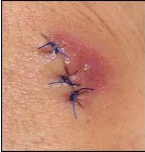
Q1: What is the diagnosis?
- Answer: Surgical Site Infection (SSI) (superficial incisional wound infection).
Q2: What is the most common causative organism?
- Answer: Staphylococcus aureus (including MRSA).
Q3: Outline the management plan.
- Answer:
- Remove sutures/staples over the fluctuant area to allow drainage.
- Evacuate purulent collection and send a swab for Gram stain and Culture & Sensitivity.
- Provide appropriate wound cleansing and an interactive dressing for secondary healing; review the wound daily.
- Select systemic antibiotics for associated cellulitis/systemic infection according to severity, local resistance and cultures. Topical antibiotics are not a substitute for drainage or treatment of spreading infection.
Operation room and surgical techniques
Station 2: Intestinal Stoma Identification, Indications, and Complications
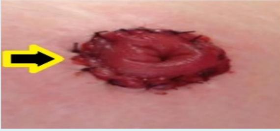

Q1: What is demonstrated in this photograph?
- Answer: Colostomy. Identify the actual lumen configuration: a loop stoma has proximal and distal lumens, an end stoma has a single exteriorized end; ileostomy output is usually liquid and colostomy output more formed.
Q2: Mention TWO surgical indications for this procedure.
- Answer:
- Relief of acute distal large bowel obstruction (e.g., obstructing rectosigmoid tumor).
- Emergency resection for perforation / diverticulitis (Hartmann’s procedure) or trauma.
Q3: Mention THREE recognized complications of an abdominal stoma.
- Answer:
- Stoma ischemia / necrosis.
- Stoma retraction.
- Parastomal hernia (or stoma prolapse, stenosis, peristomal dermatitis, or infection).
Imaging & instruments
Station 3: Nasogastric Tube (NGT)
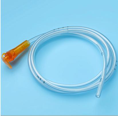
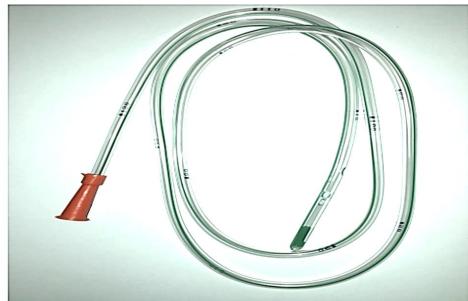
Q1: Identify this surgical instrument.
- Answer: Nasogastric Tube (NG Tube) (Levin / Salem sump tube).
Q2: Mention TWO therapeutic and TWO diagnostic uses.
- Answer:
- Therapeutic: Gastrointestinal decompression in intestinal obstruction / paralytic ileus; enteral feeding / drug administration.
- Diagnostic: Aspiration of gastric contents to confirm upper GI bleeding; administration of enteral radiographic contrast.
Q3: Mention FIVE important steps to introduce this tube safely.
- Answer: Explain/consent and check for contraindications (including suspected skull-base injury for the nasal route); inspect the nostrils and choose a suitable tube caliber/length. Position upright if safe, measure nose–earlobe–xiphoid length and mark, lubricate with water-soluble gel, and advance gently along the nasal floor, with neck flexion and small sips/swallowing only when safe. Stop for distress, secure the tube, and confirm initial placement before use with approved aspirate pH testing or an appropriately interpreted X-ray.
Q4: Mention FOUR potential complications and explain how placement is confirmed.
- Answer: Aspiration/airway misplacement, epistaxis/nasal erosion, pharyngeal or esophageal injury/perforation, and sinusitis; discomfort, gastritis/esophagitis and rare esophagotracheal fistula are additional complications. Intracranial placement is a severe hazard with skull-base injury. Initial confirmation uses aspirate pH 1–5.5 or a properly interpreted X-ray according to protocol. The air insufflation/auscultation “whoosh test” is unreliable and must not be used as confirmation.
Station 4: Closed Wound Suction Drain (Zimmer Hemovac 400 mL)
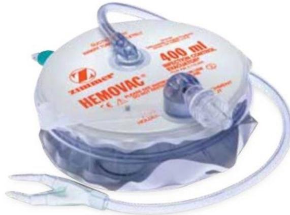
Q1: Identify the device shown.
- Answer: Zimmer Hemovac Closed Wound Suction Drain (400 mL).
Q2: Mention TWO operative indications or clinical uses of this drain.
- Answer: Evacuate postoperative blood/fluid and reduce dead-space hematoma/seroma, for example after mastectomy, neck surgery/axillary dissection, or major orthopedic surgery. Record output volume and character to support postoperative assessment.
Station 5: Foley’s Urinary Retention Catheter
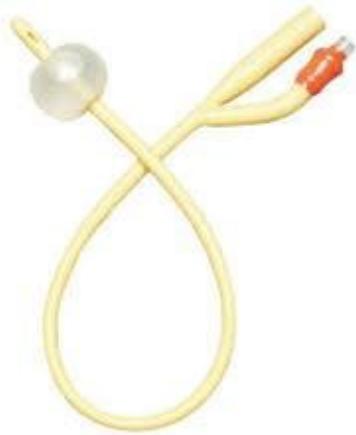
Q1: Identify this instrument.
- Answer: Two-Way Foley Retention Urinary Catheter.
Q2: Mention TWO diagnostic uses and TWO therapeutic uses.
- Answer:
- Diagnostic: Hourly urine output monitoring (in shock, ICU, major surgery); collecting sterile urine for culture.
- Therapeutic: Relief of acute or chronic urinary retention (e.g., BPH); intraoperative bladder decompression.
Station 6: Incentive Spirometer
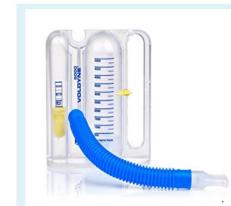
Q1: Name this instrument and mention TWO clinical uses.
- Answer: Volume-oriented incentive spirometer. It provides visual feedback for sustained deep inspiration/lung expansion and can support postoperative respiratory exercises/recovery as part of a broader program. It is not a peak-flow meter for monitoring asthma. Incentive spirometry alone is not proven routine prophylaxis for all patients; combine indicated use with deep breathing, coughing, mobilization and adequate analgesia.
Spot Dx
Station 7: Acute Paronychia
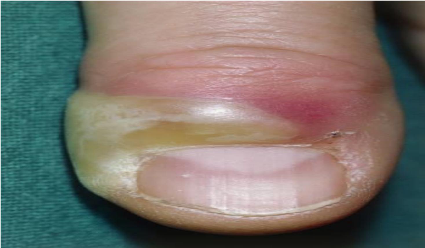
Q1: What is the diagnosis shown on the distal phalanx?
- Answer: Acute Paronychia (nail fold infection).
Q2: Outline the management.
- Answer:
- Warm soaks and oral antibiotics (if early / cellulitic).
- Surgical incision and drainage (I&D) or partial nail plate avulsion (if fluctuant abscess is present).
Station 8: Carbuncle of the Posterior Neck
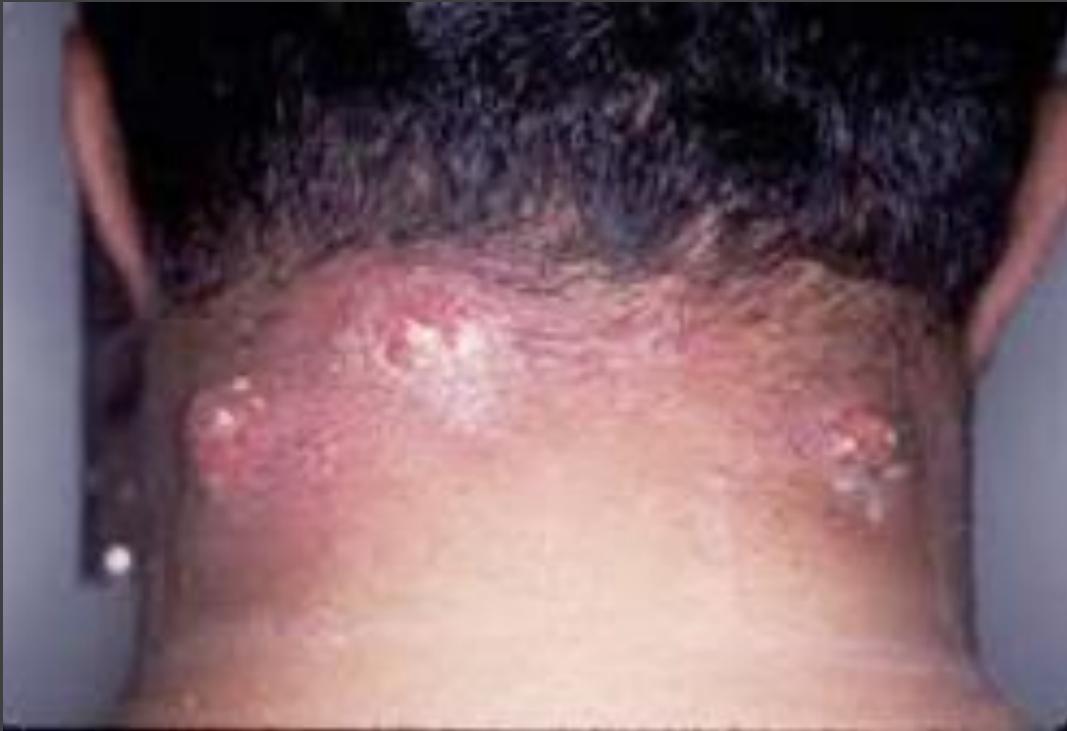
Q1: What is the diagnosis shown on the nape of the neck?
- Answer: Carbuncle of the neck (confluent subcutaneous furuncles).
Q2: Outline the treatment plan.
- Answer:
- Surgical Incision and Drainage (cruciate incision / debridement of necrotic slough).
- Systemic intravenous antibiotics (anti-staphylococcal).
- Strict blood glucose control.
Laboratory Interpretation
Station 9: Metabolic Acidosis in a Diabetic Patient
Case: An ABG in a diabetic patient shows low pH, low HCO₃⁻ and low PaCO₂.
Q1: What is the acid–base disturbance?
- Answer: Primary metabolic acidosis with respiratory compensation is suggested. Check the anion gap, lactate/ketones, clinical context and whether PaCO₂ is appropriate for compensation; without numerical values, a mixed disorder cannot be excluded. Do not assume DKA from diabetes alone.
Q2: Mention TWO tests to investigate a possible diabetic cause.
- Answer: Blood glucose and blood β-hydroxybutyrate (urine ketones if unavailable); assess electrolytes/renal function and anion gap.
Trauma & Emergency Surgery
Trauma
Station 10: Lateral Cervical Spine Radiography in Trauma
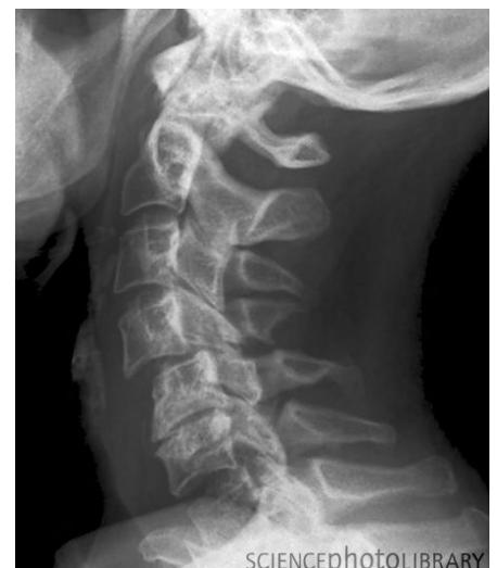
Case: An unconscious road-traffic victim has low respiratory rate and low BP. After ABC/GCS, the examiner supplies quadriplegia, asks localization and radiograph description, then neurosurgical review and MRI if stable.
Q1: What is the immediate management if cervical spine injury is suspected?
- Answer: Rigid cervical collar, sandbags, and tape (complete cervical spinal immobilization).
Q2: If a trauma patient presents with quadriplegia versus paraplegia, where is the level of cord injury?
- Answer:
- Quadriplegia (tetraplegia): Cervical spinal cord injury.
- Paraplegia: Thoracic or lumbar spinal cord injury.
Q3: What is the definitive treatment and what major complications can cause death?
- Answer:
- Definitive Treatment: Operative reduction, stabilization, and internal fixation/realignment.
- Fatal Complications: Respiratory failure/arrest (phrenic nerve involvement C3–C5), pulmonary aspiration, and severe sepsis/pneumonia.
Q4: How do you assess the unconscious trauma patient and calculate GCS?
- Answer: Use the ABCDE and GCS checklist. The illustrated radiograph shows a C5/C6 injury; describe actual alignment/vertebral/disc-space abnormalities rather than inferring the level solely from a generic picture. In adult high-risk trauma, CT is generally preferred to plain films; MRI assesses cord/ligament injury when indicated.
Station 11: Below-Knee Amputation (BKA) Stump Evaluation
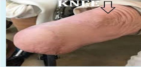
Case: Follow-up after surgery for foot pain.
Q1: What surgical procedure is demonstrated?
- Answer: Below-Knee Amputation (BKA / Transtibial Amputation).
Q2: Mention TWO likely causes/indications for amputation.
- Answer: Severe ischemia/nonviable limb (often PAD/diabetes) and trauma; severe uncontrolled infection (“toxic limb”) and selected malignancy are other indications.
Station 12: Blunt Splenic Injury on CT
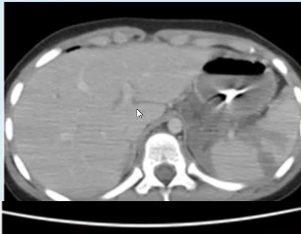
Case: A conscious car-accident victim has abdominal pain; pulse 96/min, BP 118/74 mmHg. IV fluids were started and an NG tube was placed. CT is shown.
Q1: What is the main CT finding?
- Answer: Splenic injury/rupture with a low-attenuation splenic laceration; describe associated hematoma/free fluid and contrast blush if demonstrated. Do not assign an injury grade from an isolated cropped image without full assessment.
Q2: Mention THREE important treatment steps and distinguish stable from unstable patients.
- Answer: Resuscitate/monitor with blood products when clinically indicated; stable patients suitable for nonoperative management require close observation, with splenic artery embolization for appropriate vascular injury/blush or high-grade risk. Persistent instability, peritonitis or failed nonoperative treatment requires operative control, sometimes splenectomy. A stable patient does not automatically need embolization or splenectomy.
Acute abdomen
Station 13: Pneumoperitoneum / Free Air Under Diaphragm on Plain Radiography
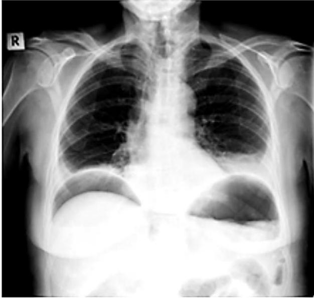
Q1: What is the radiographic finding shown?
- Answer: Pneumoperitoneum (free subdiaphragmatic air crescent under the right hemidiaphragm).
Q2: Mention TWO common surgical causes.
- Answer:
- Perforated peptic ulcer (duodenal or gastric).
- Perforated diverticulitis / bowel perforation (or trauma, recent laparotomy).
Q3: Mention FOUR immediate management steps, especially if unstable.
- Answer: ABCDE resuscitation, IV access/fluids with monitoring, broad-spectrum antibiotics and analgesia, and urgent surgical review/source control for suspected perforation; keep NPO and obtain labs/crossmatch. Recent abdominal surgery can cause residual free air, so the clinical context determines whether perforation surgery is required.
Station 14: Grey Turner’s Sign in Severe Retroperitoneal Hemorrhage
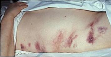
Q1: Give FOUR relevant history questions, TWO findings, and FOUR laboratory investigations.
- Answer: History: prior epigastric pain radiating to the back, relieved by leaning forward and worse supine, gallstones/previous cholecystectomy or ERCP, alcohol/drug (including steroid) exposure, and vomiting or trauma. Findings: flank ecchymosis and abdominal distension. Labs: CBC, lipase, U&E/creatinine, and LFTs/coagulation as indicated. This sign does not establish pancreatitis; investigate the suspected cause of retroperitoneal hemorrhage.
Upper GIT bleeding
Station 15: Endoscopic Diagnosis and Management of Bleeding Esophageal Varices
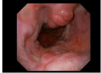
Q1: Identify the endoscopic finding shown.
- Answer: Bleeding Esophageal Varices (dilated, tortuous submucosal veins).
Q2: Mention THREE steps before endoscopy, including TWO pharmacologic treatment groups.
- Answer: Resuscitate and assess airway protection; start a vasoactive agent such as octreotide or terlipressin; give antibiotic prophylaxis, e.g. ceftriaxone according to local policy.
Q3: What is the endoscopic procedure of choice?
- Answer: Endoscopic variceal ligation (banding). Sclerotherapy is an alternative where banding is not feasible.
Q4: What rescue procedures are available if endoscopic therapy fails?
- Answer: Temporary balloon tamponade (Sengstaken–Blakemore tube) or an esophageal stent can bridge uncontrolled bleeding to definitive rescue TIPS in an appropriate center. Transplant assessment treats advanced liver disease in selected patients; transplantation is not immediate treatment for every variceal bleed.
Station 16: Coagulation Profile Interpretation in Massive Upper GI Bleeding
Case: Massive hematemesis in chronic liver disease: PT 21 s (10.5–13.5), aPTT 38 s (20–30), INR 2.6 (about 1). Platelet count and fibrinogen are not supplied; do not assume thrombocytopenia without measurement.
Q1: What do these results indicate, and do they diagnose variceal rupture or DIC?
- Answer: Prolonged clotting tests in chronic liver disease, consistent with altered liver-related coagulation. They do not alone diagnose variceal rupture, vitamin K deficiency, or DIC. Investigate the bleeding source; assess platelet count, fibrinogen and the clinical context when DIC is suspected. INR alone does not quantify bleeding risk in cirrhosis.
Q2: What urgent action and indication-dependent hemostatic treatment are appropriate?
- Answer: Resuscitate and treat the source; suspected variceal bleeding requires vasoactive medication, antibiotics and urgent endoscopic treatment. Do not routinely give FFP to normalize INR in acute variceal bleeding. Vitamin K is for suspected deficiency/appropriate reversal; platelet or fibrinogen replacement is individualized for clinically significant bleeding and measured deficits. Cryoprecipitate replaces fibrinogen; RBCs treat blood loss/anemia. Heparin is not routine treatment here.
Small Bowel Obstruction
Station 17: Intussusception in Children and Adults
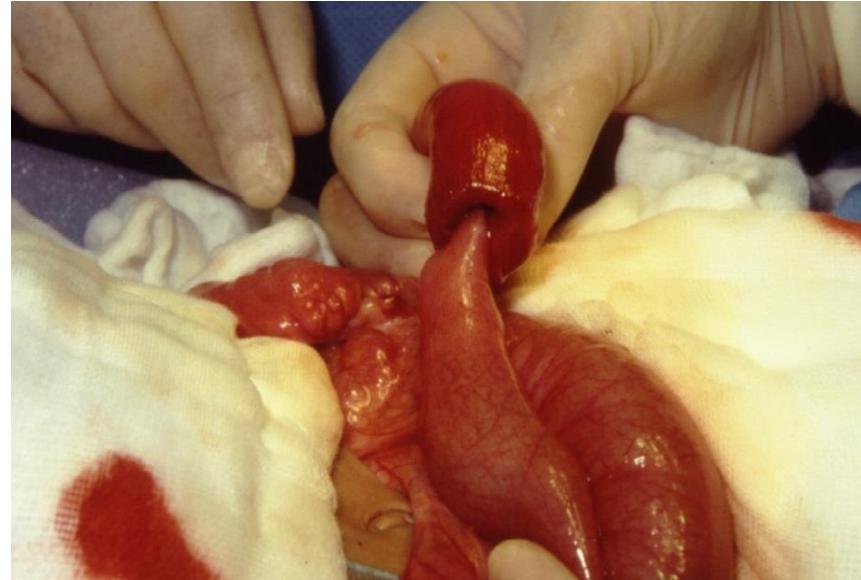
Q1: Identify the operative finding and name the characteristic ultrasound sign.
- Answer: The operative photograph shows bowel telescoping into adjacent bowel: intussusception. The target/donut or pseudokidney sign refers to a separate ultrasound examination, not this photograph.
Q2: What is the management in pediatric versus adult intussusception?
- Answer: Children: resuscitation followed by image-guided pneumatic/hydrostatic enema reduction when there is no perforation/peritonitis and the child is suitable; surgery for contraindications or failed reduction. Adults commonly need surgery; resection without reduction is favored for colonic disease or suspected malignancy, while selected benign small-bowel cases are individualized.
Station 18: Plain Abdominal Radiography: Small Bowel Obstruction
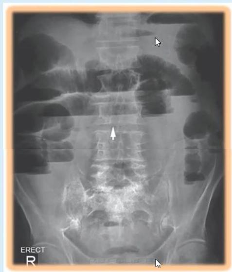
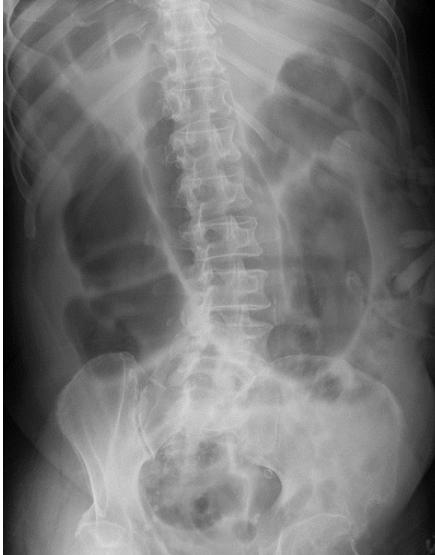
Case: Colicky pain, vomiting and no stool for 2 days, with an RLQ scar, mild non-tender distension and increased bowel sounds.
Q1: Name the modality, describe TWO findings, and give the likely diagnosis.
- Answer: Plain abdominal X-ray; identify dilated small-bowel loops, visible valvulae conniventes and multiple air–fluid levels on an erect view when visible. In this stem the likely diagnosis is SBO. Paralytic ileus is a DDx; the photograph/film does not alone prove the cause or bowel viability.
Q2: Mention TWO common causes and TWO further investigations.
- Answer: Adhesions and hernias. CT abdomen/pelvis evaluates transition point and complications; renal function/electrolytes and other labs assess dehydration. A selected water-soluble contrast challenge can assist adhesive SBO.
Q3: Mention TWO initial management steps and indications for surgery.
- Answer: NG decompression when indicated, NPO initially and monitored IV fluids/electrolyte correction. Peritonitis/ischemia/perforation requires urgent intervention; failure of an appropriate conservative trial prompts surgery.
Station 19: Strangulated Bowel at Laparotomy
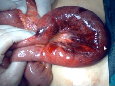
Case: Operative finding during surgery for an acute abdomen.
Q1: What is the diagnosis?
- Answer: Strangulated/ischemic bowel. Dusky congested bowel and compromised blood supply require operative viability assessment; the isolated photograph cannot distinguish every mechanism.
Q2: Mention TWO causes.
- Answer: An adhesive band (including a congenital Ladd band in the appropriate setting) and volvulus; an incarcerated/strangulated hernia is another cause.
Large Bowel Obstruction
Station 20: Hartmann’s Procedure for Obstructing / Perforated Colonic Pathology
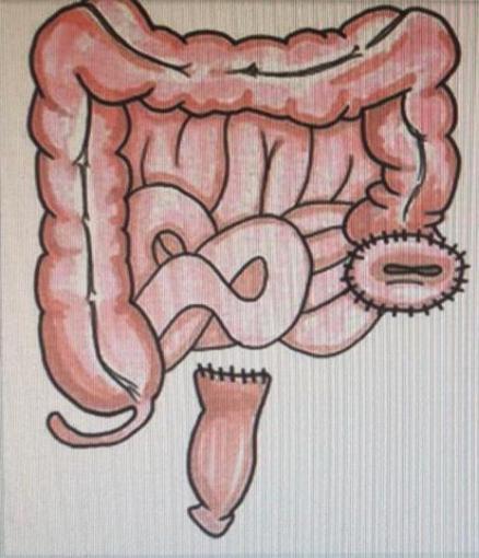
Case: A patient with abdominal pain underwent the operation illustrated in the diagram.
Q1: What surgical procedure is demonstrated?
- Answer: Hartmann’s Procedure (resection of rectosigmoid colon, end colostomy formation, and closure of the rectal stump).
Q2: Mention TWO major surgical indications.
- Answer:
- Perforated diverticulitis with feculent peritonitis (Hinchey IV).
- Obstructing rectosigmoid cancer in an emergency/unstable setting.
Station 21: Barium Enema Demonstrating “Apple-Core” Sign of Colon Cancer
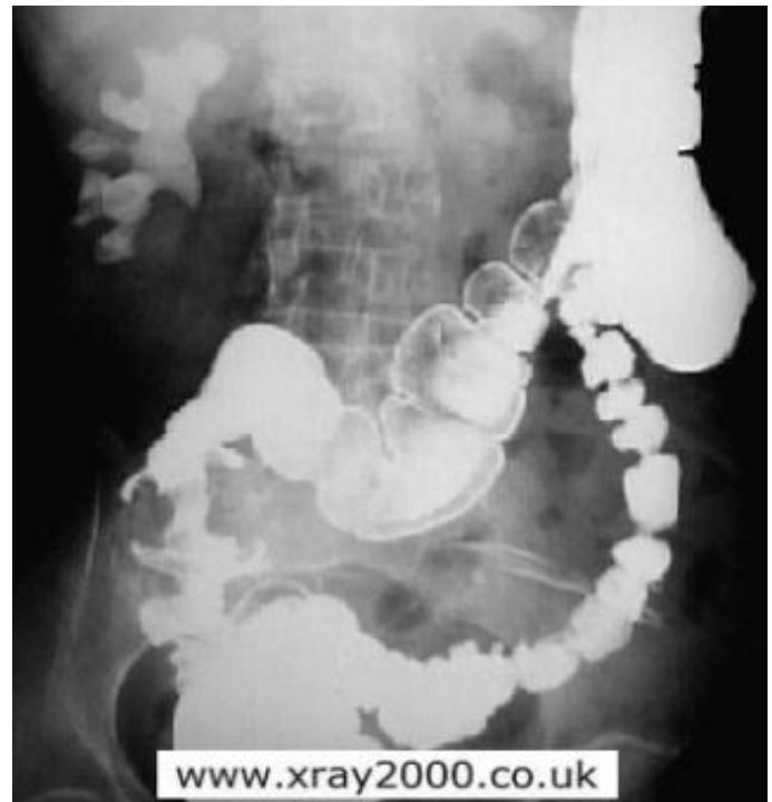
Q1: Name this radiological investigation and describe the classic sign.
- Answer: Barium Enema; classic sign: “Apple-core” lesion (annular constricting stenosis with shouldered margins).
Q2: What is the diagnosis?
- Answer: Colorectal Carcinoma (colon cancer).
Q3: What is the definitive diagnostic procedure to confirm diagnosis?
- Answer: Colonoscopy with multiple tissue biopsies.
Q4: What staging investigation and baseline marker are required, and where are full management questions covered?
- Answer: Contrast CT chest/abdomen/pelvis and baseline CEA. CEA is not an imaging investigation and does not independently determine stage.
Burns
Station 22: Split-Thickness Skin Graft
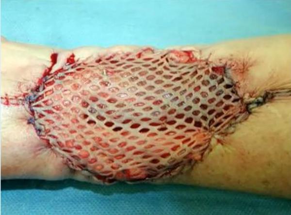
Case: A deep burn has been covered with the mesh-like graft shown.
Q1: Identify the graft and which layers of skin it contains.
- Answer: Split-thickness skin graft: epidermis and a portion of dermis. The donor site retains dermal elements that allow healing.
Q2: Mention TWO disadvantages.
- Answer: Color/texture mismatch and altered/reduced sensation; additional disadvantages include contraction, vulnerability to trauma, less satisfactory cosmetic contour and donor-site pain/scarring. General anesthesia may be needed depending on the procedure but is not inherently required for every split graft.
Head & Neck
Neck swellings
Station 23: Clinical Examination and Evaluation of Multinodular Goiter
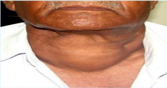

Q1: Mention FOUR specific physical examination steps performed from behind the patient.
- Answer:
- Palpate each thyroid lobe and isthmus systematically for size, consistency, and nodularity.
- Palpate for cervical lymphadenopathy (levels II–VI).
- Palpate lower border of goiter to rule out retrosternal extension.
- Auscultate over the superior thyroid poles for vascular systolic bruits.
Q2: Demonstrate the complete thyroid examination.
- Answer: Introduce/consent, position and expose the neck appropriately. Inspect from front/side for symmetry, scars, skin change and visible swelling, then observe swallowing and tongue protrusion. Palpate from behind: lobes/isthmus, size, consistency, tenderness, nodules, lower border and movement; assess trachea and cervical nodes. Assess retrosternal compression with Pemberton’s maneuver when appropriate, and auscultate a bruit if hypervascularity is suspected. Complete with pulse, hands/tremor, eye signs and thyroid-function signs.
Station 24: Parotid Gland Tumor / Pleomorphic Adenoma
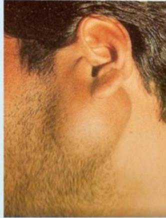
Q1: What investigations confirm the nature of the lump?
- Answer: Ultrasound with guided FNA/core sampling according to local specialist practice. MRI/CT is selected for deep-lobe disease, suspected malignancy or extent, rather than mandatory in every small superficial benign-appearing lump. Avoid an unplanned open biopsy or enucleation; definitive diagnosis requires pathological assessment. Pleomorphic adenoma is the commonest benign salivary tumor (F:M ≈ 2:1, typically 3rd–5th decade); it is slow-growing in the superficial lobe, and malignant transformation is uncommon (≈1.5% over 5 years).
Q2: What is the standard surgical procedure?
- Answer: Superficial Parotidectomy with Facial Nerve Preservation; total parotidectomy is reserved for deep-lobe or malignant disease, and submandibular gland tumors are treated by total gland excision. An impacted salivary stone that cannot be extracted may also require gland excision.
Q3: What THREE palpation signs and THREE differential diagnoses were asked?
- Answer: Palpation: tenderness, consistency and mobility/fixation/attachment. DDx: benign/malignant salivary gland tumor, parotid abscess, and a branchial cyst or regional lymph-node lesion. Examine facial nerve function and oral cavity. (Station stem: a 35-year-old male with swelling for 3 months.)
Station 25: Acute Parotid Abscess
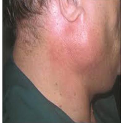
Q1: What is the diagnosis shown in this photograph?
- Answer: Acute Suppurative Parotitis / Parotid Abscess.
Q2: Outline the treatment plan.
- Answer: IV hydration, IV anti-staphylococcal antibiotics, sialogogues (lemon drops) to stimulate saliva, and surgical incision and drainage (horizontal incision parallel to facial nerve branches) if fluctuant.
Q3: Mention differential diagnoses and the role of aspiration.
- Answer: Differentials include acute suppurative parotitis without a drainable abscess, infected cyst, salivary obstruction and an inflamed/necrotic tumor. Ultrasound helps identify a collection; image-guided aspiration/drainage may be suitable in selected cases, while open drainage is planned to protect facial nerve branches.
Station 26: Hypothyroid and Hyperthyroid Signs
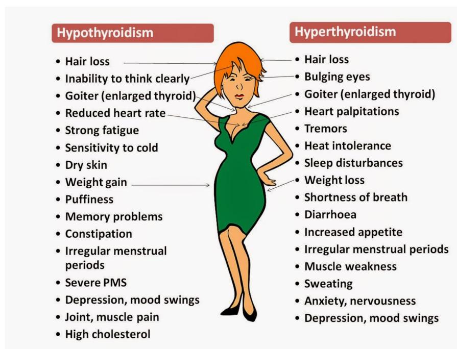
Q1: Contrast the multisystem clinical manifestations of hypothyroidism versus hyperthyroidism.
- Answer:
| System / Feature | Hypothyroidism | Hyperthyroidism |
|---|---|---|
| Metabolism & Weight | Cold intolerance, weight gain with decreased appetite | Heat intolerance, excessive sweating, weight loss despite increased appetite |
| Cardiovascular | Bradycardia, diastolic hypertension, pericardial effusion | Sinus tachycardia, palpitations, atrial fibrillation, wide pulse pressure |
| Gastrointestinal | Constipation, abdominal distension | Frequent bowel movements / diarrhea |
| Neuromuscular / CNS | Mental sluggishness, memory impairment, slow-relaxing deep tendon reflexes, fatigue | Fine resting tremor, anxiety, irritability, insomnia, proximal muscle weakness, brisk reflexes |
| Integumentary | Dry, rough skin, non-pitting myxedema, brittle hair, coarse features | Warm, moist palms, onycholysis, pretibial myxedema (Graves) |
| Ophthalmological | Periorbital puffiness | Exophthalmos, proptosis, lid lag, lid retraction (Dalrymple’s sign) |
| Reproductive | Menorrhagia, oligomenorrhea, subfertility | Oligomenorrhea, amenorrhea |
(Note: Goiter may occur in either condition. These represent characteristic multisystem symptom patterns; serum thyroid function tests [TSH, Free T3/T4] confirm the definitive biochemical diagnosis).
Station 27: Thyroid Eye Signs: Lid Retraction
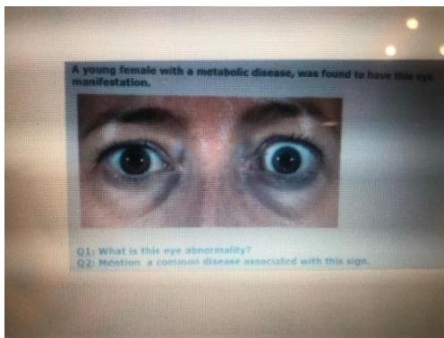
Case: A young female with a metabolic disease is found to have this eye manifestation.
Q1: Name the finding and give an associated disease.
- Answer: Lid retraction, associated with Graves disease/thyrotoxicosis. Distinguish lid lag (lag of upper lid on looking down), proptosis and ophthalmoplegia. Lid retraction is the key answer; eye signs alone do not establish thyroid hormone levels.
Neurosurgery
Brain Tumor
Station 28: Intracranial Meningioma on Brain MRI
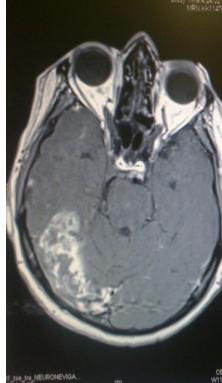
Q1: Identify the neuroimaging findings and state the diagnosis.
- Answer: Meningioma. Describe the enhancing mass and its relationship to dura/brain; extra-axial location, enhancement and a dural tail are typical supporting features, not findings guaranteed on every image.
Q2: Mention TWO treatment approaches for symptomatic or growing tumors.
- Answer: Resection where appropriate (complete removal including involved dura/bone when safely feasible), or radiotherapy/radiosurgery for selected residual, recurrent or inoperable tumors. Selected small asymptomatic lesions can be observed. Routine chemotherapy is not the standard second treatment for a typical benign meningioma. The image is MRI.
Station 29: Ring-Enhancing Cerebral Lesion on Brain MRI
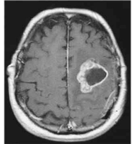
Q1: Describe the radiological appearance shown.
- Answer: Axial brain MRI with a left-hemisphere ring-enhancing lesion. Describe the visible central signal, rim and surrounding edema/mass effect. Ring enhancement alone does not prove necrosis, pathology or an extensive edema burden.
Q2: Give FOUR differential diagnoses and expand MAGIC DR correctly.
- Answer: Metastasis, Abscess, Glioblastoma, Infarction, Contusion, Demyelination, Radiation necrosis. Also consider resolving hematoma, toxoplasmosis and tuberculoma according to context. Select any four appropriate causes if the prompt requests four; the mnemonic has seven components.
Head injury
Station 30: Epidural Hematoma on Non-Contrast Head CT
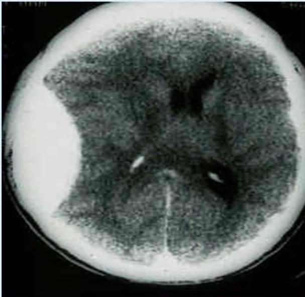
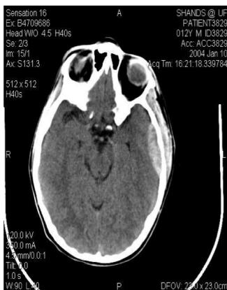
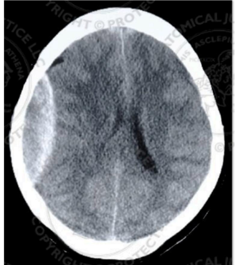
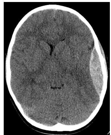
Q1: Identify the diagnostic finding on this CT scan.
- Answer: High-density (hyperdense) biconvex (lenticular), smoothly demarcated extra-axial collection that is limited by cranial suture lines.
Q2: What is the diagnosis?
- Answer: Acute Epidural Hematoma (EDH).
Q3: What is the main cause and what TWO treatment approaches were asked?
- Answer: Trauma, often involving middle meningeal artery injury. Evacuate a significant or deteriorating EDH urgently, usually by craniotomy. Selected small EDHs can undergo specialist observation/serial CT: BTF criteria include volume <30 cm³, thickness <15 mm, shift <5 mm, GCS >8 and no focal deficit. Routine endovascular embolization/burr-hole drainage is not interchangeable with definitive evacuation of a large acute EDH. IV fluids and correction/reversal with vitamin K, FFP, cryoprecipitate or protamine require the corresponding bleeding, coagulation or anticoagulant indication.
Station 31: Subdural Hematoma on Head CT
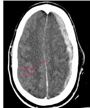
Q1: Identify the diagnostic finding on this CT scan.
- Answer: Hyperdense (or isodense/hypodense) crescentic (concave-convex) extra-axial collection that crosses cranial suture lines along the cerebral hemisphere.
Q2: What is the diagnosis?
- Answer: Subdural Hematoma (SDH).
Q3: What population is at highest risk for chronic subdural hematoma?
- Answer: Elderly patients and chronic alcoholics (cerebral atrophy stretches bridging veins, triggered by minor head trauma). Coagulopathy/anticoagulation and repeated trauma are additional risk groups.
Breast
Breast Disease
Station 32: Diagnostic Mammography of Breast Carcinoma
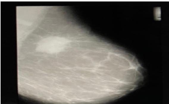
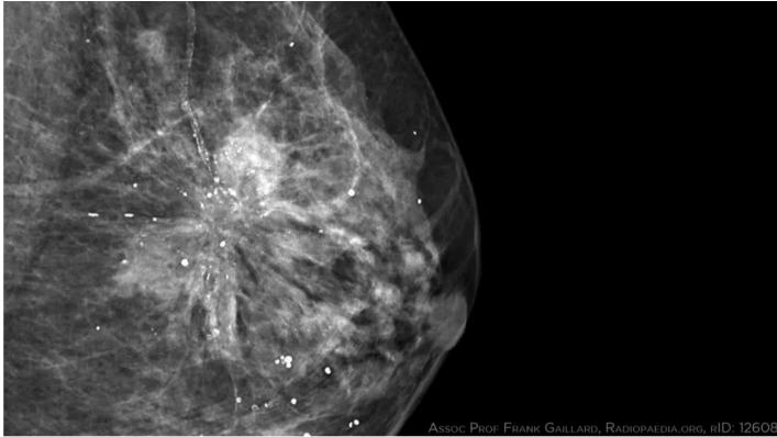
Q1: What imaging modality is shown?
- Answer: Mammography. Specify craniocaudal (CC) or mediolateral oblique (MLO) only when that view is actually shown; the collection contains individual views.
Q2: What is the likely diagnosis and which mammographic findings support malignancy?
- Answer:
The likely diagnosis is suspected breast carcinoma. Typical suspicious findings (describe only those visible) include:
- Spiculated, high-density, irregular soft tissue mass.
- Pleomorphic, clustered microcalcifications.
- Architectural distortion or skin thickening / nipple retraction.
Q3: What further test confirms the nature of a suspicious mammographic finding?
- Answer: Image-guided core needle (Tru-Cut) biopsy; choose stereotactic or ultrasound guidance according to visibility. Histology cannot be inferred from mammography alone.
Station 33: Peau d’Orange Sign and Suspected Breast Cancer
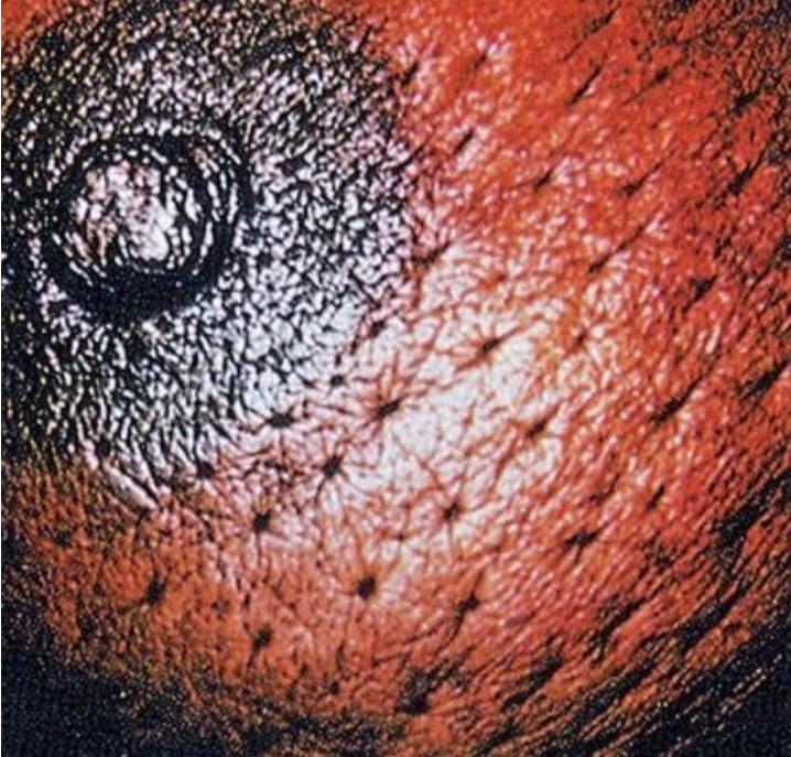
Q1: What physical sign is demonstrated on the breast skin?
- Answer: Peau d’Orange (“orange peel” skin appearance).
Q2: What is the most likely diagnosis associated with this sign?
- Answer: Breast cancer. The sign is suspicious but does not independently prove an inflammatory subtype or stage; investigate with clinical assessment, imaging and biopsy.
Station 34: Acute Lactational Breast Abscess
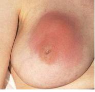
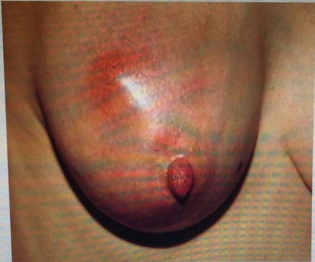
Q1: What is the diagnosis shown in this lactating female?
- Answer: Acute Lactational Breast Abscess.
Q2: What is the most common causative organism?
- Answer: Staphylococcus aureus (including MRSA).
Q3: Outline the management plan.
- Answer:
- Drainage: Ultrasound-guided needle aspiration (first-line) or surgical incision and drainage (if large/refractory).
- Systemic oral/IV anti-staphylococcal antibiotics (e.g., Flucloxacillin or Clindamycin).
- Continue breastfeeding or appropriate expression when feasible; prevent the infant’s mouth from contacting purulent drainage or infected tissue.
Q4: What specialized investigation is performed before specific treatment?
- Answer: Breast ultrasound identifies and sizes a drainable collection, guides aspiration, and assesses other lesions. Other stems: a 30-year-old with unilateral warmth/tenderness, or a lactating woman with pain/swelling for two days. Arrange follow-up for persistent or atypical masses.
Thoracic
Management of patients with lung lesions
Station 35: Pulmonary Mass on Chest Imaging (Lung Cancer)
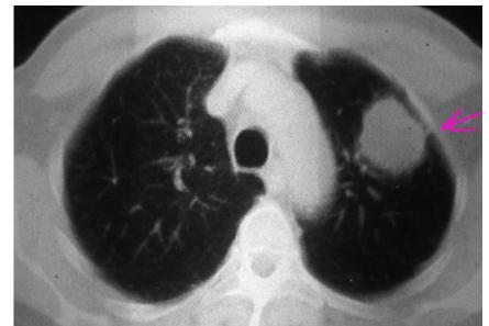
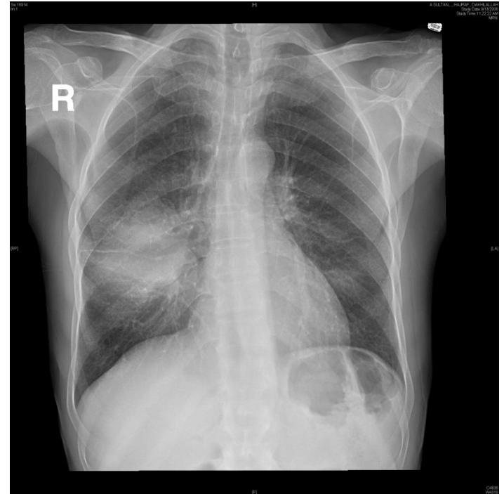
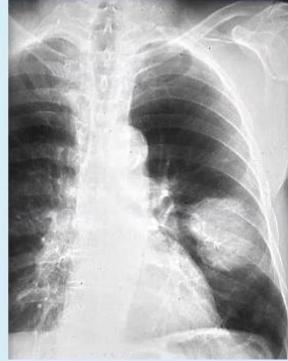
Q1: Mention TWO initial diagnostic and staging investigations.
- Answer:
- Contrast-enhanced CT of Chest and Upper Abdomen (including liver and adrenal glands).
- Bronchoscopy with endobronchial biopsy / brushings (or CT-guided percutaneous transthoracic needle biopsy; thoracoscopy is a selected alternative tissue-access route).
Q2: Which TWO diagnoses/histologies are considered, and can the image distinguish them?
- Answer: Squamous cell carcinoma and small-cell lung cancer, with stems such as a 65-year-old with cough, dyspnea and hemoptysis or a 60-year-old with cough/hemoptysis. Imaging suggests a lung malignancy but cannot establish the subtype. Histology/cytology is required. Bronchoscopy/CT-guided biopsy and appropriate staging are used; mediastinal node sampling (EBUS or selected mediastinoscopy) and selected brain MRI assess spread.
Upper GI & Abdominal Surgery
Esophageal Diseases
Station 36: Achalasia Cardia on Barium Swallow
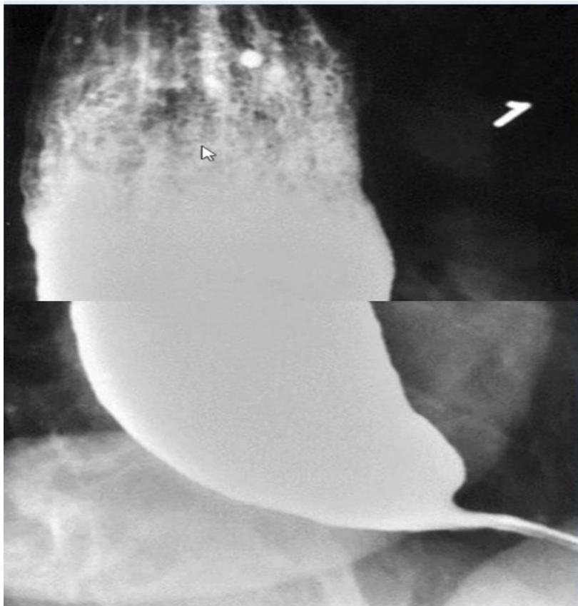
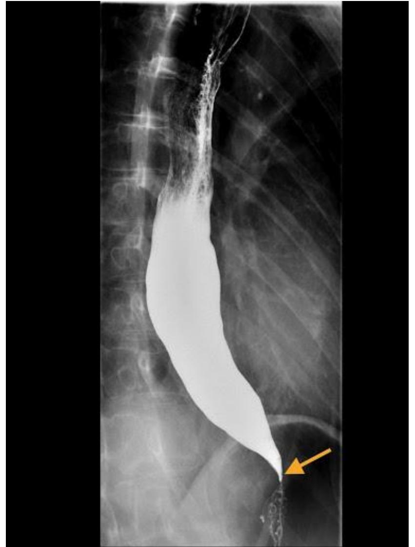
Q1: Name this radiological investigation and describe the diagnostic finding.
- Answer: Barium Swallow (Esophagram); diagnostic finding: Marked dilatation of the thoracic esophagus with smooth, symmetric, tapered narrowing at the gastroesophageal junction producing the classic “Bird’s Beak” (or “Rat’s Tail”) appearance.
Q2: What is the definitive diagnosis?
- Answer: Achalasia Cardia (primary esophageal motility disorder).
Q3: What specialized physiological investigation is the gold standard?
- Answer: High-Resolution Esophageal Manometry (HREM) (demonstrates complete aperistalsis and incomplete/absent LES relaxation).
Q4: Mention TWO definitive treatment modalities.
- Answer:
- Laparoscopic Heller Myotomy (surgical division of lower esophageal circular muscle with partial Dor/Toupet fundoplication).
- Pneumatic Balloon Dilatation (or Per-Oral Endoscopic Myotomy [POEM] / Botulinum toxin injection).
Q5: What additional investigation and treatment alternatives are relevant?
- Answer: Upper endoscopy excludes a mechanical obstruction/pseudoachalasia. Botulinum toxin is used in selected patients unfit for definitive procedures; nitrates/calcium-channel blockers have limited temporary benefit. Pneumatic dilation, Heller myotomy and POEM are definitive options selected by subtype/fitness. The illustrated esophageal study is a barium swallow/esophagram; a barium “meal” in a 40-year-old with dysphagia to solids and liquids is the same investigation.
Station 37: Esophageal Carcinoma on Barium Swallow
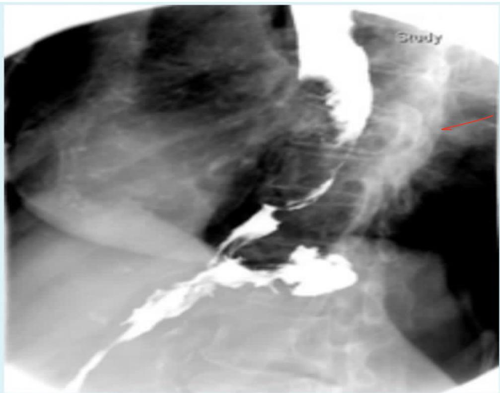
Q1: What is the most likely diagnosis in the 60-year-old with dysphagia and weight loss?
- Answer: Esophageal Carcinoma (Esophageal Cancer).
Q2: What is the gold standard diagnostic investigation to establish the diagnosis?
- Answer: Upper Endoscopy (OGD) with multiple targeted biopsies and brush cytology.
Q3: Mention TWO staging modalities.
- Answer:
- Contrast-enhanced CT of Chest and Abdomen (for distant metastases).
- Endoscopic Ultrasound (EUS) (gold standard for local T and N staging). (PET-CT for systemic staging).
Q4: What FOUR further history questions and histological types should be mentioned?
- Answer: For the 60-year-old male with dysphagia and weight loss (initial CXR normal), ask progression/solids versus liquids, odynophagia or reflux, duration/weight loss/appetite, and smoking/alcohol/family history or prior injury/stricture. Major types are squamous cell carcinoma and adenocarcinoma. Endoscopy/biopsy precedes definitive staging; a normal CXR does not exclude esophageal cancer.
Obstructive jaundice
Station 38: Clinical Recognition of Scleral Icterus / Jaundice
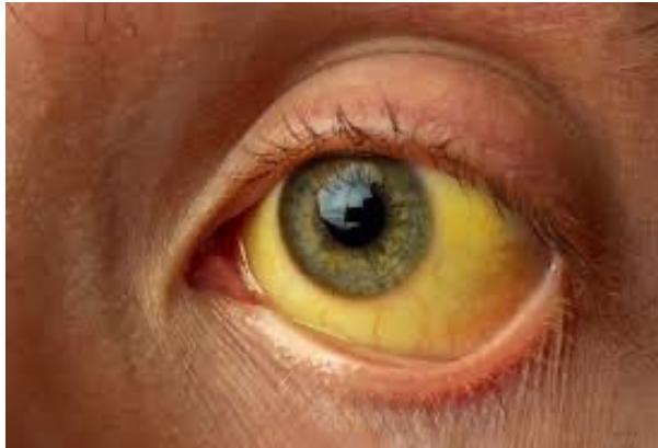
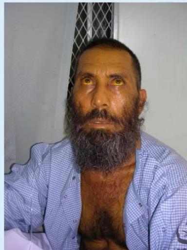
Q1: What TWO further questions and initial imaging were asked in the jaundice photograph station?
- Answer: Ask about pain and pruritus; also stool/urine color, fever, travel/transfusion/hepatitis exposure and weight loss. Initial imaging is ultrasound for suspected hepatobiliary obstruction. Another stem has yellow eyes and tea-colored urine for two weeks.
Station 39: Endoscopic Retrograde Cholangiopancreatography (ERCP) in Choledocholithiasis
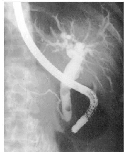
Case: A patient has abdominal pain and jaundice for seven days, with a dilated CBD containing a stone on ERCP; questions cover history, DDx, why malignancy is less typical, modality/findings, preparation, complications and management.
Q1: Identify this radiological and endoscopic procedure.
- Answer: Endoscopic Retrograde Cholangiopancreatography (ERCP).
Q2: Describe the finding and give the most likely diagnosis.
- Answer: A dilated CBD contains contrast filling defects; the diagnosis is choledocholithiasis/CBD stone. Relate the finding to symptoms and full imaging rather than inferring the exact number of stones from a cropped view.
Q3: Mention TWO therapeutic interventions performed during this procedure.
- Answer:
- Endoscopic Sphincterotomy (Papillotomy) of the ampulla of Vater.
- Stone extraction using a Dormia basket or Fogarty balloon catheter (or biliary stent insertion).
Q4: Mention TWO recognized complications of this procedure.
- Answer:
- Acute Post-ERCP Pancreatitis (most common, ).
- Duodenal perforation or post-sphincterotomy hemorrhage (or ascending cholangitis).
Q5: What must be considered before ERCP, and what other complications were asked?
- Answer: Consent, appropriate indication, ultrasound/MRCP/EUS as needed, CBC/renal function/electrolytes and coagulation review; manage relevant anticoagulation/hemostatic problems. Antibiotic prophylaxis is selective, such as anticipated incomplete biliary drainage, rather than universal for every ERCP. Discuss pancreatitis, bleeding, cholangitis/infection, perforation and sedation risks. Duct clearance is followed by cholecystectomy during the same admission when possible; actual timing depends on fitness, complications and the surgical pathway.
Q6: What history and FOUR DDx are relevant, and does pain exclude pancreatic cancer?
- Answer: Ask about dark urine, pale stool, itching, fever and weight loss. Four DDx are CBD stone, cholangitis, pancreatic-head cancer and cholangiocarcinoma. Sudden painful jaundice favors stones over a typical progressive malignant presentation, but does not exclude cancer.
Complications of Abdominal Surgery
Station 40: Pseudomembranous Colitis after Antibiotics
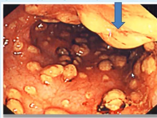
Case: After appendectomy for perforated appendicitis and broad-spectrum antibiotics, a patient develops fever and foul-smelling greenish watery diarrhea on postoperative day 5; the endoscopic image shows adherent plaques.
Q1: What is the diagnosis and how is it confirmed?
- Answer: C. difficile–associated pseudomembranous colitis is suspected. Test unformed stool for toxin/toxigenic C. difficile using the local algorithm in a symptomatic patient; assess CBC/creatinine and severity. Endoscopy is not routinely needed when stool testing and the clinical context establish the diagnosis.
Q2: What is the specific treatment?
- Answer: Stop unnecessary precipitating antibiotics when possible, institute contact precautions and fluids/electrolyte support, and give oral fidaxomicin (preferred where available) or oral vancomycin for an initial nonfulminant episode. Fulminant disease needs a dedicated regimen and urgent specialist/surgical assessment. “Stop/change the antibiotic” alone is not adequate specific therapy.
Abdominal Wall & Hernias
Station 41: Incisional Hernia
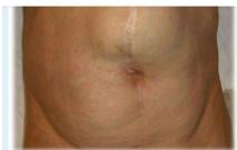
Case: An older male has abdominal pain and repeated vomiting with a postoperative abdominal-wall swelling.
Q1: What is the abnormality and the definitive treatment?
- Answer: Incisional hernia. Assess reducibility, pain, skin changes, obstruction/strangulation and the surgical scar. Appropriate repair often uses mesh, with timing/approach selected by symptoms, contamination, anatomy and patient fitness. Pain and vomiting require urgent assessment for a complication rather than routine elective repair advice.
Station 42: Adult Groin Hernia Identification
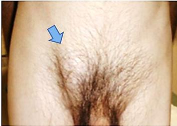
Case: A 55-year-old male has a right groin swelling for 6 months; consider an indirect inguinal hernia and the deep-ring occlusion test.
Q1: What is the likely diagnosis, definitive treatment and special test?
- Answer: A suspected inguinal hernia; assess cough impulse, reducibility and relation to the pubic tubercle. Definitive symptomatic adult treatment is appropriate hernia repair, usually mesh-based when suitable. The deep-ring occlusion test is a historical clinical maneuver; a photograph alone cannot prove direct versus indirect type.
Station 43: Adult Paraumbilical Hernia
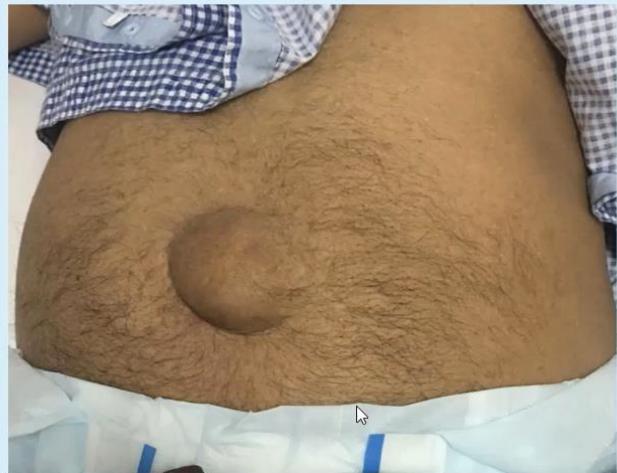
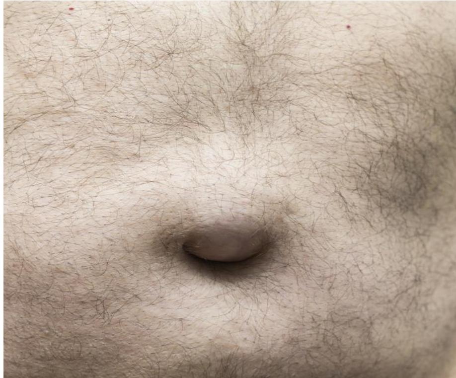
Q1: What is the diagnosis, and which TWO complications are important?
- Answer: The 40-year-old male has painless abdominal-wall swelling that enlarges during activity and becomes smaller after overnight rest. Diagnosis: paraumbilical hernia; complications: incarceration and strangulation. The adult photo should not be presented as a pediatric umbilical hernia.
Q2: What is the definitive treatment?
- Answer: Hernia repair. Assess symptoms, reducibility and possible obstruction/strangulation before choosing timing and technique.
Esophageal & Gastric Diseases
Station 44: Gastric Lesion on Upper GI Endoscopy
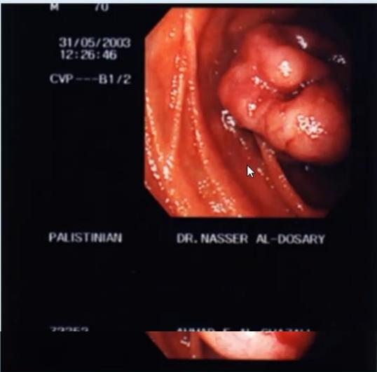
Case: Melena with stable vitals; an upper-GI endoscopic lesion is shown. Histology is not established.
Q1: Mention TWO differential diagnoses.
- Answer: Gastric carcinoma and GIST; a gastric polyp/subepithelial lesion is another possibility according to morphology. Gastrinoma/Zollinger–Ellison syndrome is considered for the relevant clinical ulcer/hypergastrinemia syndrome, not diagnosed from the lesion’s appearance alone.
Q2: What TWO further specialized investigations should be performed in order?
- Answer: Endoscopic tissue assessment/biopsy when appropriate, followed by EUS for a subepithelial lesion and/or contrast CT for staging. A suspected GIST may require EUS-guided tissue acquisition rather than an uninformative superficial mucosal biopsy. Fasting gastrin is selective for suspected gastrinoma and requires interpretation with acid-suppression/gastric-acidity context.
Biliary & Pancreatic Disease
Station 45: Gallstones and Chronic Calculous Cholecystitis
Case: A gallbladder/gallstones specimen or ultrasound; identify the organ and disease, request laboratory tests and list four complications.
Q1: Identify the organ and likely disease.
- Answer: Gallbladder with gallstones (cholelithiasis). The ultrasound may be labelled chronic calculous cholecystitis; chronic inflammation needs clinical/pathological context, not just the presence of stones.
Q2: What relevant laboratory tests would you request?
- Answer: CBC, LFTs/bilirubin/ALP and U&E/renal function; lipase if pancreatitis is suspected.
Q3: Mention FOUR complications of gallstone disease.
- Answer: Acute/chronic cholecystitis, CBD obstruction/obstructive jaundice, cholangitis and pancreatitis. Empyema, perforation and gallstone ileus can occur. Gallbladder cancer is associated with longstanding gallstones/chronic inflammation but is not inevitable.
Station 46: Pancreatitis: Laboratory Interpretation
Case: A 40-year-old female has sudden severe epigastric pain. Amylase 1920 U/L (normal <100), ALT 39 U/L (5–30), AST 48 U/L (10–40), ALP 145 U/L (25–120); total bilirubin 17 is printed with mmol/L units/reference <17 mmol/L, an apparent unit error. Verify the original laboratory unit (commonly μmol/L), rather than interpreting 17 mmol/L as a valid result. Another stem describes amylase over 2000 U/L with raised LFTs.
Q1: What is the most likely diagnosis and TWO common causes?
- Answer: Acute pancreatitis is strongly supported by typical pain and pancreatic enzymes >3× upper limit; common causes are gallstones and alcohol. Raised LFTs can suggest a biliary cause but do not prove it.
Q2: What is the first imaging study and what alternatives may be used?
- Answer: Ultrasound assesses gallstones/duct dilatation. MRCP is selected for suspected duct stones; CT is used for diagnostic uncertainty or complications rather than automatically at admission in every straightforward mild case. AXR may show ileus/sentinel loop but is not definitive.
Colorectal Surgery
Station 47: Sessile Colonic Polyp
Case: Colonoscopy in a 55-year-old male with rectal bleeding; another image shows a sessile polyp.
Q1: What is the abnormality and its clinical significance?
- Answer: A sessile colonic polyp. Some polyps (adenomas/selected serrated lesions) are premalignant, but sessile shape alone does not prove high malignant potential. Risk depends on histology, size, dysplasia and other features.
Urology
Upper UT
Station 48: Renal Cell Carcinoma on Contrast-Enhanced Abdominal CT
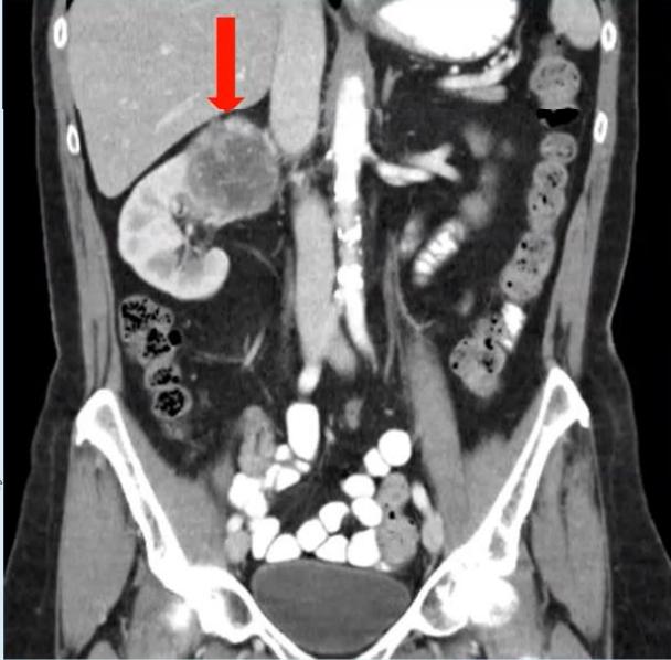
Case: A 60-year-old male has a right-kidney mass on CT; another stem describes a 62-year-old male with right loin pain and hematuria.
Q1: Mention primary treatment options for localized RCC and distinguish a renal-pelvis tumor.
- Answer: Partial nephrectomy is preferred for suitable localized small/T1 tumors when feasible; radical nephrectomy is selected for larger/complex lesions. Adrenalectomy is not routine without evidence of adrenal involvement. Selected small masses may undergo surveillance or percutaneous ablation (radiofrequency or cryoablation) according to patient/tumor factors. Nephroureterectomy is treatment for appropriate upper-tract urothelial carcinoma, not a synonym for RCC surgery.
Q2: Mention TWO differential diagnoses and TWO risk/pathogenesis factors.
- Answer: For a solid renal mass, RCC and angiomyolipoma are key alternatives; also consider oncocytoma/other benign adenoma, lymphoma, metastasis or abscess in context. A collecting-system lesion raises urothelial carcinoma. Risk factors include smoking, obesity, hypertension and genetic syndromes (e.g. VHL/tumor-suppressor dysfunction in clear-cell RCC). Conventional chemotherapy is not a routine localized-RCC treatment. Hematuria, flank pain and a palpable mass form the classic triad, but most patients do not have all three.
Lower UT
Station 49: Urinary Bladder Tumor on Cystoscopy
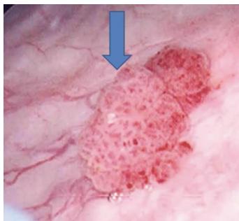
Q1: In a 60-year-old male presenting with painless gross hematuria, what is the finding and diagnosis?
- Answer: Papillary, frond-like, exophytic mucosal mass inside the urinary bladder lumen; Diagnosis: Urinary Bladder Cancer (Urothelial / Transitional Cell Carcinoma).
Q2: What initial diagnostic and staging operation must be performed?
- Answer: Transurethral Resection of Bladder Tumor (TURBT) (provides complete tumor resection and depth of invasion assessment: lamina propria vs detrusor muscularis propria).
Q3: Contrast definitive treatment for Non-Muscle Invasive versus Muscle-Invasive Bladder Cancer.
- Answer:
- Non-Muscle Invasive (Ta, T1): Complete TURBT followed by Intravesical BCG or Chemotherapy (Mitomycin C) instillation.
- Muscle-Invasive (T2–T4): Radical Cystectomy with Pelvic Lymph Node Dissection and urinary diversion (Ileal Conduit / Neobladder) neoadjuvant chemotherapy. Partial cystectomy is suitable only for selected disease; these operations are not interchangeable.
External Genitalia
Station 50: Acute Testicular Torsion
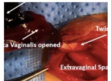
Q1: What is the intraoperative surgical diagnosis shown?
- Answer: Testicular Torsion (torsion of the spermatic cord with acute testicular ischemia/infarction).
Q2: Mention ONE risk factor for testicular torsion.
- Answer: Trauma or a family history; torsion can also occur without any trigger. A bell-clapper deformity is the classic anatomical predisposition.
Station 51: Hydrocele and the Transillumination Test
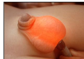
Case: A swollen scrotum transmits light during a bedside examination.
Q1: What is the diagnosis, name of the test, and positive finding?
- Answer: Hydrocele; a congenital type is possible, but the type cannot be proved from the image alone. The test is transillumination: light passes through a fluid-filled swelling; it is not completely specific.
Q2: Mention THREE further examinations/investigations.
- Answer: Assess whether the examiner can get above the swelling, fluctuation, reducibility and cough impulse, palpate the testis when possible, and examine the opposite side. Ultrasound/Doppler evaluates uncertainty or an underlying testicular lesion. A noncommunicating hydrocele is usually irreducible without a cough impulse; a communicating congenital hydrocele may vary in size, so irreducibility is not universal.
Vascular
Arterial Diseases
Station 52: Ischemic Toe Lesion and Claudication
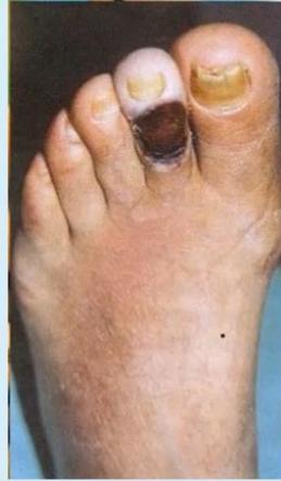
Q1: Describe the clinical features of an Arterial Ischemic Ulcer.
- Answer:
- Location: Distal extremities (tips of toes, heel, over bony pressure points, lateral malleolus).
- Base: Pale, dry, “punched-out” well-demarcated edges, necrotic/gray base with little or no granulation tissue.
- Surrounding skin: Shiny, hairless, atrophic, cool to touch with dystrophic thickened nails.
- Pulses: Absent or severely diminished distal pulses; accompanied by severe ischemic pain worse at night.
Station 53: Diabetic Foot Ulceration and Neuro-Ischemic Pathogenesis
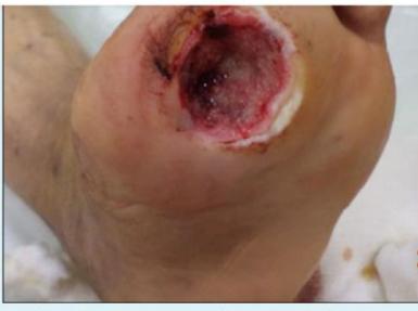

Q1: What are the TWO primary pathogenic factors contributing to this condition?
- Answer:
- Diabetic Peripheral Neuropathy (loss of protective sensation leading to repetitive unnoticed mechanical trauma, autonomic neuropathy causing dry cracked skin).
- Peripheral Arterial Disease (Macro- and Microvascular Ischemia) (impaired perfusion and delayed wound healing).
Q2: Mention TWO initial imaging investigations.
- Answer:
- Plain Radiograph (X-ray) of the Foot (detects osteomyelitis, cortical erosion, soft tissue gas, or Charcot arthropathy).
- Arterial Duplex Ultrasound (evaluates vascular run-off).
Q3: Mention FIVE important steps in initial treatment.
- Answer:
- Strict non-weight-bearing / pressure offloading (specialized footwear/cast).
- Surgical debridement of necrotic tissue and callous.
- Deep tissue culture and targeted systemic antibiotics for clinically infected wounds; avoid relying on superficial swabs.
- Optimized glycemic control (insulin therapy).
- Vascular assessment and revascularization if ischemia is present.
Q4: What FOUR further history questions and TWO laboratory investigations are required?
- Answer: Ask onset/course (growth, color, discharge, odor, healing/re-ulceration), pain or sensory loss, trauma/burn/barefoot injury and smoking, and diabetes duration/control/medication/follow-up. Include other ulcers, previous treatment and prior amputation/intervention. Labs: glucose/HbA1c and CBC/inflammatory markers; deep-tissue culture is obtained from clinically infected wounds, with ketones for hyperglycemia/systemic illness.
Q5: What additional palpation findings, complications and treatment details apply?
- Answer: Assess tenderness/temperature, pulses/capillary refill, ulcer depth/base and surrounding swelling; avoid indiscriminate squeezing. Complications include osteomyelitis, Charcot change, gangrene and sepsis. Admit and stabilize a septic/unwell patient first. Use multidisciplinary care, wound cleansing with saline, debridement, appropriate moist dressings with regular/daily review and offloading, infection treatment and vascular assessment; negative-pressure therapy is selected after adequate assessment/source control. Review tetanus for relevant wounds. Antiseptics can be toxic to healthy tissue; avoid indiscriminate use. Infection alone does not mandate amputation: reserve it for appropriate nonviable tissue or otherwise uncontrolled life-threatening disease.
Q6: What additional investigations are relevant?
- Answer: Relevant investigations include CBC, kidney-function tests, albumin, CRP/ESR, wound culture, X-ray, MRI, ABI and angiography. Correlate CBC with leukocytosis, renal tests with nephropathy, albumin with nutritional/healing status, CRP/ESR with inflammation, X-ray with subcutaneous gas and MRI with osteomyelitis. Use deep tissue sampling for a clinically infected wound where appropriate; select perfusion/advanced imaging according to the clinical question rather than ordering the whole panel for every ulcer.
Venous Lymphatic Diseases
Station 54: Lower Extremity Varicose Veins
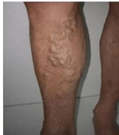
Q1: What is the primary diagnostic imaging investigation?
- Answer: Venous Duplex Ultrasound of the lower extremities in the standing position (maps incompetent saphenofemoral/saphenopopliteal junctions and perforators, rules out DVT).
Q2: Mention FOUR recognized complications of chronic untreated varicose veins.
- Answer:
- Venous stasis dermatitis and eczema.
- Lipodermatosclerosis (“inverted champagne bottle” leg).
- Venous stasis ulceration (medial malleolus).
- Superficial thrombophlebitis — which can extend to deep vein thrombosis, especially above the knee — or bleeding from ruptured varices.
Q3: What FOUR relevant history questions and TWO investigations were requested?
- Answer: Ask about pain/standing-related symptoms, family history/previous varicosities, previous DVT/immobility/trauma, and pregnancies/OCP exposure, ulcers, edema or thrombophlebitis. Duplex maps reflux/deep-system patency. MR venography is selected for suspected proximal obstruction; plethysmography is a specialist alternative. Perthes’ test is a historical clinical test, not an imaging study; routine arterial angiography is not a varicose-vein investigation.
Station 55: Venous Stasis Ulceration
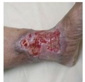
Q1: Mention TWO inspection signs.
- Answer: An ulcer in the medial-malleolar/gaiter region and visible granulation tissue are the key inspection signs.
Q2: Mention TWO definitive treatment modalities for this condition.
- Answer:
- Multilayer Compression Bandaging (four-layer compression system) after ruling out arterial disease ().
- Endovenous ablation of incompetent superficial veins (to correct underlying reflux and prevent recurrence).
Station 56: Lower Extremity Lymphedema
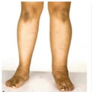
Q1: What is the diagnosis shown in this photograph?
- Answer: Chronic Lymphedema of the lower extremity.
Q2: List TWO causes and distinguish primary from secondary lymphedema.
- Answer: Infection and trauma, including filariasis. These are secondary causes. Additional secondary causes include surgery/lymph-node dissection, radiotherapy and malignant obstruction. Primary examples include Milroy disease, Meige disease/lymphedema praecox and lymphedema tarda.
Station 57: Red Swollen Lower Limb: Examination and Differential Diagnosis
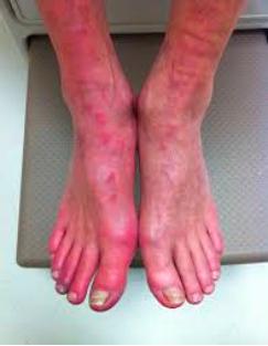
Q1: What FOUR clinical examinations were requested for the red swollen limb?
- Answer: Compare both limbs for temperature/tenderness, arterial pulses, capillary refill and edema/distribution; assess sensation and systemic sepsis. ABI/arterial assessment, Buerger’s test and femoral auscultation are relevant if arterial disease is suspected, not mandatory tests of cellulitis. Consider DVT, acute ischemia and necrotizing infection in the differential diagnosis.
Station 58: Postoperative Deep Vein Thrombosis
Case: After abdominal surgery, a patient has left-leg pain and swelling.
Q1: What is the likely diagnosis and how is it confirmed?
- Answer: Suspected DVT. Assess clinical probability (e.g. Wells), obtain compression venous ultrasound and use D-dimer within the appropriate pathway. D-dimer is less specific postoperatively; angiography is not a routine first test.
Skin & Soft Tissue
Skin, & Subcutaneous Tumors
Station 59: Cutaneous Malignant Melanoma Evaluation (ABCDE Criteria)
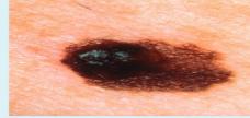

Q1: Apply the ABCDE criteria to this photograph, noting what cannot be measured from it.
- Answer: Assess asymmetry, irregular border, color variation and apparent lesion extent; the image shows irregular pigmentation. Exact diameter and evolution need a scale/history and cannot be established from the photograph alone.
Q2: What is the clinical diagnosis and how is it confirmed?
- Answer: Suspicious pigmented lesion / suspected melanoma. Confirm by appropriate full-thickness excisional biopsy; histological subtype cannot be established from a photograph.
Q3: Mention TWO microscopic factors that determine the T stage of the primary tumor.
- Answer:
- Breslow thickness (in mm).
- Presence or absence of histological ulceration.
Station 60: Pilonidal Sinus Disease
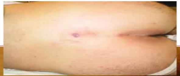
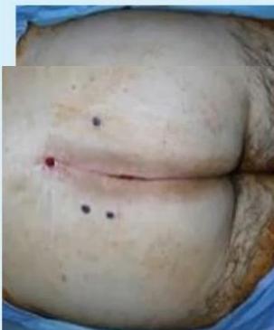
Case: A young male has recurrent purulent discharge and discomfort at the lower back; contrast clean sinus excision/closure with infected disease left open for daily dressings (hygiene/hair removal, acute drainage, chronic definitive treatment).
Q1: What is the diagnosis shown in these photographs?
- Answer: Pilonidal Sinus Disease (Sacrococcygeal Pilonidal Sinus).
Q2: Contrast management of an Acute Pilonidal Abscess versus Chronic Pilonidal Sinus.
- Answer:
- Acute Abscess: Surgical incision and drainage, with removal of hair/debris and appropriate wound care/dressing; packing is selected rather than universally required.
- Chronic Sinus: Elective excision: Off-midline flap reconstruction (e.g., Bascom cleft lift procedure, Karydakis flap, or Limberg flap) to flatten the natal cleft and move the scar away from the midline; permanent laser hair depilation.
Station 61: Acute Perianal Abscess
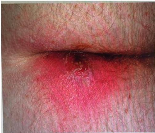
Q1: What is the diagnosis shown in this male with perianal pain and swelling for 2 days?
- Answer: Acute Perianal Abscess.
Q2: Mention TWO treatment steps and state the role of antibiotics.
- Answer: Prompt adequate incision and drainage is definitive source control, with wound care and follow-up. Antibiotics are selected for associated cellulitis, systemic infection or immunosuppression rather than routinely required after every uncomplicated drained abscess. EUA, incision approach, loculation management and packing are selected by anatomy/severity and surgical practice; routine continued packing is not essential for every patient.
MIS / Laparoscopy
Laparoscopic Instruments
Station 62: Veress Needle
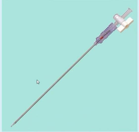
Station: Identify the laparoscopic instrument and its use.
Q1: Name the instrument and state its use.
- Answer: Veress needle: a spring-loaded blunt-tipped insufflation needle used to establish CO₂ pneumoperitoneum for closed laparoscopic entry. It is not a trocar.
Pediatric Surgery
Common pediatric surgical problems
Station 63: Congenital Pediatric Inguinal Hernia
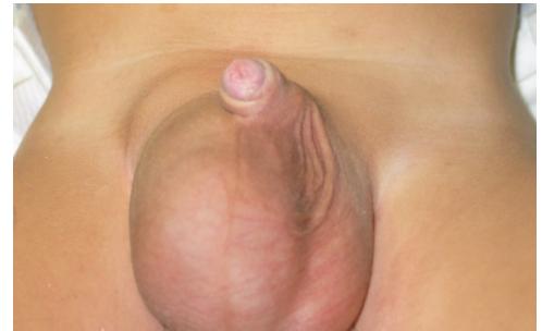
Case: A one-year-old boy has a swelling that becomes smaller during sleep.
Q1: What is the diagnosis shown in this 1-year-old boy?
- Answer: Congenital inguinal hernia, usually indirect. State the side shown.
Q2: Mention TWO recognized complications if left untreated.
- Answer:
- Incarceration / Irreducibility.
- Strangulation with bowel ischemia and secondary testicular infarction.
Q3: What is the surgical procedure of choice in children, and how does it fundamentally differ from adult hernia repair?
- Answer:
- Procedure: High Ligation of the Hernia Sac (Herniotomy).
- Fundamental Difference: Children have normal abdominal wall musculature and internal ring floor; therefore, herniotomy alone (ligating the sac at the internal ring) is curative. Routine infant/child congenital indirect hernia repair does not require mesh; adolescent or atypical defects need individualized specialist assessment rather than an absolute “never” rule.
Station 64: Undescended Testis (Cryptorchidism)
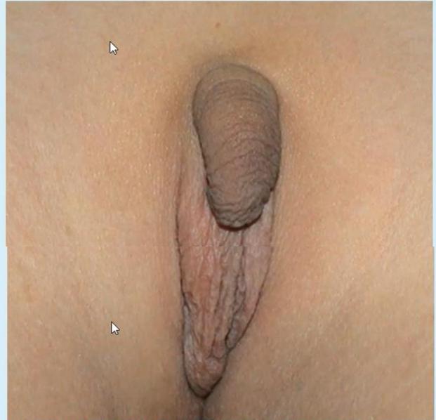
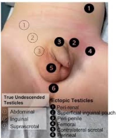
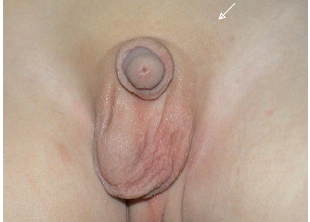
Q1: Describe the physical finding shown in the clinical photograph.
- Answer: Empty left hemiscrotum. Describe the asymmetry, scrotal development and skin appearance.
Q2: Mention TWO major long-term complications of untreated cryptorchidism.
- Answer:
- Subfertility / Infertility (due to thermal degenerative injury to germ cells).
- Testicular Malignancy (primarily Seminoma, risk remains elevated even after orchiopexy). (Also increased risk of testicular torsion and trauma).
Q3: What is the recommended surgical operation and optimal timing?
- Answer:
- Operation: Surgical Orchidopexy (Orchiopexy) (mobilization of spermatic cord and fixation of testis in a subdartos scrotal pouch; laparoscopic Fowler-Stephens for intra-abdominal testis).
- Optimal Timing: Performed between 6 and 12 months of age (and completed before 18 months).
Q4: Mention TWO inspection findings and differentiate UDT from other causes of an empty hemiscrotum.
- Answer: Empty/underdeveloped hemiscrotum and asymmetry; note whether there is an inguinal swelling and whether overlying skin is normal. Differential possibilities include retractile/ascending testis, ectopic testis (perirenal, superficial inguinal pouch, peripenile, femoral, contralateral scrotal or perineal) and absent/atrophic testis. Palpate in a warm relaxed child and examine both sides.
Q5: What is the diagnostic approach to a nonpalpable testis and why not request routine CT/US?
- Answer: Clinical assessment and examination under anesthesia guide surgery; if still nonpalpable, specialist diagnostic laparoscopy can locate an intra-abdominal testis or confirm absent/vanishing anatomy. Routine ultrasound/CT cannot reliably exclude an intra-abdominal testis and may delay treatment. Imaging is reserved for selected circumstances; bilateral nonpalpable testes/DSD features need endocrine/genetic assessment.
Additional Image Stations
Station 65: Proctoscope

Station: Identify the instrument shown and describe its use.
Q1: Identify the instrument.
- Answer: Proctoscope (rigid anoscope/proctoscope) with its obturator and light carrier. It provides direct vision of the anal canal and distal rectum.
Q2: Mention TWO clinical uses.
- Answer: Assessment of anal/rectal bleeding, hemorrhoids, fissure, fistula or prolapse; evaluation of a low rectal mass/polyp and guided biopsy or polypectomy of accessible lesions. It can also assist rectal examination and assessment of rectal injuries.
Q3: Describe the correct technique and its limitations.
- Answer: Consent and chaperone; left lateral position; inspect the anus, lubricate the assembled scope with obturator and introduce gently, following the anal canal axis; remove the obturator, insufflate air and inspect the mucosa while slowly withdrawing. The rigid proctoscope reaches only the distal rectum (~15 cm); use flexible sigmoidoscopy/colonoscopy for more proximal assessment.
Q4: Mention TWO possible complications.
- Answer: Discomfort/pain and minor bleeding; rarely perforation or vasovagal reaction. Adequate lubrication and gentle technique reduce risk.
Station 66: Plain Abdominal Radiograph: Dilated Bowel Loops (Obstruction vs Ileus)
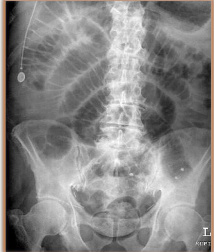
Case: An unwell patient has abdominal distension; an NG tube has been inserted and a supine abdominal film obtained.
Q1: Name the modality and describe the findings.
- Answer: Supine plain abdominal radiograph. There is marked gaseous distension of large and small bowel loops with haustral markings visible; a nasogastric tube is in situ. An erect film is needed to show air–fluid levels.
Q2: What is the differential diagnosis and next investigation?
- Answer: Distal bowel obstruction versus paralytic ileus. Correlate with bowel sounds, stool/flatus, previous surgery and medications. Contrast-enhanced CT of the abdomen/pelvis identifies a transition point, cause and any ischemia when the diagnosis is unclear.
Q3: Outline initial management.
- Answer: NPO, NG decompression, IV fluids and electrolyte correction, analgesia and serial abdominal examinations. Operative intervention is indicated for peritonitis, ischemia/perforation or failure of appropriate conservative management.
Station 67: Duodenal Ulcer on Upper GI Endoscopy
Q1: Identify the lesion shown.
- Answer: Duodenal ulcer at endoscopy (deep crater with necrotic slough and surrounding inflammation).
Q2: Mention TWO complications.
- Answer: Bleeding and perforation; also obstruction (gastric outlet) and penetration into adjacent structures.
Q3: How is a bleeding duodenal ulcer managed?
- Answer: ABC/resuscitation, NPO, IV PPI, crossmatch as needed, and urgent endoscopy with dual hemostasis (adrenaline injection plus clips or thermal therapy). Test and eradicate H. pylori, review NSAIDs/anticoagulants; surgery or interventional radiology for failed endoscopic control.
Station 68: Open Wound: Dehiscence and Infection

Case: A postoperative patient has a gaping wound with slough and surrounding inflammation.
Q1: Describe the finding.
- Answer: An open wound with gaping edges showing granulation tissue and fibrinous slough, with surrounding inflammation; assess size, depth, discharge (colour/odour), change in size, granulation quality and signs of infection. Palpate the wound base, squeeze for discharge, and check tenderness, local temperature and capillary refill.
Q2: How do you assess and manage it?
- Answer: Swab/deep tissue culture only if infected, CBC/CRP and imaging for a deep collection. Cleanse and debride slough/necrotic tissue, dress for secondary healing (or repair when appropriate), treat spreading infection with systemic antibiotics, and correct contributing factors (infection, tension, diabetes, malnutrition, steroids, raised intra-abdominal pressure). Suspected deep dehiscence/evisceration (burst abdomen) requires theatre.
Q3: Mention TWO preventive measures.
- Answer: Aseptic technique and tension-free closure; optimize glycaemic control/nutrition, treat cough/constipation, and review steroids and other healing-impairing drugs.
Station 69: Chest Tube (Tube Thoracostomy)

Station: Identify the device and describe its safe use.
Q1: Identify the device.
- Answer: Intercostal chest drain (tube thoracostomy) secured with an occlusive dressing, connected to an underwater-seal drainage system.
Q2: Mention TWO indications.
- Answer: Pneumothorax (especially after needle decompression of tension pneumothorax), hemothorax, empyema and pleural effusion drainage; also after thoracic surgery/trauma.
Q3: Describe safe insertion and aftercare.
- Answer: Use the safe triangle (5th intercostal space, mid-axillary line; 4th–6th space) and insert just above the rib below to avoid the neurovascular bundle. Sterile technique, local anaesthetic, blunt dissection, finger sweep, tube directed apically for air or basally for fluid, then secure and dress; confirm position with CXR. Keep the drain below chest level, never clamp a bubbling drain, and monitor swing/bubbling/output.
Q4: Mention TWO complications.
- Answer: Bleeding and infection/empyema; organ injury (lung, diaphragm, liver, spleen), subcutaneous emphysema, tube blockage or dislodgement, and re-expansion pulmonary edema.
Station 70: Laparoscopic L-Hook Electrode

Station: Identify the laparoscopic instrument shown.
Q1: Identify the instrument and its use.
- Answer: Monopolar laparoscopic hook (L-hook) electrode used through a port for dissection, tissue division and coagulation (e.g., adhesiolysis, gallbladder dissection, hemostasis) under direct camera vision.
Q2: Mention TWO safety points.
- Answer: Check insulation and avoid off-screen activation or direct/capacitive coupling to adjacent structures; use the lowest effective power, evacuate smoke, and be aware of thermal spread near bowel, vessels and bile ducts.
Station 71: Fournier’s Gangrene (Clinical Photograph and Perineal CT)


Case: A patient presents with a swollen, discolored scrotum and systemic illness.
Q1: What is the diagnosis shown in the photograph?
- Answer: Fournier’s gangrene — necrotizing fasciitis of the perineum/scrotum, with necrotic skin, blistering and extension along fascial planes (a urinary catheter is visible).
Q2: What does the CT/soft-tissue image show, and which structures are relevant?
- Answer: A soft-tissue reconstruction of the male perineum/scrotum showing extensive soft-tissue gas around the genital structures; the ventral midline structure is the corpus spongiosum (with the corpora cavernosa). Gas in the perineal soft tissues is a surgical emergency until necrotizing infection is excluded.
Q3: Outline the management.
- Answer: Resuscitate, broad-spectrum IV antibiotics and urgent radical surgical debridement of all necrotic tissue, with repeated debridements as needed; urinary diversion (suprapubic catheter) and faecal diversion in selected cases, wound care and later reconstruction. Treat predisposing conditions (diabetes, immunosuppression), involve urology/critical care, and counsel about high mortality.
Q4: Mention TWO risk factors/predisposing conditions.
- Answer: Diabetes mellitus and immunosuppression; also alcohol excess, perianal/urinary infection and local trauma.