collected from Faisal Alkharji, Faisal Alturki, Haneen H. Alsultan, Khalid Alanazi, Lina Serhan, Moath Bin Abdullah, Sultan AlDajani, Talal M. Ak.
اللهم يا معلّم موسى علّمني، ويا مفهم سليمان فهّمني، ويا مؤتي لقمان الحكمة وفصل الخطاب آتني الحكمة وفصل الخطاب اللهم اجعل ألستنا عامرة بذكرك، وقلوبنا بخشيتك، وأسرارنا بطاعتك، إنك على كل شيء قدير، حسبنا الله ونعم الوكيل
Table of Contents
- General Principles & Perioperative Care
- Trauma & Emergency Surgery
- Head & Neck
- Neurosurgery
- Breast
- Thoracic
- Upper GI & Abdominal Surgery
- Urology
- Vascular
- Skin & Soft Tissue
- MIS / Laparoscopy
- Metabolic Surgery
- Transplant Surgery
- Pediatric Surgery
General Principles & Perioperative Care
Preoperative & Postoperative care
Clinical Scenarios (CS)
Case 1: Blood Transfusion Requirements and Adverse Reactions in Acute Surgical Bleeding
A 58-year-old surgical patient presented with acute upper gastrointestinal hemorrhage. Admission laboratory results:
- WBC:
- Hemoglobin (Hb): (Normal: )
- Platelets:
Q1: How many units of packed red blood cells (PRBCs) are required to raise hemoglobin to ?
- Answer: 3 units (each unit of PRBCs raises adult hemoglobin by approximately ).
Q2: Mention FOUR recognized adverse effects of blood transfusion.
- Answer:
- Febrile non-hemolytic transfusion reaction (fever, chills).
- Acute hemolytic transfusion reaction (ABO incompatibility).
- Allergic / anaphylactic reaction (urticaria, bronchospasm).
- Transfusion-transmitted infection (viral hepatitis, HIV, bacterial sepsis). (Also acceptable: TRALI, TACO, hypocalcemia/citrate toxicity).
Case 2: Clinical Demonstration and Bedside Assessment of Ascites
A 52-year-old male with chronic liver cirrhosis is suspected of having mild-to-moderate ascites.
Q1: Mention TWO physical findings expected on inspection of the abdomen.
- Answer:
- Generalized abdominal distension with flank fullness (bulging flanks).
- Everted or flattened umbilicus.
Q2: Describe STEPWISE how to clinically demonstrate the presence of mild-to-moderate ascites.
- Answer: Shifting Dullness:
- Percuss from the resonant midline laterally toward the flank until dullness is reached; keep the pleximeter finger at the transition point.
- Roll the patient onto the opposite side (lateral decubitus) and wait 15–30 seconds.
- Percuss again at the stationary finger: the previous dullness becomes resonant, confirming shifting fluid.
-OSPE Station 1: Superficial Incisional Surgical Site Infection (SSI)
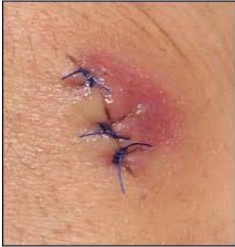
Q1: What is the diagnosis?
- Answer: Surgical Site Infection (SSI) (superficial incisional wound infection).
Q2: What is the most common causative organism?
- Answer: Staphylococcus aureus (including MRSA).
Q3: Outline the management plan.
- Answer:
- Remove sutures/staples over the fluctuant area to allow drainage.
- Evacuate purulent collection and send a swab for Gram stain and Culture & Sensitivity.
- Irrigate wound with sterile normal saline and pack loosely with saline gauze (secondary intention).
- Prescribe systemic antibiotics only if surrounding cellulitis () or systemic signs (fever) are present.
Day surgery
(No past exam questions recorded in source)
Operation room and surgical techniques
-OSPE Station 1: Intestinal Stoma Identification, Indications, and Complications
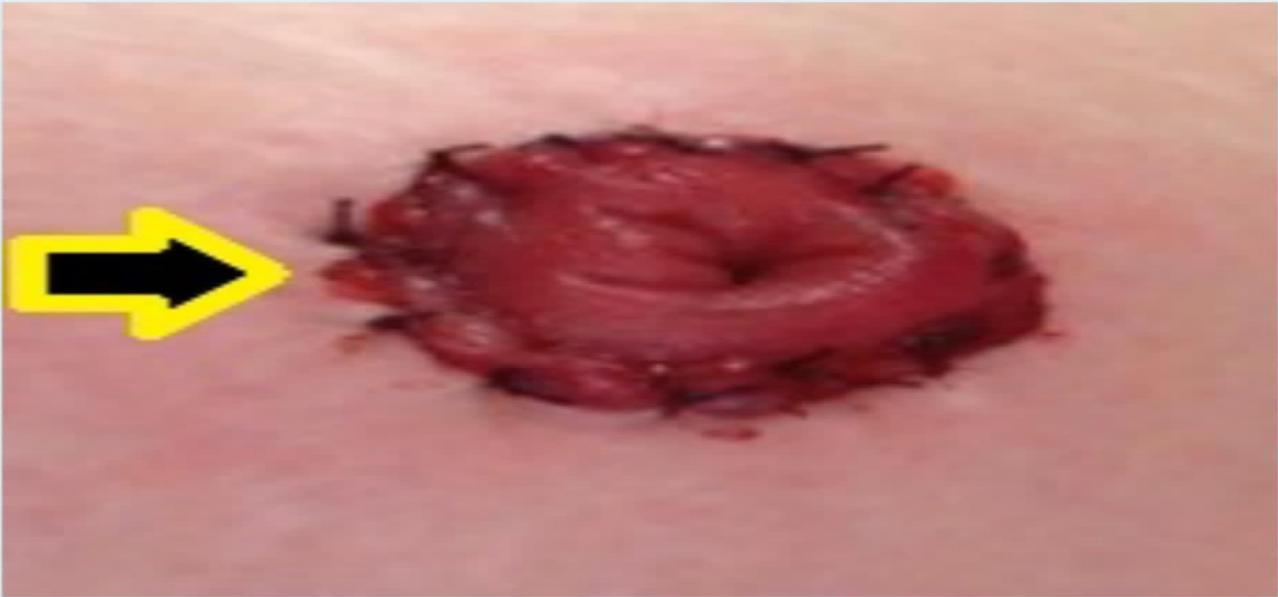
Q1: What is demonstrated in this photograph?
- Answer: End Colostomy (abdominal intestinal stoma).
Q2: Mention TWO surgical indications for this procedure.
- Answer:
- Relief of acute distal large bowel obstruction (e.g., obstructing rectosigmoid tumor).
- Emergency resection for perforation / diverticulitis (Hartmann’s procedure) or trauma.
Q3: Mention THREE recognized complications of an abdominal stoma.
- Answer:
- Stoma ischemia / necrosis.
- Stoma retraction.
- Parastomal hernia (or stoma prolapse, stenosis, peristomal dermatitis).
Imaging & instruments
-OSPE Station 1: Nasogastric Tube (NGT)
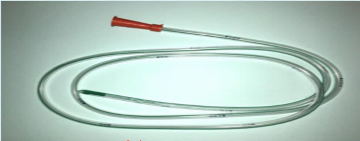
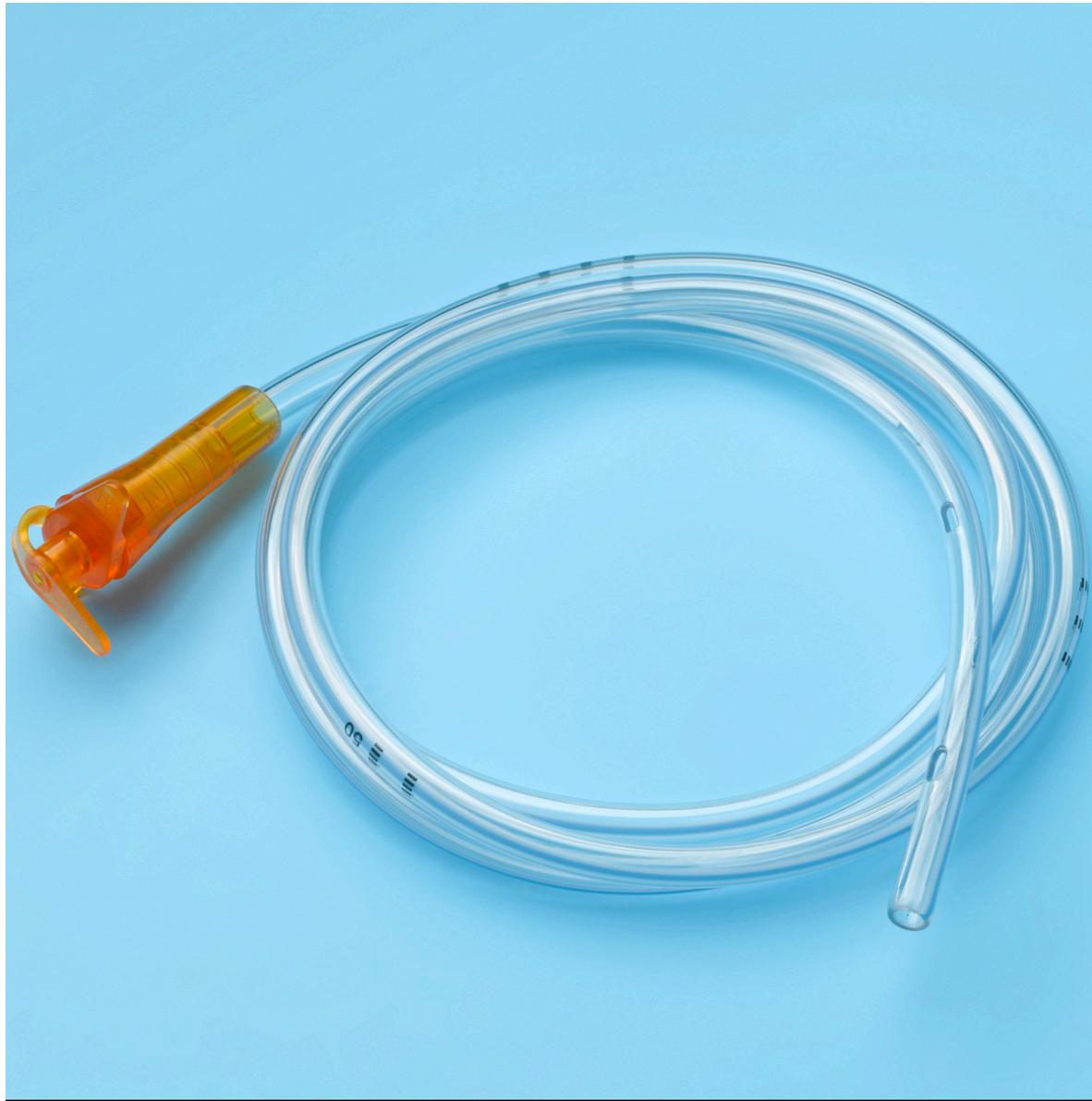
Q1: Identify this surgical instrument.
- Answer: Nasogastric Tube (NG Tube) (Levin / Salem sump tube).
Q2: Mention TWO therapeutic and TWO diagnostic uses.
- Answer:
- Therapeutic: Gastrointestinal decompression in intestinal obstruction / paralytic ileus; enteral feeding / drug administration.
- Diagnostic: Aspiration of gastric contents to confirm upper GI bleeding; administration of enteral radiographic contrast.
Q3: Mention FIVE important steps to introduce this tube safely.
- Answer:
- Position patient upright / High Fowler’s with neck slightly flexed.
- Measure insertion length using the NEX measurement (Nose Earlobe Xiphoid process) and mark depth.
- Lubricate the distal tip with water-soluble gel.
- Insert gently along nasal floor; advance into esophagus while asking patient to swallow sips of water.
- Secure tube to nose and verify intragastric placement (aspirate pH or chest X-ray).
Q4: Mention THREE potential complications of placement.
- Answer:
- Tracheobronchial misplacement / pulmonary aspiration.
- Epistaxis and nasal mucosal trauma.
- Esophageal or pharyngeal perforation.
-OSPE Station 2: Closed Wound Suction Drain (Zimmer Hemovac 400 mL)
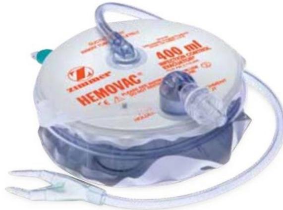
Q1: Identify the device shown.
- Answer: Zimmer Hemovac Closed Wound Suction Drain (400 mL).
Q2: Mention TWO surgical indications for placing this drain.
- Answer:
- Obliteration of dead space and evacuation of hematoma/seroma (e.g., post-mastectomy, neck surgery, axillary dissection).
- Evacuation of postoperative blood in major musculoskeletal / orthopedic surgery.
Q3: Mention TWO potential complications of surgical drains.
- Answer:
- Retrograde / ascending surgical site infection.
- Tissue erosion or visceral injury (or retained drain fragment).
-OSPE Station 3: Foley’s Urinary Retention Catheter
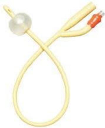
Q1: Identify this instrument.
- Answer: Two-Way Foley Retention Urinary Catheter.
Q2: Mention TWO diagnostic uses and TWO therapeutic uses.
- Answer:
- Diagnostic: Hourly urine output monitoring (in shock, ICU, major surgery); collecting sterile urine for culture.
- Therapeutic: Relief of acute or chronic urinary retention (e.g., BPH); intraoperative bladder decompression.
Q3: Mention THREE recognized complications of catheterization.
- Answer:
- Catheter-Associated Urinary Tract Infection (CAUTI).
- Urethral trauma and false passage formation.
- Urethral stricture (or accidental balloon inflation in the urethra).
Spot Dx
-OSPE Station 1: Acute Paronychia
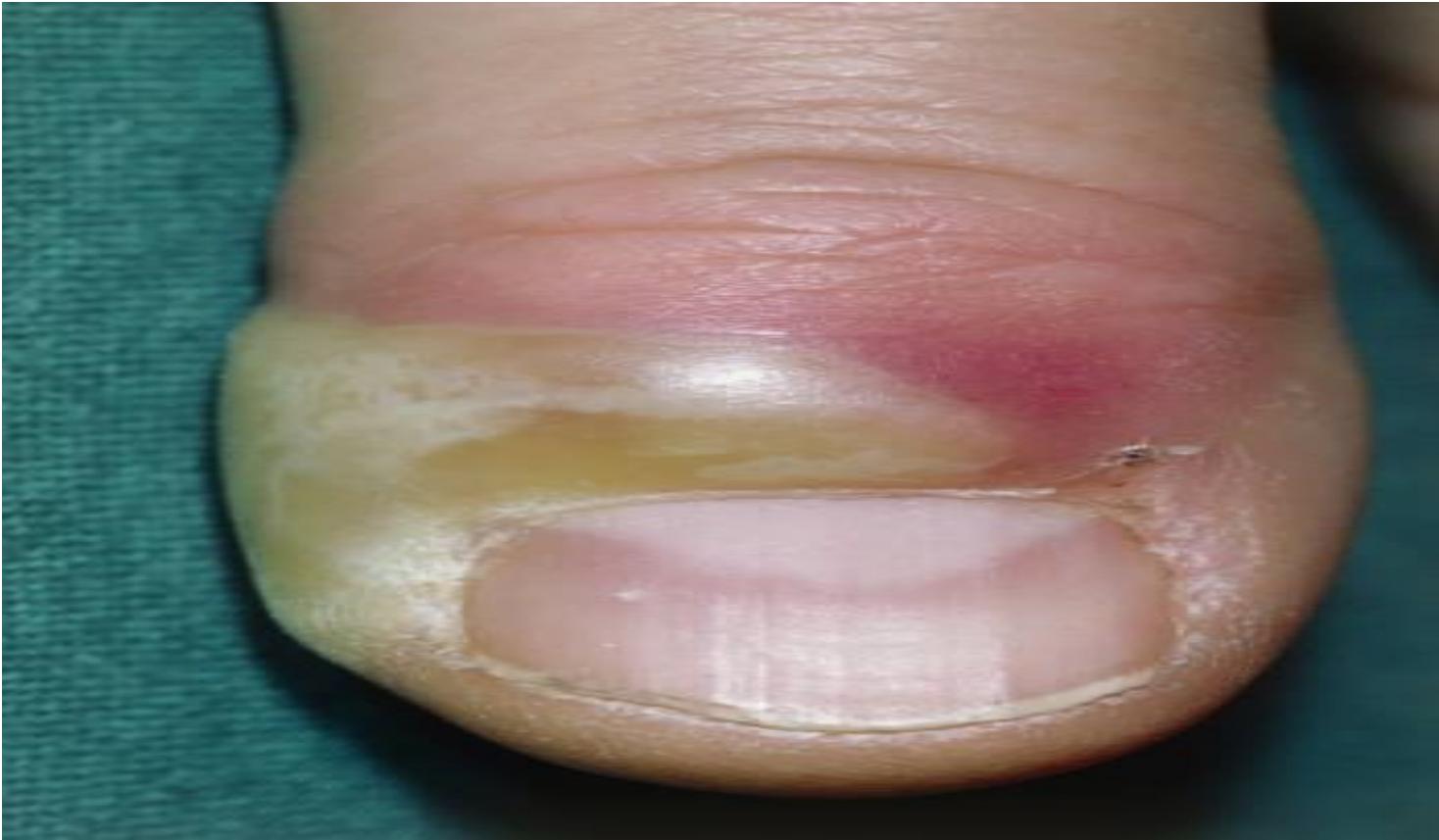
Q1: What is the diagnosis shown on the distal phalanx?
- Answer: Acute Paronychia (nail fold infection).
Q2: What is the most common causative organism?
- Answer: Staphylococcus aureus.
Q3: Outline the management.
- Answer:
- Warm soaks and oral antibiotics (if early / cellulitic).
- Surgical incision and drainage (I&D) or partial nail plate avulsion (if fluctuant abscess is present).
-OSPE Station 2: Carbuncle of the Posterior Neck
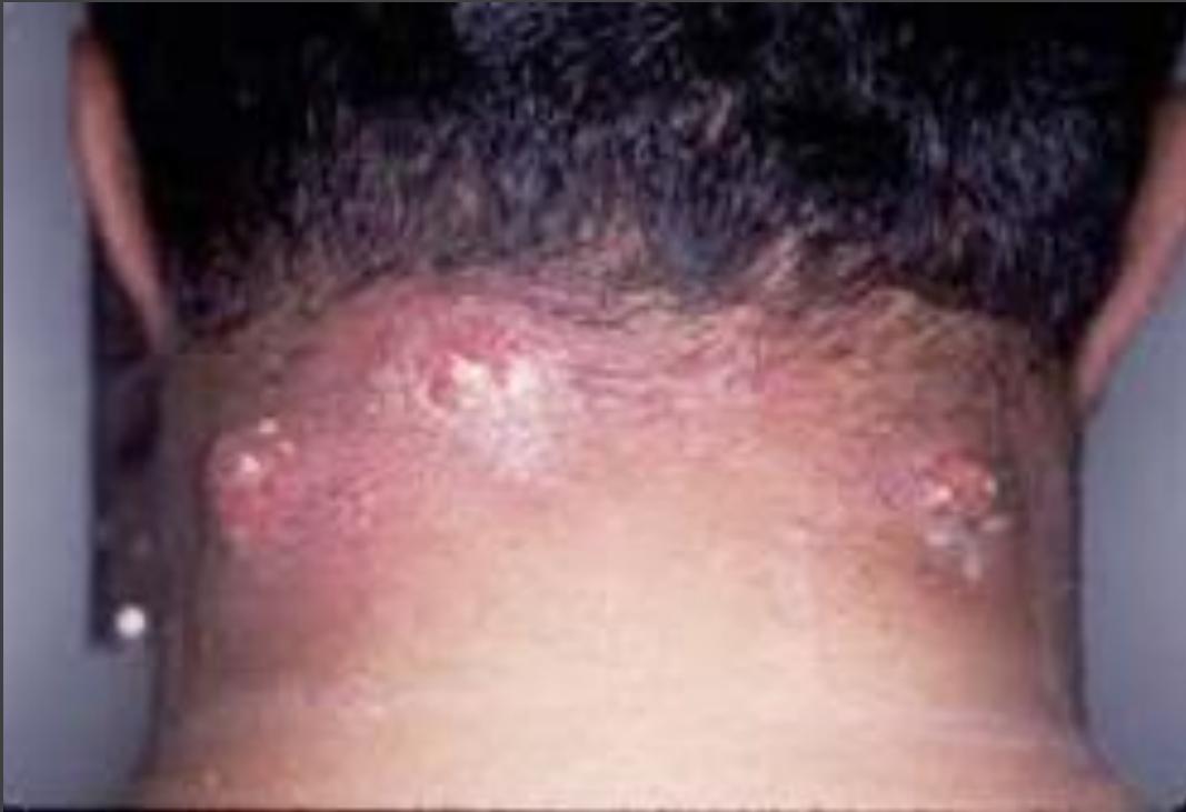
Q1: What is the diagnosis shown on the nape of the neck?
- Answer: Carbuncle of the neck (confluent subcutaneous furuncles).
Q2: What is the most common causative organism?
- Answer: Staphylococcus aureus.
Q3: What underlying systemic disease must be actively screened for?
- Answer: Diabetes Mellitus.
Q4: Outline the treatment plan.
- Answer:
- Surgical Incision and Drainage (cruciate incision / debridement of necrotic slough).
- Systemic intravenous antibiotics (anti-staphylococcal).
- Strict blood glucose control.
Trauma & Emergency Surgery
Trauma
Clinical Scenarios (CS)
Case 1: Penetrating Spinal Cord Injury (Brown-Séquard Syndrome)
A 28-year-old male presents to the Emergency Department following a penetrating stab wound to the mid-thoracic back.
Q1: What is the clinical syndrome?
- Answer: Brown-Séquard Syndrome (hemisection of the spinal cord).
Q2: What neurological deficits are expected on physical examination?
- Answer:
- Ipsilateral (same side as lesion): Loss of motor function (spastic paralysis/weakness) and loss of vibration/proprioception sense below the level of injury.
- Contralateral (opposite side): Loss of pain and temperature sensation beginning 1–2 segments below the lesion.
Q3: What is the initial emergency imaging investigation?
- Answer: Non-contrast CT scan of the thoracic spine (followed by MRI to assess cord compression).
-OSPE Station 1: Lateral Cervical Spine Radiography in Trauma
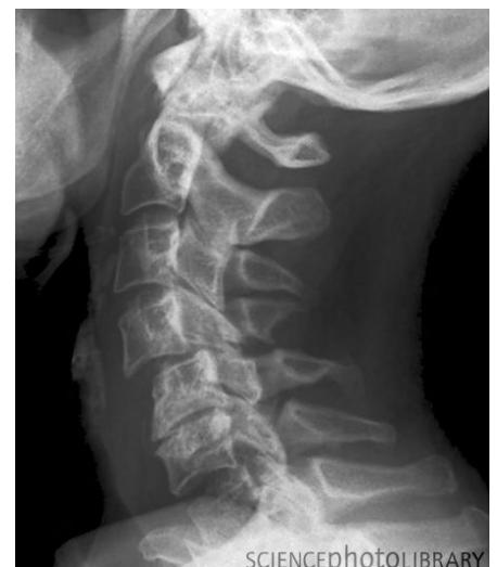
Q1: Identify this radiographic view and what anatomical landmark MUST be visualized for adequacy.
- Answer: Lateral Cervical Spine Radiograph; must visualize down to the C7–T1 junction (if not seen, order Swimmer’s view or CT cervical spine).
Q2: Name the FOUR continuous anatomical lines evaluated on this view.
- Answer:
- Anterior vertebral line.
- Posterior vertebral line.
- Spinolaminar line.
- Tips of spinous processes line.
Q3: What is the immediate management if cervical spine injury is suspected?
- Answer: Rigid cervical collar, sandbags, and tape (complete cervical spinal immobilization).
Q4: If a trauma patient presents with quadriplegia versus paraplegia, where is the level of cord injury?
- Answer:
- Quadriplegia (tetraplegia): Cervical spinal cord injury.
- Paraplegia: Thoracic or lumbar spinal cord injury.
Q5: What is the definitive treatment and what major complications can cause death?
- Answer:
- Definitive Treatment: Operative reduction, stabilization, and internal fixation/realignment.
- Fatal Complications: Respiratory failure/arrest (phrenic nerve involvement C3–C5), pulmonary aspiration, and severe sepsis/pneumonia.
-OSPE Station 2: Below-Knee Amputation (BKA) Stump Evaluation
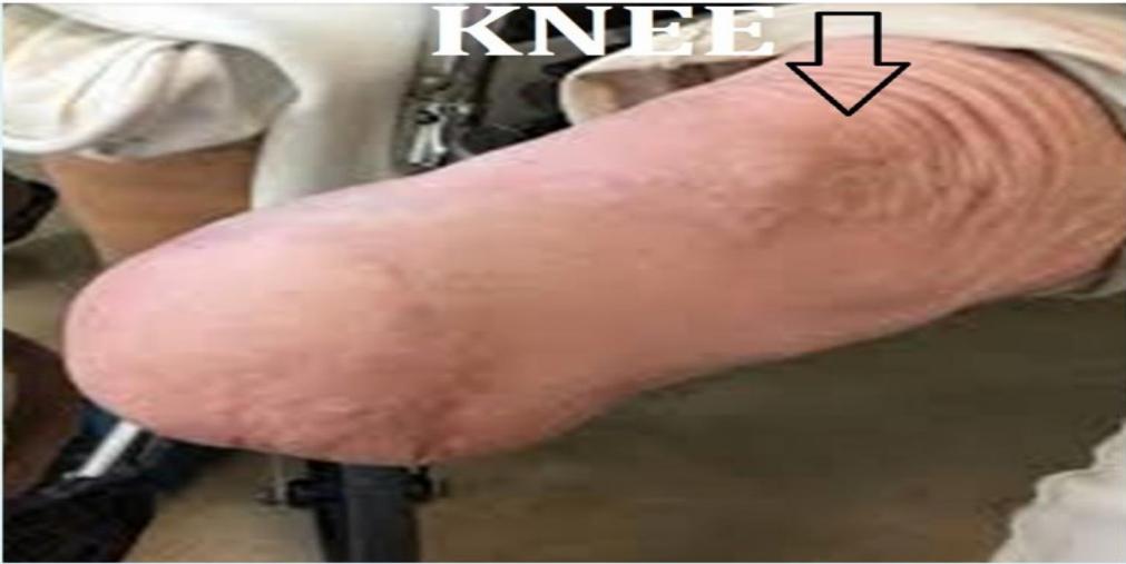
Q1: What surgical procedure is demonstrated?
- Answer: Below-Knee Amputation (BKA / Transtibial Amputation).
Q2: Mention THREE common complications of an amputation stump.
- Answer:
- Phantom limb pain / sensation.
- Stump infection / wound breakdown / ulceration.
- Flexion contracture of the knee joint (or stump neuroma).
Q3: Mention TWO measures to prepare the stump for prosthesis fitting.
- Answer:
- Stump shaping and elastic compression bandaging (shrinker sock).
- Physical therapy and range-of-motion exercises to prevent knee contracture.
Poly trauma patient
(No past exam questions recorded in source)
burns
(No past exam questions recorded in source)
Acute abdomen
Clinical Scenarios (CS)
Case 1: Acute Cholecystitis
A 45-year-old female presents with a 3-day history of severe right upper quadrant (RUQ) abdominal pain and fever.
Q1: What FIVE further relevant questions will you ask regarding the presenting complaint?
- Answer:
- History of jaundice, dark urine, or pale stools.
- Radiation of pain to the right shoulder or back (scapula).
- Association of pain with fatty or greasy meals.
- Previous similar episodes of biliary pain or known gallstones.
- Associated chills, rigors, or vomiting.
Q2: What is the most likely diagnosis?
- Answer: Acute Calculous Cholecystitis.
Q3: What FIVE laboratory investigations will you request at admission?
- Answer:
- CBC (leukocytosis with left shift).
- Liver function tests: ALT, AST.
- Alkaline Phosphatase (ALP) and Bilirubin.
- Serum Amylase / Lipase (to rule out secondary gallstone pancreatitis).
- Serum Electrolytes / Renal function tests.
Q4: What TWO imaging investigations will you initially request?
- Answer:
- Abdominal Ultrasound (investigation of choice: gallstones, thickened gallbladder wall , pericholecystic fluid, sonographic Murphy’s sign).
- Contrast-enhanced CT abdomen (or MRCP).
Q5: Mention FIVE initial management steps after admission.
- Answer:
- NPO (bowel rest).
- IV fluid resuscitation.
- IV analgesics (NSAIDs or opioids).
- IV broad-spectrum antibiotics.
- Early Laparoscopic Cholecystectomy (within 72 hours of admission).
Case 2: Acute Pancreatitis
A 50-year-old male presents with severe epigastric pain radiating to the back and multiple vomiting for 1 day; sitting and leaning forward eases the pain.
Q1: What is the most likely clinical diagnosis?
- Answer: Acute Pancreatitis.
Q2: What are the FOUR most common causes of this illness?
- Answer:
- Gallstones (biliary pancreatitis).
- Chronic alcohol consumption.
- Hypertriglyceridemia / Hypercalcemia.
- Post-ERCP / Trauma / Drugs.
Q3: What laboratory investigations will you request to establish the diagnosis?
- Answer:
- Serum Lipase (more sensitive and specific; remains elevated longer).
- Serum Amylase (elevated upper limit of normal).
- CBC, LFTs, BUN/creatinine, and serum calcium.
Q4: Name TWO clinical scoring systems used to assess disease severity.
- Answer: Ranson criteria and Glasgow-Imrie score (or APACHE II).
Q5: What imaging investigations should be requested?
- Answer:
- Abdominal Ultrasound at admission (to detect gallstones or biliary duct dilatation).
- Contrast-Enhanced CT (CECT) abdomen after 48–72 hours (to evaluate pancreatic necrosis and fluid collections).
Q6: Outline initial management.
- Answer: NPO, aggressive IV crystalloid resuscitation, adequate analgesia (opioids), antiemetics, and monitoring of urine output.
Case 3: Acute Perforated Peptic Ulcer
A 35-year-old male presents with sudden, severe, generalized abdominal pain and a rigid, board-like abdomen.
Q1: What is the most likely diagnosis?
- Answer: Perforated Peptic Ulcer (with generalized peritonitis).
Q2: What initial imaging investigation should be ordered and what finding is diagnostic?
- Answer: Erect Chest X-ray (or erect abdominal X-ray); diagnostic finding: Pneumoperitoneum (free air / gas crescent under the diaphragm).
Q3: What are the immediate resuscitation steps?
- Answer: NPO, 2 large-bore IV lines, aggressive IV fluid resuscitation, IV broad-spectrum antibiotics, IV PPI bolus, and NG tube decompression.
Q4: What is the definitive surgical treatment?
- Answer: Emergency exploratory laparotomy (or laparoscopy) and Graham patch (omental patch) closure of the perforation with peritoneal lavage.
Case 4: Acute RLQ Abdominal Pain in a Reproductive-Age Female
A 24-year-old female presents with 12 hours of worsening RLQ pain, temperature , dysuria, and missed menses for 5 days.
Q1: Mention FOUR differential diagnoses.
- Answer:
- Acute appendicitis.
- Ectopic pregnancy (ruptured or unruptured).
- Ruptured ovarian cyst / Mittelschmerz.
- Pelvic Inflammatory Disease (PID) / Salpingitis (or ovarian torsion).
Q2: What MANDATORY initial investigation must be performed first?
- Answer: Urine or Serum -hCG (pregnancy test) (to definitively rule out ectopic pregnancy).
Q3: Mention TWO imaging modalities to help differentiate the cause.
- Answer:
- Pelvic / Transvaginal Ultrasound (TVUS).
- Abdominal Ultrasound (or CT abdomen/pelvis if non-pregnant).
Q4: Mention TWO supportive laboratory tests.
- Answer: CBC (leukocytosis) and Urinalysis (to rule out UTI).
Case 5: Complicated Ruptured Appendicitis with Appendiceal Abscess / Phlegmon
A 40-year-old male presents with 5 days of worsening RLQ pain and fever; CT abdomen shows perforated appendicitis with an inflammatory phlegmon and localized fluid collection.
Q1: What is the diagnosis?
- Answer: Ruptured Appendicitis with Appendiceal Phlegmon / Abscess.
Q2: Outline the management plan (Ochsner-Sherren regimen).
- Answer:
- Conservative medical management: NPO, IV fluid resuscitation, IV broad-spectrum antibiotics.
- Percutaneous image-guided (US or CT) drainage of the abscess if .
- Close monitoring (vitals, WBC count, serial abdominal exams).
- Interval appendectomy after 6–8 weeks (elective).
Case 6: Right Upper Quadrant (RUQ) Abdominal Mass Evaluation
A 53-year-old female presents with a palpable, non-tender right upper quadrant mass.
Q1: Mention FOUR differential diagnoses for a RUQ abdominal mass.
- Answer:
- Enlarged gallbladder (mucocele, empyema, or gallbladder carcinoma).
- Hepatomegaly / Liver mass (hepatoma, hepatic cyst, pyogenic abscess).
- Colonic mass (hepatic flexure carcinoma).
- Right renal mass (renal cell carcinoma, hydronephrosis).
Q2: What TWO initial imaging investigations should be requested?
- Answer:
- Abdominal Ultrasound (initial non-invasive imaging).
- Contrast-enhanced CT (CECT) of the abdomen.
Q3: Mention THREE relevant laboratory investigations.
- Answer: CBC, Liver Function Tests (LFTs), and Renal Function Tests (BUN/Cr).
-OSPE Station 1: Pneumoperitoneum / Free Air Under Diaphragm on Plain Radiography
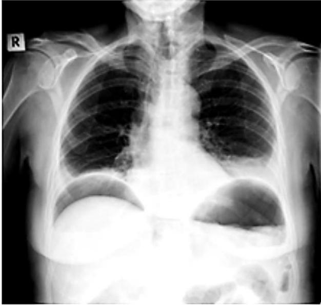
Q1: What is the radiographic finding shown?
- Answer: Pneumoperitoneum (free subdiaphragmatic air crescent under the right hemidiaphragm).
Q2: Mention TWO common surgical causes.
- Answer:
- Perforated peptic ulcer (duodenal or gastric).
- Perforated diverticulitis / bowel perforation (or trauma, recent laparotomy).
Q3: What is the next management step?
- Answer: Emergency surgical consultation, IV resuscitation, NG decompression, IV antibiotics, and urgent exploratory laparotomy.
-OSPE Station 2: Grey Turner’s Sign in Severe Retroperitoneal Hemorrhage
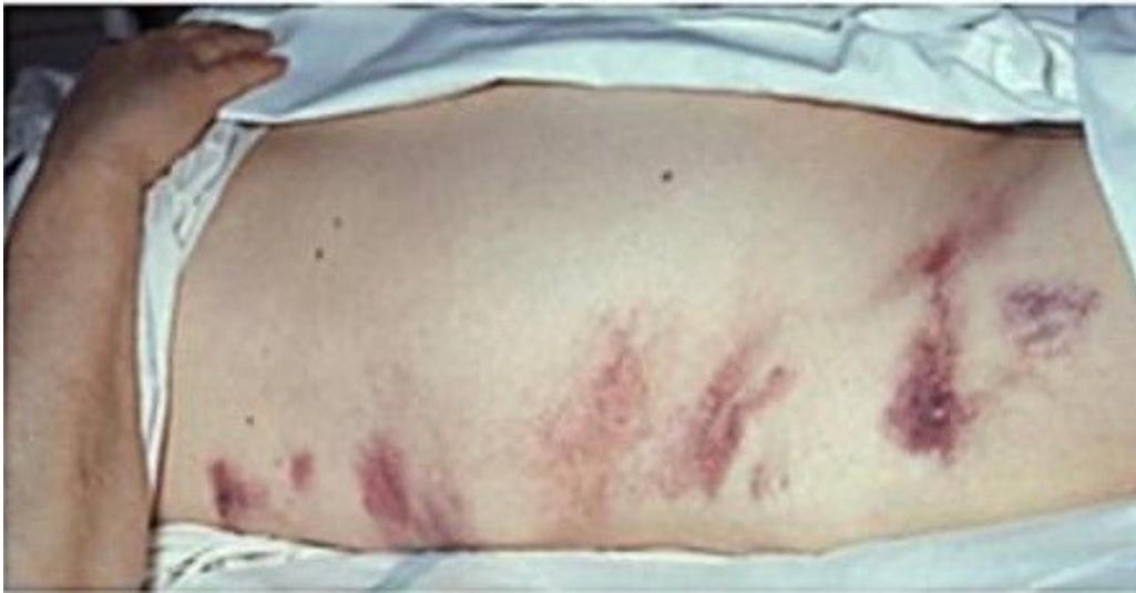
Q1: What is the clinical sign shown?
- Answer: Grey Turner’s sign (flank ecchymosis / bruising).
Q2: What underlying pathological process does it signify?
- Answer: Retroperitoneal hemorrhage.
Q3: Mention TWO important surgical causes.
- Answer:
- Severe acute necrotizing pancreatitis.
- Ruptured abdominal aortic aneurysm (AAA) (or retroperitoneal trauma).
Upper GIT bleeding
Clinical Scenarios (CS)
Case 1: Bleeding Peptic Ulcer Disease
A 60-year-old male presents with vomiting dark coffee-ground blood and passing melena, tachycardia, and hypotension.
Q1: What are the initial FIVE resuscitation steps on arrival?
- Answer:
- ABCDE resuscitation (Airway protection, high-flow oxygen).
- Establish 2 large-bore IV lines (16–18G).
- IV crystalloid fluid resuscitation.
- Blood crossmatch and transfusion as indicated.
- High-dose IV Proton Pump Inhibitor (PPI) bolus and infusion.
Q2: What is the diagnostic and therapeutic modality of choice?
- Answer: Urgent Upper Endoscopy (OGD) (within 24 hours).
Q3: Mention TWO endoscopic hemostasis modalities used to treat active bleeding ulcers.
- Answer:
- Injection therapy (epinephrine / adrenaline).
- Mechanical therapy (hemoclips) or thermal coagulation.
Q4: In an uncomplicated presentation with epigastric hunger pain relieved by meals, what are the primary differential diagnoses and medical therapy?
- Answer:
- Differential Diagnoses: Gastric ulcer, gastritis, GERD, or esophagitis.
- Medical Therapy: Proton Pump Inhibitors (PPIs) or H2-blockers, Helicobacter pylori eradication therapy, and dietary/lifestyle modifications.
-OSPE Station 1: Endoscopic Diagnosis and Management of Bleeding Esophageal Varices
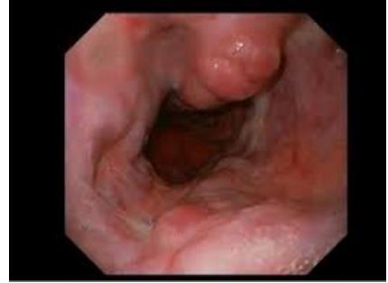
Q1: Identify the endoscopic finding shown.
- Answer: Bleeding Esophageal Varices (dilated, tortuous submucosal veins).
Q2: Mention TWO pharmacologic agents started immediately.
- Answer:
- IV Vasoactive drugs: Octreotide or Terlipressin.
- Prophylactic IV Antibiotics: Ceftriaxone.
Q3: What is the endoscopic procedure of choice?
- Answer: Endoscopic Variceal Ligation (EVL / Banding).
Q4: What rescue procedure can be performed if endoscopic therapy fails?
- Answer: Balloon Tamponade (Sengstaken-Blakemore tube) or Transjugular Intrahepatic Portosystemic Shunt (TIPS).
-OSPE Station 2: Coagulation Profile Interpretation in Massive Upper GI Bleeding
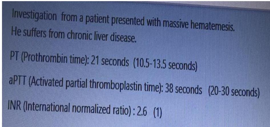
A cirrhotic patient with massive hematemesis has elevated PT/INR, prolonged aPTT, and thrombocytopenia.
Q1: Identify the clinical problem.
- Answer: Coagulopathy of chronic liver disease with bleeding.
Q2: Mention THREE blood products or pharmacologic agents used to correct this coagulopathy.
- Answer:
- Fresh Frozen Plasma (FFP).
- Platelet concentrate.
- Vitamin K (IV) (or Cryoprecipitate).
Lower GIT bleeding
Clinical Scenarios (CS)
Case 1: Acute Severe Lower Gastrointestinal Bleeding in an Elderly Patient
A 65-year-old male presents with two episodes of painless massive bright red rectal bleeding (hematochezia).
Q1: What FIVE relevant questions will you ask?
- Answer:
- Color and quantity of blood (bright red vs dark maroon).
- Presence of abdominal pain or cramping.
- History of constipation, straining, or altered bowel habits.
- Previous episodes of GI bleeding or known diverticulosis/polyps.
- Medication history (anticoagulants, antiplatelets, NSAIDs).
Q2: Mention THREE most likely differential diagnoses.
- Answer:
- Diverticular bleeding (diverticulosis).
- Angiodysplasia (arteriovenous malformation).
- Colorectal cancer / polyps (or brisk upper GI bleed).
Q3: Mention FOUR initial investigations.
- Answer:
- CBC (Hb/Hct) and Blood crossmatch.
- Coagulation profile and electrolytes.
- Upper endoscopy (OGD) to rule out brisk upper GI source.
- Colonoscopy (or CT angiography if actively bleeding ).
Q4: Outline initial management steps.
- Answer: ABC resuscitation, 2 large-bore IV lines, IV crystalloid fluids, blood transfusion if indicated, Foley catheter, and monitored unit admission.
Small Bowel Obstruction
Clinical Scenarios (CS)
Case 1: Adhesive Small Bowel Obstruction (SBO)
A 45-year-old female presents with 2 days of colicky abdominal pain, multiple vomiting, abdominal distension, and obstipation; she had an appendectomy 2 years ago.
Q1: What FOUR clinical signs would you look for on physical examination?
- Answer:
- Abdominal distension.
- Surgical scar (previous appendectomy).
- High-pitched, tinkling bowel sounds (or absent bowel sounds in late stage).
- Abdominal tenderness and check for incarcerated groin hernias.
Q2: What is the most likely diagnosis and underlying cause?
- Answer: Small Bowel Obstruction (SBO) secondary to postoperative peritoneal adhesions.
Q3: What are the cardinal radiographic findings on plain abdominal X-ray?
- Answer: Dilated small bowel loops (), multiple air-fluid levels in a “step-ladder” pattern, and absence of colonic gas.
Q4: Outline the initial conservative management plan (“Drip and Suck”).
- Answer:
- NPO (bowel rest).
- Nasogastric (NG) tube decompression to suction.
- IV fluid resuscitation with crystalloids and electrolyte correction.
- Foley catheter for hourly urine output monitoring.
- Serial abdominal examinations.
Q5: Mention TWO absolute clinical indications for emergency surgical exploration.
- Answer:
- Signs of bowel strangulation / ischemia / perforation (fever, tachycardia, localized peritonitis, leukocytosis, acidosis).
- Failure of conservative management after 48–72 hours.
-OSPE Station 1: Intussusception in Children and Adults
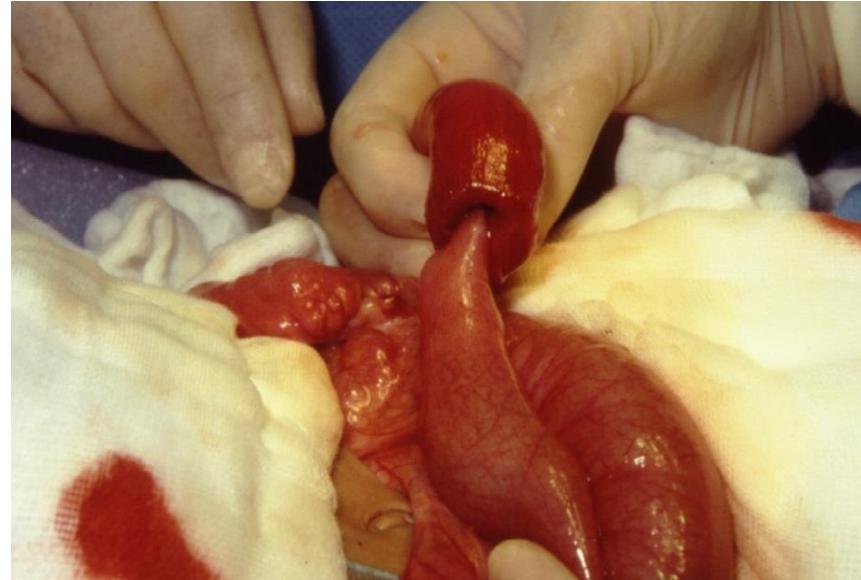
Q1: What is the diagnosis and characteristic ultrasound sign?
- Answer: Intussusception; characteristic sign: “Target sign” (or “donut sign” / pseudokidney sign).
Q2: Differentiate the etiology between infants and adults.
- Answer:
- Infants ( years): Usually idiopathic (hypertrophied lymphoid Peyer’s patches post-viral infection).
- Adults: Usually secondary to a pathological lead point (colorectal adenocarcinoma, polyp, lipoma, or Meckel’s diverticulum).
Q3: What is the primary management in pediatric vs adult intussusception?
- Answer:
- Pediatric: Non-operative pneumatic or hydrostatic (air/saline) enema reduction under fluoroscopy/ultrasound.
- Adult: Surgical resection (without prior reduction due to high malignancy risk).
Large Bowel Obstruction
-OSPE Station 1: Plain Radiography of Large Bowel Obstruction
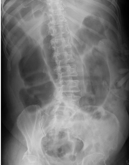
Q1: Identify the diagnostic radiographic finding.
- Answer: “Coffee bean” sign (inverted-U sign) arising from the pelvis and pointing to the RUQ.
Q2: What is the diagnosis?
- Answer: Sigmoid Volvulus.
Q3: What is the initial therapeutic management?
- Answer: Rigid sigmoidoscopy (or colonoscopy) with flatus tube insertion for endoscopic detorsion and decompression.
Q4: What is the definitive management to prevent recurrence?
- Answer: Elective sigmoid colectomy with primary anastomosis.
-OSPE Station 2: Hartmann’s Procedure for Obstructing / Perforated Colonic Pathology
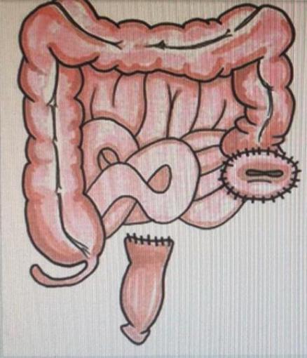
Q1: What surgical procedure is demonstrated?
- Answer: Hartmann’s Procedure (resection of rectosigmoid colon, end colostomy formation, and closure of the rectal stump).
Q2: Mention TWO major surgical indications.
- Answer:
- Perforated diverticulitis with feculent peritonitis (Hinchey IV).
- Obstructing rectosigmoid cancer in an emergency/unstable setting.
Q3: What is the advantage over primary anastomosis in the emergency setting?
- Answer: Avoids high risk of anastomotic leak and breakdown in the presence of fecal contamination, sepsis, and hemodynamic instability.
-OSPE Station 3: Barium Enema Demonstrating “Apple-Core” Sign of Colon Cancer
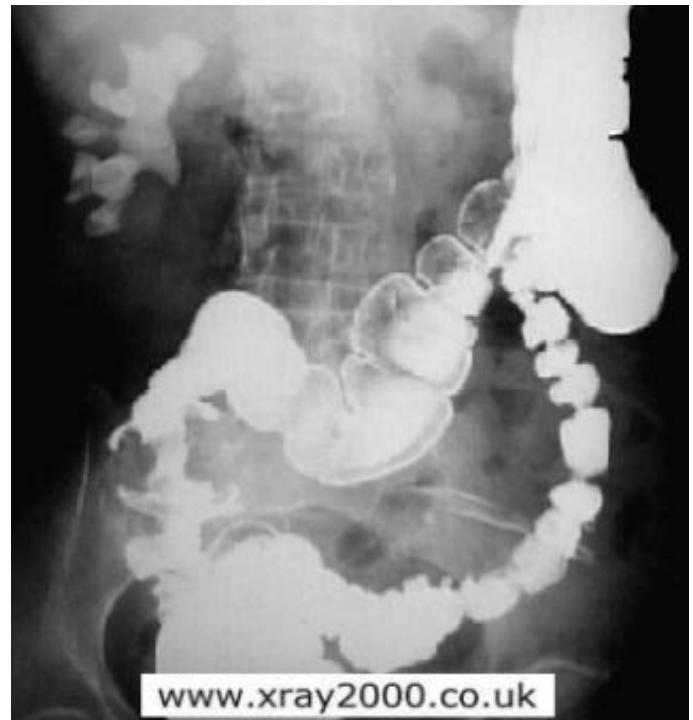
Q1: Name this radiological investigation and describe the classic sign.
- Answer: Barium Enema; classic sign: “Apple-core” lesion (annular constricting stenosis with shouldered margins).
Q2: What is the diagnosis?
- Answer: Colorectal Carcinoma (colon cancer).
Q3: What is the definitive diagnostic procedure to confirm diagnosis?
- Answer: Colonoscopy with multiple tissue biopsies.
Q4: Mention TWO staging investigations.
- Answer:
- Contrast-enhanced CT of Chest, Abdomen, and Pelvis (CT CAP).
- Serum Carcinoembryonic Antigen (CEA) level.
Head & Neck
Neck swellings
Clinical Scenarios (CS)
Case 1: Graves’ Disease (Toxic Diffuse Goiter)
A 32-year-old female presents with an anterior neck swelling, weight loss despite increased appetite, palpitations, heat intolerance, tremor, and prominent eyes.
Q1: What is the most likely clinical diagnosis?
- Answer: Graves’ Disease (toxic diffuse goiter / hyperthyroidism).
Q2: What physical findings support this diagnosis?
- Answer: Diffuse, smooth, non-tender goiter with thyroid vascular bruit; warm moist palms; fine resting finger tremor; tachycardia; and ocular signs (exophthalmos / proptosis, lid lag).
Q3: What diagnostic laboratory investigations will you request?
- Answer:
- Serum TSH (suppressed / undetectable) and Free T3 / Free T4 (markedly elevated).
- TSH receptor antibodies (TRAb) (pathognomonic).
Q4: Mention THREE treatment modalities available for Graves’ disease.
- Answer:
- Antithyroid medications: Thionamides (Methimazole / Carbimazole, or Propylthiouracil).
- Radioactive Iodine Ablation ().
- Surgery: Total or near-total thyroidectomy (after achieving euthyroid state with medications).
Case 2: Thyroid Malignancy (Suspected Papillary Thyroid Carcinoma)
A 42-year-old female presents with a hard, solitary thyroid nodule present for 2 years, with recent onset of hoarseness and ipsilateral hard cervical lymphadenopathy.
Q1: What is the most likely diagnosis?
- Answer: Thyroid Carcinoma (most commonly Papillary Thyroid Carcinoma).
Q2: What symptom indicates local extrathyroidal invasion?
- Answer: Hoarseness of voice (indicates invasion of the ipsilateral recurrent laryngeal nerve).
Q3: What diagnostic procedure is the gold standard to establish the cytological diagnosis?
- Answer: Ultrasound-guided Fine-Needle Aspiration Cytology (FNAC) (Bethesda reporting system).
Q4: What is the primary surgical management?
- Answer: Total Thyroidectomy with central compartment neck lymph node dissection (Level VI) selective lateral neck dissection.
Case 3: Approach to Anterior Neck Swellings and Retrosternal Goiter
A 45-year-old male presents with a painless midline anterior neck swelling present for 2 years that moves upward with deglutition.
Q1: What is the anatomical structure involved?
- Answer: The Thyroid Gland (thyroid swellings move upward on deglutition due to attachment of the pretracheal fascia to the larynx).
Q2: What clinical examination sign evaluates retrosternal extension?
- Answer: Pemberton’s sign (raising both arms above the head for 1 minute produces facial congestion, cyanosis, and respiratory stridor from thoracic inlet compression).
Q3: Mention THREE initial diagnostic investigations.
- Answer:
- Thyroid Function Tests (TSH, free T4/T3).
- Thyroid and Neck Ultrasound.
- Fine-Needle Aspiration Cytology (FNAC) of suspicious nodules. (CT neck/chest without IV contrast if retrosternal goiter is suspected).
Q4: Mention TWO indications for surgical intervention.
- Answer:
- Compressive symptoms (dyspnea, dysphagia, stridor).
- Suspected or confirmed malignancy (or retrosternal extension, cosmetic deformity).
Case 4: Rubbery Cervical Neck Mass in a Young Adult (Hodgkin Lymphoma)
A 22-year-old male presents with a painless, rubbery, discrete, non-tender cervical lymph node mass, accompanied by drenching night sweats and weight loss.
Q1: What is the most likely diagnosis?
- Answer: Hodgkin Lymphoma (lymphoma).
Q2: What “B symptoms” should be specifically inquired about?
- Answer:
- Unintentional weight loss ( over 6 months).
- Unexplained persistent fever ().
- Drenching night sweats.
Q3: What is the definitive diagnostic procedure?
- Answer: Excisional Lymph Node Biopsy (histopathology demonstrating Reed-Sternberg cells; FNAC is insufficient for definitive lymphoma subtyping).
Q4: What initial staging investigation is indicated?
- Answer: PET-CT scan (whole-body FDG-PET/CT) or CT chest-abdomen-pelvis.
-OSPE Station 1: Clinical Examination and Evaluation of Multinodular Goiter
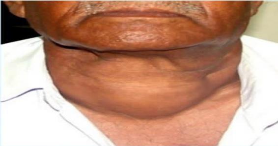
Q1: How do you clinically verify that this mass arises from the thyroid gland?
- Answer: Ask the patient to swallow sips of water; the thyroid gland moves upward on deglutition because it is enclosed within the pretracheal fascia attached to the thyroid and cricoid cartilages.
Q2: Mention FOUR specific physical examination steps performed from behind the patient.
- Answer:
- Palpate each thyroid lobe and isthmus systematically for size, consistency, and nodularity.
- Palpate for cervical lymphadenopathy (levels II–VI).
- Palpate lower border of goiter to rule out retrosternal extension.
- Auscultate over the superior thyroid poles for vascular systolic bruits.
Q3: What is the gold standard imaging modality?
- Answer: High-resolution Neck Ultrasound.
-OSPE Station 2: Parotid Gland Tumor / Pleomorphic Adenoma
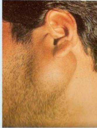
Q1: What is the most likely diagnosis for a slow-growing, painless mass below and in front of the earlobe?
- Answer: Pleomorphic Adenoma (benign mixed tumor of the parotid gland).
Q2: What cranial nerve must be meticulously examined?
- Answer: Cranial Nerve VII (Facial Nerve) (examine forehead wrinkling, eye closure, smiling, puffing cheeks). Facial weakness strongly suggests malignancy (e.g., adenoid cystic carcinoma).
Q3: What diagnostic investigation is indicated to confirm the nature of the lump?
- Answer: Ultrasound-guided FNAC and MRI/CT of the parotid gland. (Open biopsy is strictly contraindicated due to tumor seeding risk).
Q4: What is the standard surgical procedure?
- Answer: Superficial Parotidectomy with Facial Nerve Preservation.
-OSPE Station 3: Acute Parotid Abscess
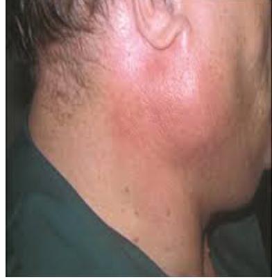
Q1: What is the diagnosis shown in this photograph?
- Answer: Acute Suppurative Parotitis / Parotid Abscess.
Q2: What is the most common causative organism?
- Answer: Staphylococcus aureus.
Q3: Mention TWO predisposing risk factors.
- Answer: Dehydration, poor oral hygiene, elderly debilitated postoperative patients, or anticholinergic drugs.
Q4: Outline the treatment plan.
- Answer: IV hydration, IV anti-staphylococcal antibiotics, sialogogues (lemon drops) to stimulate saliva, and surgical incision and drainage (horizontal incision parallel to facial nerve branches) if fluctuant.
-OSPE Station 4: Thyroid Symptoms & Complications of Total Thyroidectomy
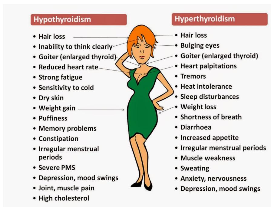
A 40-year-old female undergoes total thyroidectomy for multinodular goiter.
Q1: What are the TWO cardinal early postoperative life-threatening complications?
- Answer:
- Reactionary Neck Hematoma: Compresses trachea causing stridor and acute airway obstruction.
- Bilateral Recurrent Laryngeal Nerve (RLN) Injury: Causes bilateral vocal cord adduction and acute asphyxia.
Q2: How do you immediately manage an expanding tense neck hematoma at the bedside?
- Answer: Immediately open all wound skin sutures/clips at the bedside, evacuate the hematoma to relieve tracheal compression, and return the patient to the OR for formal hemostasis.
Q3: What complication presents with perioral tingling and carpopedal spasm on post-op day 2?
- Answer: Hypocalcemia secondary to accidental removal or devascularization of the parathyroid glands (positive Chvostek’s and Trousseau’s signs; treat with IV calcium gluconate).
Neurosurgery
Brain Tumor
Clinical Scenarios (CS)
Case 1: Intracranial Space-Occupying Lesion and Motor System Examination
A 54-year-old male presents with morning headaches, projectile vomiting, and progressive right-sided arm and leg weakness.
Q1: What triad indicates raised intracranial pressure (ICP)?
- Answer: Headache (worse in early morning/bending forward), projectile vomiting (without nausea), and papilledema on fundoscopy.
Q2: What FIVE components comprise a complete bedside Motor System Examination?
- Answer:
- Inspection: Muscle bulk, wasting, asymmetry, fasciculations, or involuntary movements.
- Muscle Tone: Passive flexion/extension (spasticity / clasp-knife vs flaccidity).
- Muscle Power: Graded on MRC scale () across major joints.
- Deep Tendon Reflexes (DTRs): Biceps, triceps, brachioradialis, knee, and ankle reflexes (hyperreflexia indicates UMN lesion).
- Plantar Response (Babinski sign): Upgoing great toe / fanning of toes indicates UMN lesion. (Coordination: Finger-nose test and heel-to-shin test).
Q3: What diagnostic imaging modality of choice should be ordered?
- Answer: Contrast-enhanced Brain MRI (or non-contrast head CT as initial emergency scan).
Case 2: Malignant Spinal Cord Compression (MSCC) from Spinal Metastases
A 62-year-old male with known prostate cancer presents with severe thoracic back pain, progressive lower limb weakness, sensory loss below the nipples (T4), and urinary retention.
Q1: What is the diagnosis?
- Answer: Malignant Spinal Cord Compression (MSCC).
Q2: What is the gold standard diagnostic investigation?
- Answer: Urgent Whole-Spine MRI (within 24 hours).
Q3: Outline immediate emergency management.
- Answer:
- Immediate high-dose IV Dexamethasone (16 mg IV bolus, then 8 mg BID) with gastric protection.
- Urgent neurosurgical / radiation oncology consultation for surgical decompression spine stabilization or emergency external beam radiotherapy.
- Foley catheter insertion for bladder drainage.
Case 3: Pituitary Adenoma / Cushing’s Disease (Endocrine Neurosurgery & Intracranial Mass)
A 38-year-old female presents with progressive weight gain, moon facies, buffalo hump, purple abdominal striae, amenorrhea, and bitemporal hemianopia.
Q1: What is the most likely diagnosis?
- Answer: Cushing’s Disease (ACTH-secreting Pituitary Adenoma).
Q2: What visual field defect is characteristically produced by sellar masses and why?
- Answer: Bitemporal Hemianopia; caused by suprasellar tumor extension upward compressing the crossing nasal retinal fibers at the Optic Chiasm.
Q3: Mention TWO initial screening tests to confirm hypercortisolemia.
- Answer:
- 24-hour urinary free cortisol (UFC).
- Overnight 1 mg Low-Dose Dexamethasone Suppression Test (LDDST) (or late-night salivary cortisol).
Q4: What is the definitive diagnostic imaging modality and primary surgical approach?
- Answer:
- Imaging: Pituitary MRI with IV Gadolinium.
- Surgery: Endoscopic Transsphenoidal Pituitary Resection (Transsphenoidal Hypophysectomy).
-OSPE Station 1: Intracranial Meningioma on Brain MRI
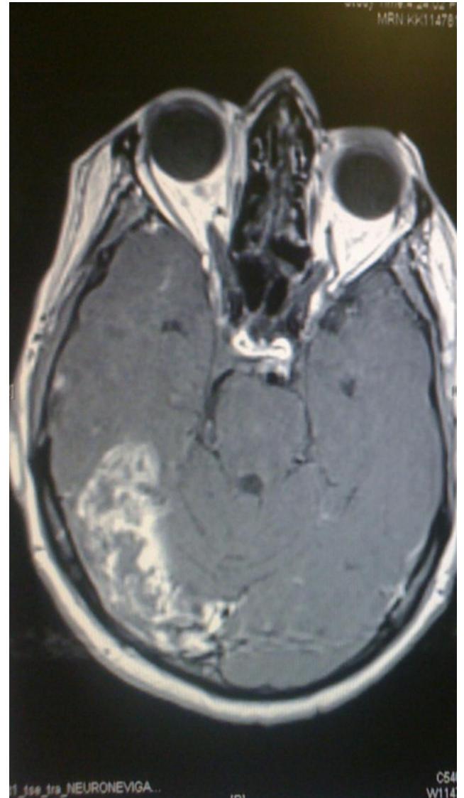
Q1: Identify the neuroimaging findings and state the diagnosis.
- Answer: Well-circumscribed, extra-axial, dural-based mass with intense homogeneous contrast enhancement and a “dural tail” sign; Diagnosis: Intracranial Meningioma.
Q2: What is the biological behavior of this lesion?
- Answer: Predominantly benign (WHO Grade I in ) with slow expansile growth compressing adjacent brain parenchyma.
Q3: What is the definitive treatment for symptomatic or growing tumors?
- Answer: Elective Surgical Resection (Simpson Grade I complete resection including dural attachment and involved bone) or stereotactic radiosurgery (Gamma Knife).
-OSPE Station 2: Ring-Enhancing Cerebral Lesion on Brain MRI
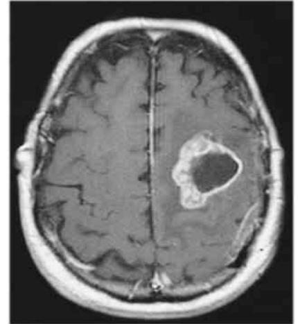
Q1: Describe the radiological appearance shown.
- Answer: An intra-axial cerebral lesion demonstrating central non-enhancing hypointense necrosis surrounded by a smooth/irregular rim of peripheral contrast enhancement with extensive surrounding vasogenic edema (Ring-enhancing lesion).
Q2: Mention FOUR differential diagnoses (mnemonic: MAGIC DR).
- Answer:
- Metastasis (cerebral metastasis from lung, breast, melanoma, renal cancer).
- Abscess (pyogenic brain abscess).
- Glioblastoma (GBM) (high-grade glioma).
- Infarct (subacute ischemic stroke).
- Contusion / Hematoma (resolving).
- Toxoplasmosis / TB tuberculoma (especially in immunocompromised/HIV patients).
Q3: What medical therapy is immediately initiated to reduce cerebral edema?
- Answer: High-dose IV Dexamethasone (corticosteroid) IV Mannitol or Hypertonic Saline.
Brain abscess
(Cross-referenced with ring-enhancing lesions: Classic clinical triad [headache, fever, focal neurological deficit], etiologies [contiguous spread from chronic otitis media/sinusitis, hematogenous spread from cyanotic heart disease or bronchiectasis, post-neurosurgical/trauma], and management: surgical burr-hole aspiration or excision + prolonged targeted IV antibiotics for 4–6 weeks).
Head injury
Clinical Scenarios (CS)
Case 1: Traumatic Acute Epidural (Extradural) Hematoma with Lucid Interval
A 20-year-old male sustained a blow to the pterion during a soccer match. He lost consciousness briefly, regained full consciousness, and 3 hours later developed severe headache, vomiting, progressive drowsiness, and a dilated unreactive left pupil.
Q1: What is the classic clinical course described?
- Answer: The “Lucid Interval” (temporary recovery of consciousness followed by rapid secondary neurological deterioration).
Q2: What is the most likely diagnosis?
- Answer: Acute Epidural (Extradural) Hematoma (EDH).
Q3: What blood vessel is torn, and what anatomical landmark is injured?
- Answer: Middle Meningeal Artery (or its anterior branch) deep to the Pterion (thin skull junction of frontal, parietal, temporal, and sphenoid bones).
Q4: What does the dilated, unreactive ipsilateral pupil signify?
- Answer: Uncal (Transtentorial) Herniation compressing the ipsilateral Cranial Nerve III (Oculomotor Nerve).
Q5: What emergency surgical intervention is indicated?
- Answer: Emergency Craniotomy and surgical evacuation of the hematoma with bipolar cauterization / ligation of the bleeding vessel.
-OSPE Station 1: Epidural Hematoma on Non-Contrast Head CT
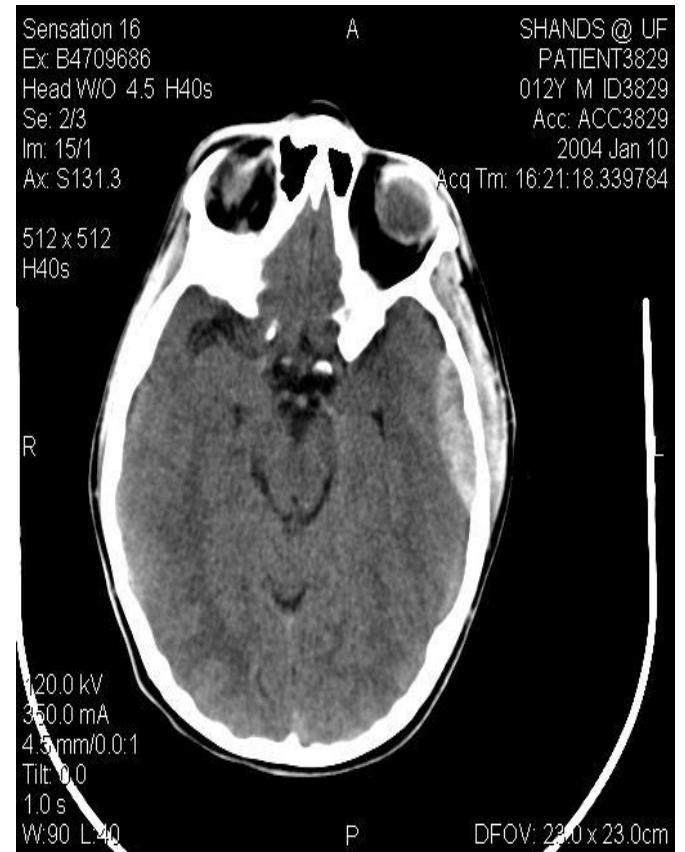
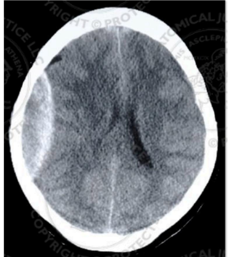
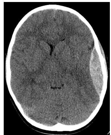
Q1: Identify the diagnostic finding on this CT scan.
- Answer: High-density (hyperdense) biconvex (lenticular), smoothly demarcated extra-axial collection that is limited by cranial suture lines.
Q2: What is the diagnosis?
- Answer: Acute Epidural Hematoma (EDH).
Q3: Why does this lesion not cross cranial suture lines?
- Answer: The dura mater is tightly anchored to the skull at the cranial suture lines, confining the collection.
-OSPE Station 2: Subdural Hematoma on Head CT
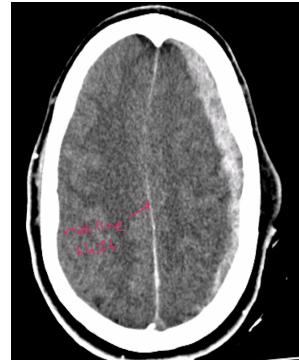
Q1: Identify the diagnostic finding on this CT scan.
- Answer: Hyperdense (or isodense/hypodense) crescentic (concave-convex) extra-axial collection that crosses cranial suture lines along the cerebral hemisphere.
Q2: What is the diagnosis?
- Answer: Subdural Hematoma (SDH).
Q3: What is the anatomical source of bleeding?
- Answer: Tearing of the Bridging Cortical Veins traversing the subdural space into the superior sagittal sinus.
Q4: What population is at highest risk for chronic subdural hematoma?
- Answer: Elderly patients and chronic alcoholics (due to cerebral atrophy stretching bridging veins, triggered by minor head trauma).
Breast
Breast Disease
Clinical Scenarios (CS)
Case 1: Triple Assessment and Management of Invasive Breast Carcinoma
A 56-year-old postmenopausal female presents with a painless, hard, irregular, non-tender lump in the upper outer quadrant of her left breast with tethering to the overlying skin and palpable ipsilateral axillary lymphadenopathy.
Q1: What are the THREE components of Triple Assessment for breast lumps?
- Answer:
- Clinical Examination: Detailed bilateral breast and regional nodal palpation.
- Radiological Imaging: Bilateral Mammography and targeted Breast Ultrasound (breast MRI in select cases).
- Pathological / Histological Biopsy: Core Needle Biopsy (CNB) (preferred over FNAC for tissue architecture, ER/PR/HER2 receptor status).
Q2: What is the most likely diagnosis?
- Answer: Invasive Breast Carcinoma (most commonly Invasive Ductal Carcinoma [IDC]).
Q3: What receptor status MUST be evaluated on core biopsy?
- Answer: Estrogen Receptor (ER), Progesterone Receptor (PR), HER2/neu receptor status, and Ki-67 proliferation index.
Q4: Mention TWO surgical options for local tumor control.
- Answer:
- Breast-Conserving Surgery (BCS / Wide Local Excision / Lumpectomy) with clear margins followed by mandatory adjuvant whole-breast radiotherapy.
- Mastectomy (Simple / Total Mastectomy immediate reconstruction).
Q5: How is the clinically node-negative axilla staged versus clinically node-positive axilla?
- Answer:
- Clinically node-negative: Sentinel Lymph Node Biopsy (SLNB) using radiotracer and/or blue dye.
- Clinically node-positive: Axillary Lymph Node Dissection (ALND) (Levels I and II).
Case 2: Benign Breast Disease (Fibroadenoma)
A 22-year-old female presents with a painless, highly mobile, firm lump in the upper outer quadrant of her left breast.
Q1: What is the most likely diagnosis?
- Answer: Fibroadenoma of the breast (“breast mouse”).
Q2: What is the single best initial imaging modality in a woman under 30–35 years?
- Answer: Targeted Breast Ultrasound (dense glandular tissue reduces mammographic sensitivity; ultrasound clearly distinguishes solid fibroadenoma from cystic lesions).
Q3: What biopsy modality confirms the diagnosis?
- Answer: Core Needle Biopsy (CNB) (or FNAC).
Q4: Mention TWO indications for surgical excision of a fibroadenoma.
- Answer:
- Rapid growth or large size ( / giant fibroadenoma, suspicion of phyllodes tumor).
- Patient anxiety / request or cosmetic concern (otherwise conservative observation is standard).
-OSPE Station 1: Diagnostic Mammography of Breast Carcinoma
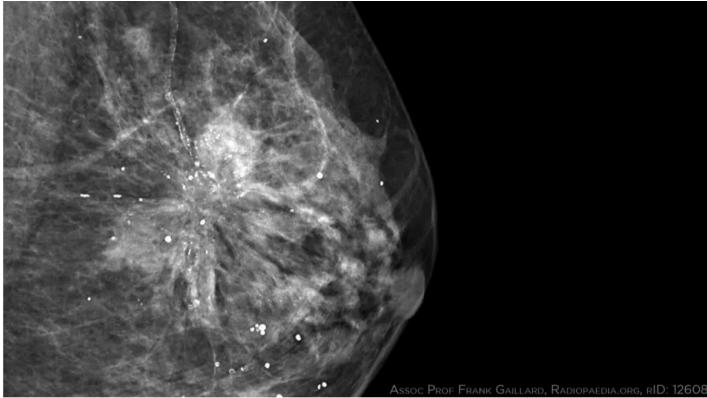
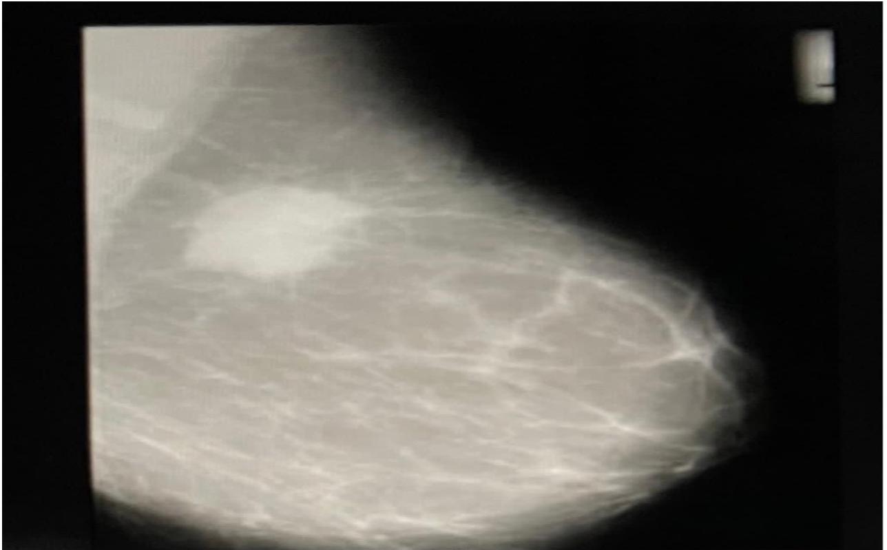
Q1: What imaging modality is shown?
- Answer: Diagnostic Bilateral Mammography (Craniocaudal [CC] and Mediolateral Oblique [MLO] views).
Q2: What are the cardinal mammographic signs of breast malignancy?
- Answer:
- Spiculated, high-density, irregular soft tissue mass.
- Pleomorphic, clustered microcalcifications.
- Architectural distortion or skin thickening / nipple retraction.
Q3: What classification system reports mammographic findings?
- Answer: BI-RADS (Breast Imaging-Reporting and Data System) (Category 0 to 6).
-OSPE Station 2: Peau d’Orange Sign in Advanced / Inflammatory Breast Cancer
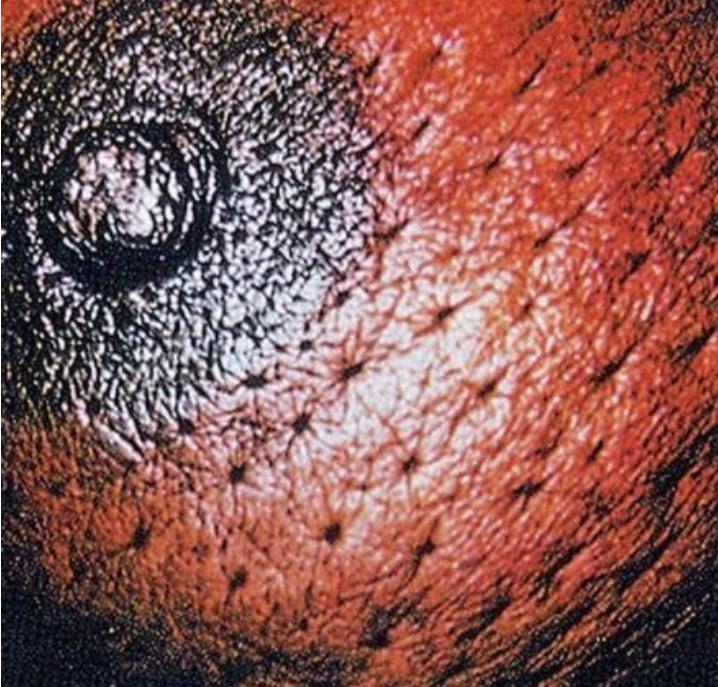
Q1: What physical sign is demonstrated on the breast skin?
- Answer: Peau d’Orange (“orange peel” skin appearance).
Q2: What is the underlying pathophysiological mechanism?
- Answer: Malignant tumor emboli causing obstruction of subdermal cutaneous lymphatics, producing localized lymphedema with tethering at the dermal hair follicles and sweat glands.
Q3: What does this sign signify regarding tumor staging?
- Answer: Indicates locally advanced breast cancer (at least T4b; or T4d if Inflammatory Breast Cancer).
-OSPE Station 3: Acute Lactational Breast Abscess
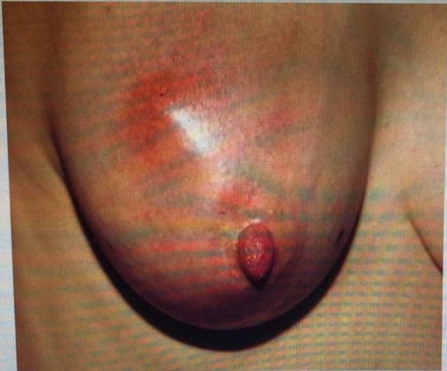
Q1: What is the diagnosis shown in this lactating female?
- Answer: Acute Lactational Breast Abscess.
Q2: What is the most common causative organism?
- Answer: Staphylococcus aureus (including MRSA).
Q3: Outline the management plan.
- Answer:
- Drainage: Ultrasound-guided needle aspiration (first-line) or surgical incision and drainage (if large/refractory).
- Systemic oral/IV anti-staphylococcal antibiotics (e.g., Flucloxacillin or Clindamycin).
- Continue breastfeeding or expression/pumping from the affected breast (milk is safe and prevents further engorgement).
Thoracic
Management of patients with lung lesions
-OSPE Station 1: Solitary Pulmonary Mass on Plain Chest Radiograph (Lung Cancer)
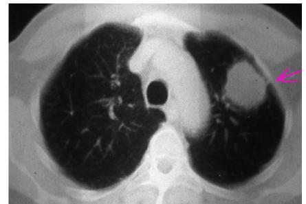
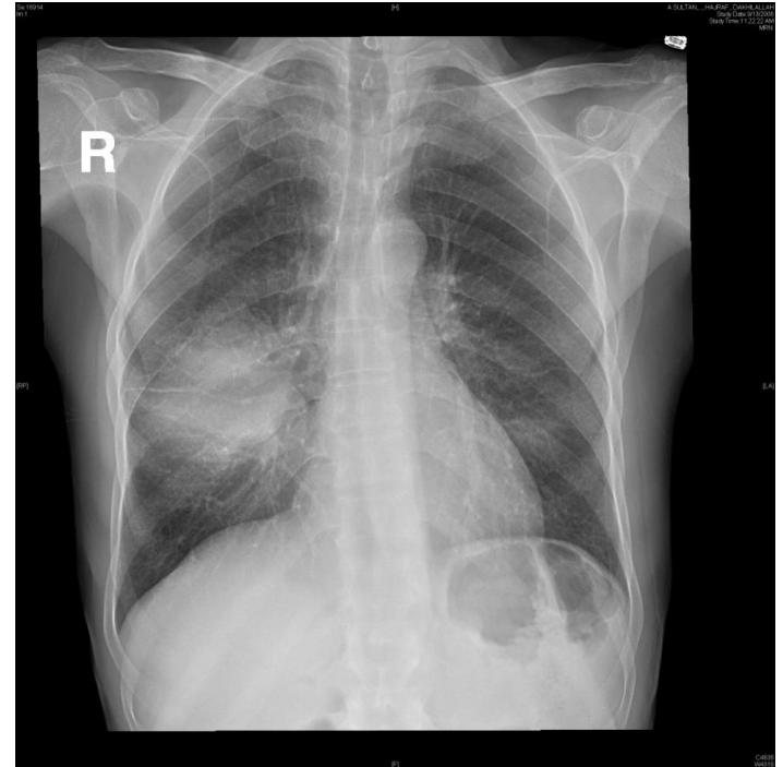
Q1: Describe the radiological finding on this chest X-ray.
- Answer: A solitary, round, dense, well-demarcated opacity/mass with irregular/spiculated borders in the lung field (Solitary Pulmonary Nodule / Mass).
Q2: What is the most probable diagnosis in an elderly smoker?
- Answer: Bronchogenic Carcinoma (Primary Lung Cancer).
Q3: Mention TWO initial diagnostic and staging investigations.
- Answer:
- Contrast-enhanced CT of Chest and Upper Abdomen (including liver and adrenal glands).
- Bronchoscopy with endobronchial biopsy / brushings (or CT-guided percutaneous transthoracic needle biopsy).
Q4: Mention TWO common histological types of non-small cell lung cancer (NSCLC).
- Answer:
- Adenocarcinoma (most common, typically peripheral).
- Squamous Cell Carcinoma (typically central, cavitating).
Upper GI & Abdominal Surgery
Esophageal Diseases
-OSPE Station 1: Achalasia Cardia on Barium Swallow
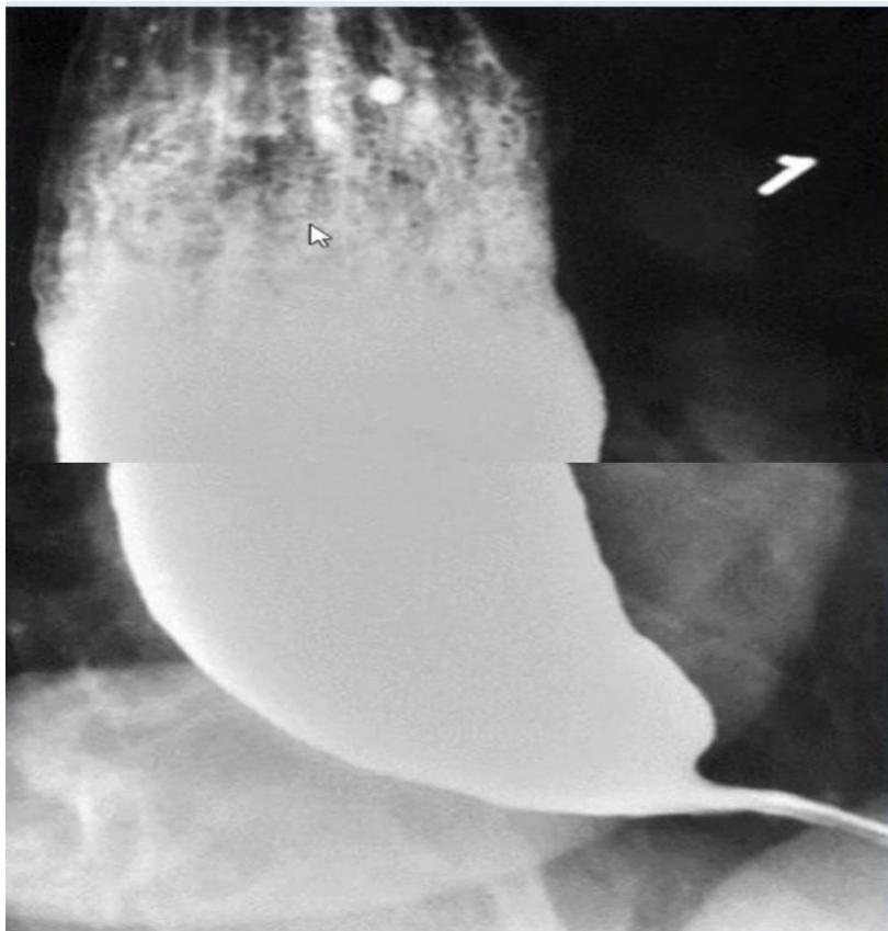
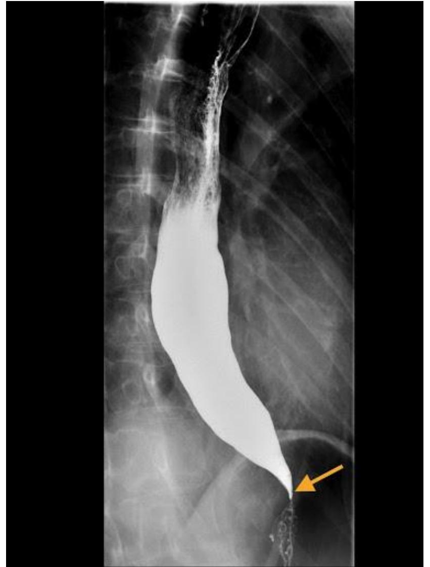
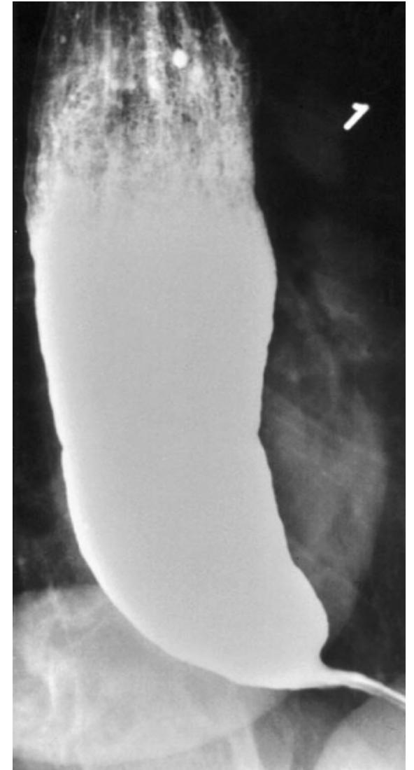
Q1: Name this radiological investigation and describe the diagnostic finding.
- Answer: Barium Swallow (Esophagram); diagnostic finding: Marked dilatation of the thoracic esophagus with smooth, symmetric, tapered narrowing at the gastroesophageal junction producing the classic “Bird’s Beak” (or “Rat’s Tail”) appearance.
Q2: What is the definitive diagnosis?
- Answer: Achalasia Cardia (primary esophageal motility disorder).
Q3: What specialized physiological investigation is the gold standard?
- Answer: High-Resolution Esophageal Manometry (HREM) (demonstrates complete aperistalsis and incomplete/absent LES relaxation).
Q4: Mention TWO definitive treatment modalities.
- Answer:
- Laparoscopic Heller Myotomy (surgical division of lower esophageal circular muscle with partial Dor/Toupet fundoplication).
- Pneumatic Balloon Dilatation (or Per-Oral Endoscopic Myotomy [POEM] / Botulinum toxin injection).
-OSPE Station 2: Esophageal Carcinoma on Barium Swallow
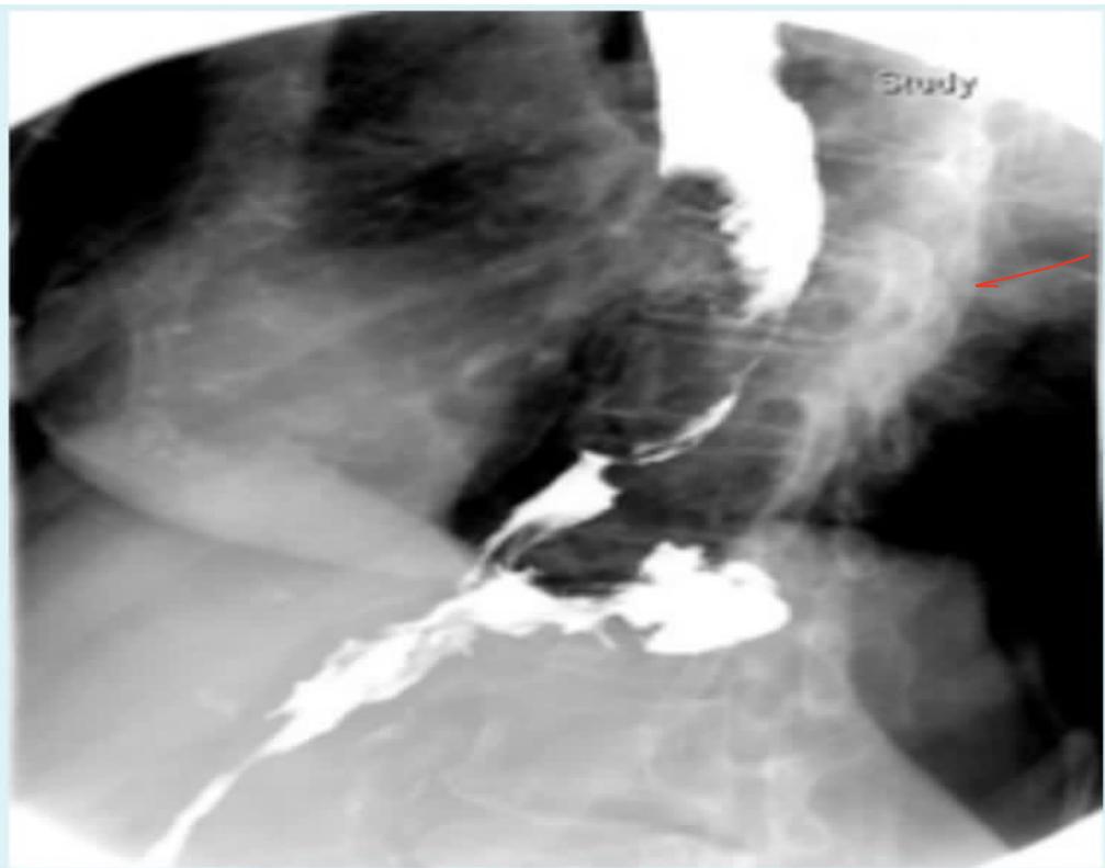
Q1: Describe the radiological finding on this barium swallow.
- Answer: Irregular, asymmetric, shouldered narrowing / filling defect with mucosal destruction in the mid-to-distal esophagus (“Apple-Core” sign / malignant stricture).
Q2: What is the most likely diagnosis in an elderly patient with progressive dysphagia?
- Answer: Esophageal Carcinoma (Esophageal Cancer).
Q3: What is the gold standard diagnostic investigation to establish the diagnosis?
- Answer: Upper Endoscopy (OGD) with multiple targeted biopsies and brush cytology.
Q4: Mention TWO staging modalities.
- Answer:
- Contrast-enhanced CT of Chest and Abdomen (for distant metastases).
- Endoscopic Ultrasound (EUS) (gold standard for local T and N staging). (PET-CT for systemic staging).
Complications of Abdominal Surgery
Clinical Scenarios (CS)
Case 1: Postoperative Intra-Abdominal Abscess and Wound Dehiscence
A 58-year-old female was readmitted 7 days after elective open abdominal surgery complaining of high swinging fevers, tachycardia, and a painful, tense, fluctuant abdominal mass measuring deep to the surgical scar.
Q1: What is the most likely diagnosis?
- Answer: Postoperative Intra-Abdominal Abscess / Deep Surgical Site Infection (with impending wound dehiscence).
Q2: Mention TWO differential diagnoses.
- Answer:
- Wound hematoma or infected seroma.
- Incisional hernia / acute fascial dehiscence (burst abdomen).
Q3: What imaging modality is the gold standard to locate and evaluate this fluid collection?
- Answer: Contrast-Enhanced CT (CECT) of the Abdomen and Pelvis.
Q4: What is the primary minimally invasive management of choice?
- Answer: Percutaneous Image-Guided (US or CT) Catheter Drainage under local anesthesia, with broad-spectrum IV antibiotics and culture of aspirated fluid.
Obstructive jaundice
Clinical Scenarios (CS)
Case 1: Clinical Evaluation of Extrahepatic Biliary Obstruction
A 55-year-old male presents with painless progressive yellowish discoloration of the eyes and skin, dark urine, pale/clay-colored stools, and generalized pruritus.
Q1: Mention FIVE relevant questions regarding the history of present illness.
- Answer:
- Any abdominal pain (biliary colic vs painless jaundice).
- Change in urine color (tea-colored/dark) or stool color (pale/acholic/clay-colored).
- Generalized itching / pruritus.
- Constitutional symptoms (anorexia, early satiety, unintentional weight loss).
- History of gallstones, alcohol intake, viral hepatitis exposure, or blood transfusions.
Q2: Mention FOUR differential diagnoses.
- Answer:
- Choledocholithiasis (common bile duct stones).
- Carcinoma of the head of the pancreas.
- Cholangiocarcinoma (bile duct cancer / Klatskin tumor).
- Periampullary carcinoma (or benign biliary stricture).
Q3: What FIVE initial laboratory investigations should be requested?
- Answer:
- Total and Direct (conjugated) Bilirubin (predominantly direct hyperbilirubinemia).
- Alkaline Phosphatase (ALP) and Gamma-Glutamyl Transferase (GGT) (markedly elevated).
- AST and ALT (mild-to-moderate elevation).
- Coagulation profile: PT/INR and aPTT (prolonged PT corrected by Vitamin K).
- CBC and Renal Function Tests.
Q4: Mention TWO initial imaging modalities to establish the cause.
- Answer:
- Abdominal Ultrasound (initial non-invasive imaging: demonstrates biliary duct dilatation and gallstones).
- Magnetic Resonance Cholangiopancreatography (MRCP) or Contrast-Enhanced CT abdomen.
Case 2: Carcinoma of the Head of the Pancreas (Whipple Procedure)
A 64-year-old male presents with painless progressive obstructive jaundice, significant weight loss, anorexia, and a palpable, non-tender gallbladder on physical examination.
Q1: What is the clinical sign and what does it indicate (Courvoisier’s Law)?
- Answer: Courvoisier’s Sign; in the presence of painless jaundice, an enlarged, palpable, non-tender gallbladder is unlikely to be due to gallstones and strongly points to a malignant obstruction of the distal biliary tree (e.g., Carcinoma of the Head of the Pancreas).
Q2: What is the most likely diagnosis?
- Answer: Adenocarcinoma of the Head of the Pancreas.
Q3: What tumor marker is commonly elevated in pancreatic carcinoma?
- Answer: CA 19-9 (Carbohydrate Antigen 19-9).
Q4: What is the gold standard imaging modality for diagnosis and resectability staging?
- Answer: Triple-phase Pancreatic Protocol Contrast-Enhanced CT (CECT) of the abdomen.
Q5: What is the standard curative surgical resection?
- Answer: Pancreaticoduodenectomy (Whipple Procedure) (resection of pancreatic head, duodenum, gallbladder, distal bile duct, and partial gastrectomy, reconstructed via pancreaticojejunostomy, hepaticojejunostomy, and gastrojejunostomy).
-OSPE Station 1: Clinical Recognition of Scleral Icterus / Jaundice
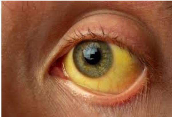
Q1: What clinical sign is demonstrated in this examination?
- Answer: Scleral Icterus (Jaundice).
Q2: What anatomical tissue has the highest affinity for bilirubin?
- Answer: The Sclera (due to its high elastin content; clinically detectable when serum total bilirubin exceeds []).
Q3: Name the THREE pathophysiological classifications of jaundice.
- Answer:
- Pre-hepatic (Hemolytic) (unconjugated).
- Hepatic (Hepatocellular) (mixed).
- Post-hepatic (Obstructive / Cholestatic) (conjugated).
-OSPE Station 2: Endoscopic Retrograde Cholangiopancreatography (ERCP) in Choledocholithiasis
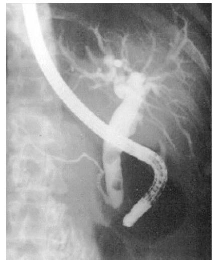
Q1: Identify this radiological and endoscopic procedure.
- Answer: Endoscopic Retrograde Cholangiopancreatography (ERCP).
Q2: Describe the diagnostic finding.
- Answer: Dilated Common Bile Duct (CBD) containing single or multiple well-defined radiolucent filling defects (Choledocholithiasis / CBD stones).
Q3: Mention TWO therapeutic interventions performed during this procedure.
- Answer:
- Endoscopic Sphincterotomy (Papillotomy) of the ampulla of Vater.
- Stone extraction using a Dormia basket or Fogarty balloon catheter (or biliary stent insertion).
Q4: Mention TWO recognized complications of this procedure.
- Answer:
- Acute Post-ERCP Pancreatitis (most common, ).
- Duodenal perforation or post-sphincterotomy hemorrhage (or ascending cholangitis).
Urology
Evaluation of the urologic patient
Clinical Scenarios (CS)
Case 1: Lower Urinary Tract Symptoms (LUTS) and Prostate Evaluation in an Elderly Male
A 68-year-old male presents with a 1-year history of progressive urinary hesitancy, weak stream, intermittency, nocturia (4 times/night), and a sensation of incomplete bladder emptying.
Q1: Classify these symptoms into Voiding (Obstructive) versus Storage (Irritative).
- Answer:
- Voiding (Obstructive): Hesitancy, weak urinary stream, straining, terminal dribbling, intermittency.
- Storage (Irritative): Frequency, nocturia, urgency, urge incontinence.
Q2: What essential physical examination must be performed?
- Answer: Digital Rectal Examination (DRE) (evaluates prostate size, symmetry, consistency, sulcus patency, tenderness, and presence of nodules).
Q3: Contrast the DRE findings of Benign Prostatic Hyperplasia (BPH) versus Prostate Cancer.
- Answer:
- BPH: Smooth, symmetrically enlarged, firm/elastic, rubbery consistency, well-defined borders, preserved median sulcus, non-tender, freely movable rectal mucosa.
- Prostate Cancer: Asymmetrical, hard (“stony hard”), irregular, nodular prostate, obliterated median sulcus, fixed overlying rectal mucosa.
Q4: Mention TWO first-line medical drug classes used to manage BPH.
- Answer:
- Alpha-1 Adrenergic Receptor Antagonists (e.g., Tamsulosin, Alfuzosin) (rapid relief of dynamic smooth muscle tone).
- 5-Alpha Reductase Inhibitors (e.g., Finasteride, Dutasteride) (blocks DHT synthesis, shrinks static prostate volume over 6 months).
Q5: What is the gold standard surgical intervention for refractory BPH?
- Answer: Transurethral Resection of the Prostate (TURP) (or Holmium Laser Enucleation of the Prostate [HoLEP]).
Upper UT
-OSPE Station 1: Renal Cell Carcinoma on Contrast-Enhanced Abdominal CT
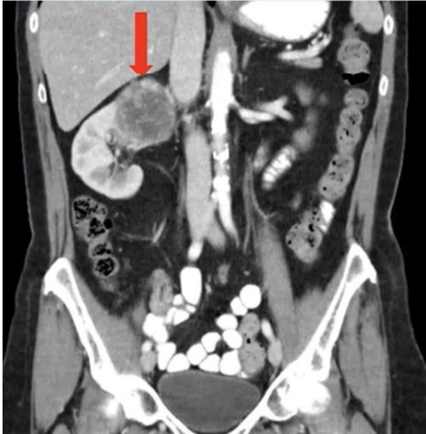
Q1: Identify the imaging modality and describe the diagnostic finding.
- Answer: Coronal Contrast-Enhanced Abdominal CT (CECT); diagnostic finding: Large, heterogeneous, hypervascular, exophytic enhancing mass arising from the right kidney with central necrosis (Renal Cell Carcinoma [RCC]).
Q2: What is the classic clinical triad of RCC, and how frequently does it occur?
- Answer: Hematuria, Flank Pain, and Palpable Flank Mass (present simultaneously in only of patients, indicating advanced disease).
Q3: What is the most common histological subtype?
- Answer: Clear Cell Renal Cell Carcinoma (75–80%).
Q4: Mention the primary surgical treatment for localized disease.
- Answer: Radical Nephrectomy (removal of kidney, Gerota’s fascia, perinephric fat, and ipsilateral adrenal gland) OR Partial Nephrectomy (Nephron-sparing surgery) for small renal masses ( / T1a).
Lower UT
-OSPE Station 1: Urinary Bladder Tumor on Cystoscopy
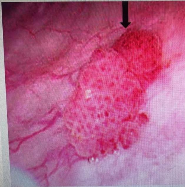
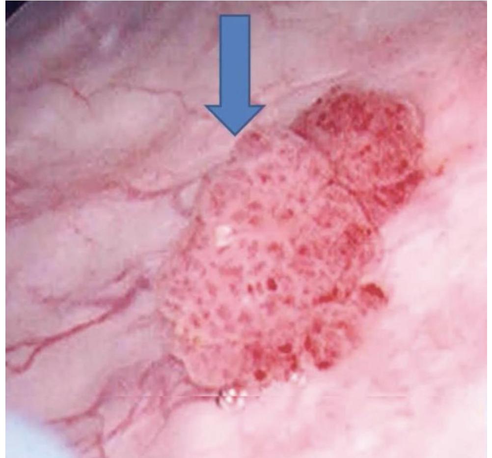
Q1: In a 60-year-old male presenting with painless gross hematuria, what is the finding and diagnosis?
- Answer: Papillary, frond-like, exophytic mucosal mass inside the urinary bladder lumen; Diagnosis: Urinary Bladder Cancer (Urothelial / Transitional Cell Carcinoma).
Q2: What is the single most important lifestyle risk factor?
- Answer: Cigarette Smoking (responsible for of cases; aromatic amines/industrial dyes is occupational cause).
Q3: What initial diagnostic and staging operation must be performed?
- Answer: Transurethral Resection of Bladder Tumor (TURBT) (provides complete tumor resection and depth of invasion assessment: lamina propria vs detrusor muscularis propria).
Q4: Contrast definitive treatment for Non-Muscle Invasive versus Muscle-Invasive Bladder Cancer.
- Answer:
- Non-Muscle Invasive (Ta, T1): Complete TURBT followed by Intravesical BCG or Chemotherapy (Mitomycin C) instillation.
- Muscle-Invasive (T2–T4): Radical Cystectomy with Pelvic Lymph Node Dissection and urinary diversion (Ileal Conduit / Neobladder) neoadjuvant chemotherapy.
External Genitalia
-OSPE Station 1: Acute Testicular Torsion
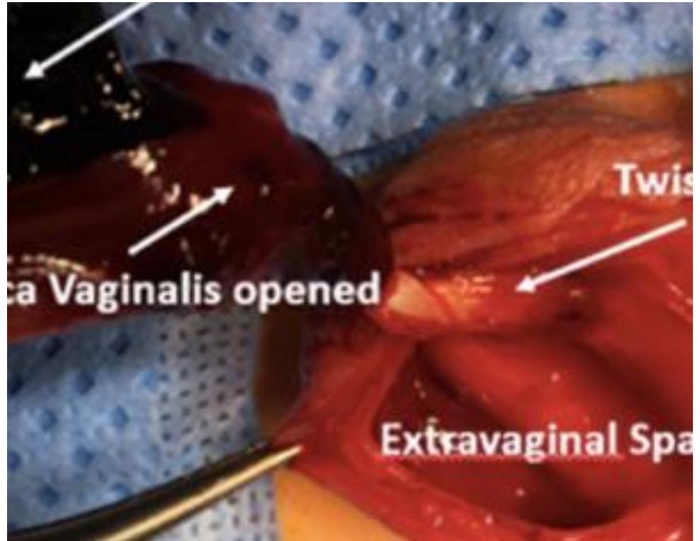
Q1: What is the intraoperative surgical diagnosis shown?
- Answer: Testicular Torsion (torsion of the spermatic cord with acute testicular ischemia/infarction).
Q2: What anatomical anomaly classically predisposes to this condition?
- Answer: “Bell-Clapper” Deformity (high insertion of the tunica vaginalis allowing the testis to rotate freely horizontally like a clapper inside a bell).
Q3: What are the cardinal clinical examination signs?
- Answer:
- High-riding, transverse-lying, exquisitely tender, swollen testis.
- Absent Cremasteric Reflex on the affected side.
- Negative Prehn’s sign (elevation of the scrotum does NOT relieve pain; relieves pain in epididymitis).
Q4: What is the critical time window for testicular salvage?
- Answer: Within 6 hours of pain onset ( salvage rate; drops to after 24 hours).
Q5: Outline the emergency surgical management.
- Answer: Emergency surgical exploration: detorsion of the cord, assessment of testicular viability (warm pads/oxygen), bilateral orchidopexy (fixation of both testes to prevent contralateral torsion); orchidectomy if frankly necrotic.
-OSPE Station 2: Scrotal Ultrasound in Acute Scrotal Pain
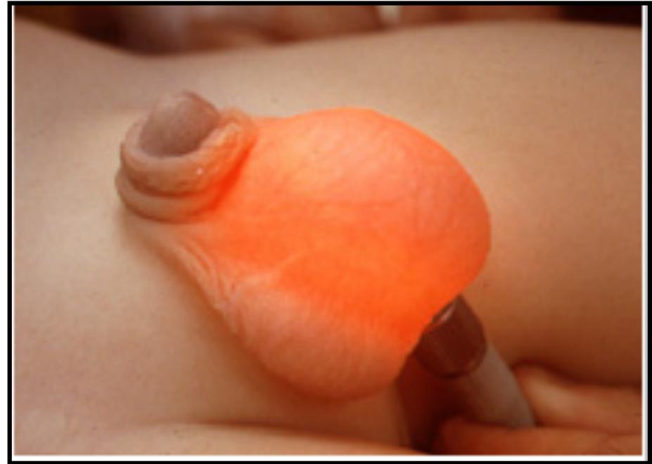
Q1: What is the role of Color Doppler Ultrasound in the evaluation of acute scrotum?
- Answer: Evaluates intratesticular blood flow:
- Testicular Torsion: Marked reduction or complete absence of intratesticular vascular flow.
- Epididymo-orchitis: Hyperemia with markedly increased blood flow.
Q2: What cardinal surgical rule must be observed regarding ultrasound in suspected torsion?
- Answer: Ultrasound must NEVER delay emergency surgical exploration; if testicular torsion is clinically suspected, proceed immediately to the operating room.
Vascular
Arterial Diseases
Clinical Scenarios (CS)
Case 1: Critical Limb Ischemia and Arterial Ischemic Foot Ulcer
A 65-year-old diabetic smoker presents with a painful, non-healing ulcer over the dorsum of his right foot and severe rest pain waking him at night, relieved by hanging the leg over the edge of the bed.
Q1: What FOUR relevant questions should you ask regarding the presenting complaint?
- Answer:
- Characteristics of pain: intermittent claudication distance, onset of rest pain.
- Ulcer duration, progression, and history of minor trauma.
- Cardiovascular risk factors: smoking history, diabetes, hypertension, dyslipidemia.
- History of coronary artery disease, stroke, or previous vascular interventions.
Q2: What FIVE clinical examination steps should you perform on palpation of this limb?
- Answer:
- Temperature gradient (coolness of distal foot compared to contralateral limb).
- Capillary refill time (prolonged ).
- Palpation of peripheral arterial pulses: femoral, popliteal, posterior tibial, and dorsalis pedis.
- Sensory examination (light touch, vibration, pinprick, 10g monofilament for diabetic neuropathy).
- Buerger’s test (vascular angle of pallor on elevation and rubor on dependency).
Q3: Mention THREE non-laboratory specialized investigations.
- Answer:
- Ankle-Brachial Index (ABI) (critical ischemia: or absolute ankle pressure ).
- Arterial Duplex Ultrasound of lower extremities.
- CT Angiography (CTA) or MR Angiography (MRA) / Digital Subtraction Angiography (DSA).
Q4: Outline the comprehensive management.
- Answer:
- Risk factor modification: smoking cessation, antiplatelet therapy (Aspirin/Clopidogrel), high-intensity statin, strict glycemic and BP control.
- Wound care: local non-weight bearing debridement, infection control with broad-spectrum antibiotics.
- Revascularization: Endovascular intervention (balloon angioplasty stenting) OR Surgical bypass (e.g., femoropopliteal or femorodistal bypass with saphenous vein).
-OSPE Station 1: Arterial Ischemic Ulcer of the Great Toe
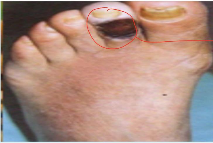
Q1: Describe the clinical features of an Arterial Ischemic Ulcer.
- Answer:
- Location: Distal extremities (tips of toes, heel, over bony pressure points, lateral malleolus).
- Base: Pale, dry, “punched-out” well-demarcated edges, necrotic/gray base with little or no granulation tissue.
- Surrounding skin: Shiny, hairless, atrophic, cool to touch with dystrophic thickened nails.
- Pulses: Absent or severely diminished distal pulses; accompanied by severe ischemic pain worse at night.
Q2: Contrast the features of an Arterial Ulcer versus a Venous Stasis Ulcer.
- Answer:
- Arterial Ulcer: Painful, relieved by dependency; distal toes/pressure points; “punched-out” dry base; cold skin, absent pulses; no edema.
- Venous Ulcer: Mildly painful, relieved by elevation; gaiter area (medial malleolus); irregular shallow borders, pink/sloughy exuding base; warm skin, normal pulses, stasis dermatitis, hemosiderin pigmentation, and chronic edema.
-OSPE Station 2: Diabetic Foot Ulceration and Neuro-Ischemic Pathogenesis
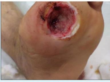
Q1: What are the TWO primary pathogenic factors contributing to this condition?
- Answer:
- Diabetic Peripheral Neuropathy (loss of protective sensation leading to repetitive unnoticed mechanical trauma, autonomic neuropathy causing dry cracked skin).
- Peripheral Arterial Disease (Macro- and Microvascular Ischemia) (impaired perfusion and delayed wound healing).
Q2: What THREE physical examination steps should be performed on this foot?
- Answer:
- Neurological sensory testing with a 10g Semmes-Weinstein monofilament and tuning fork.
- Palpation of dorsalis pedis and posterior tibial arterial pulses.
- Probe-to-bone test (if positive, highly predictive of underlying osteomyelitis).
Q3: Mention TWO initial imaging investigations.
- Answer:
- Plain Radiograph (X-ray) of the Foot (detects osteomyelitis, cortical erosion, soft tissue gas, or Charcot arthropathy).
- Arterial Duplex Ultrasound (evaluates vascular run-off).
Q4: Mention FIVE important steps in initial treatment.
- Answer:
- Strict non-weight-bearing / pressure offloading (specialized footwear/cast).
- Surgical debridement of necrotic tissue and callous.
- Deep tissue culture (avoid superficial swabs) and targeted systemic antibiotics.
- Optimized glycemic control (insulin therapy).
- Vascular assessment and revascularization if ischemia is present.
Venous Lymphatic Diseases
Clinical Scenarios (CS)
Case 1: Clinical Approach to Unilateral Lower Extremity Swelling
A 50-year-old female presents with acute swelling of her entire left lower limb of 3 days duration.
Q1: Mention FOUR questions to ask in history to differentiate potential causes.
- Answer:
- Onset and duration of swelling (acute sudden vs chronic insidious).
- Associated pain, redness, or warmth in the calf/thigh.
- Risk factors for DVT: recent surgery, prolonged immobilization, travel, malignancy, oral contraceptive use.
- Associated chest pain, dyspnea, or hemoptysis (screening for Pulmonary Embolism).
Q2: Mention FOUR clinical differential diagnoses for unilateral lower extremity swelling.
- Answer:
- Deep Vein Thrombosis (DVT).
- Acute Cellulitis.
- Ruptured Baker’s (Popliteal) Cyst.
- Lymphedema (or superficial thrombophlebitis).
Q3: What is the diagnostic imaging investigation of choice?
- Answer: Venous Compression Duplex Ultrasound of the lower extremity (loss of vein compressibility and absent respiratory phasicity confirms DVT).
-OSPE Station 1: Lower Extremity Varicose Veins
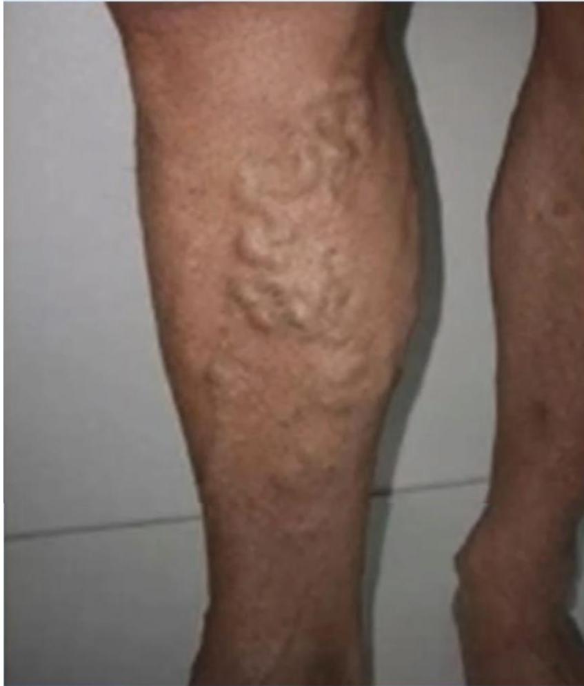
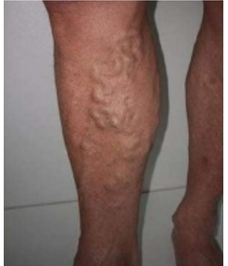
Q1: Describe the physical findings shown in these photographs.
- Answer: Dilated, elongated, tortuous superficial subcutaneous veins raised above the skin surface along the medial aspect of the leg and calf in the Great Saphenous Vein (GSV) distribution, accompanied by stasis hyperpigmentation.
Q2: What is the primary diagnostic imaging investigation?
- Answer: Venous Duplex Ultrasound of the lower extremities in the standing position (maps incompetent saphenofemoral/saphenopopliteal junctions and perforators, rules out DVT).
Q3: Mention FOUR recognized complications of chronic untreated varicose veins.
- Answer:
- Venous stasis dermatitis and eczema.
- Lipodermatosclerosis (“inverted champagne bottle” leg).
- Venous stasis ulceration (medial malleolus).
- Superficial thrombophlebitis (or bleeding from ruptured varices).
Q4: Mention TWO conservative and TWO modern interventional treatment options.
- Answer:
- Conservative: Graduated compression stockings (Class 2), leg elevation, avoidance of prolonged standing.
- Interventional: Endovenous Thermal Ablation (Radiofrequency Ablation [RFA] or Endovenous Laser Ablation [EVLA]), Ultrasound-guided foam sclerotherapy, or open surgical saphenofemoral ligation and stripping.
-OSPE Station 2: Venous Stasis Ulceration
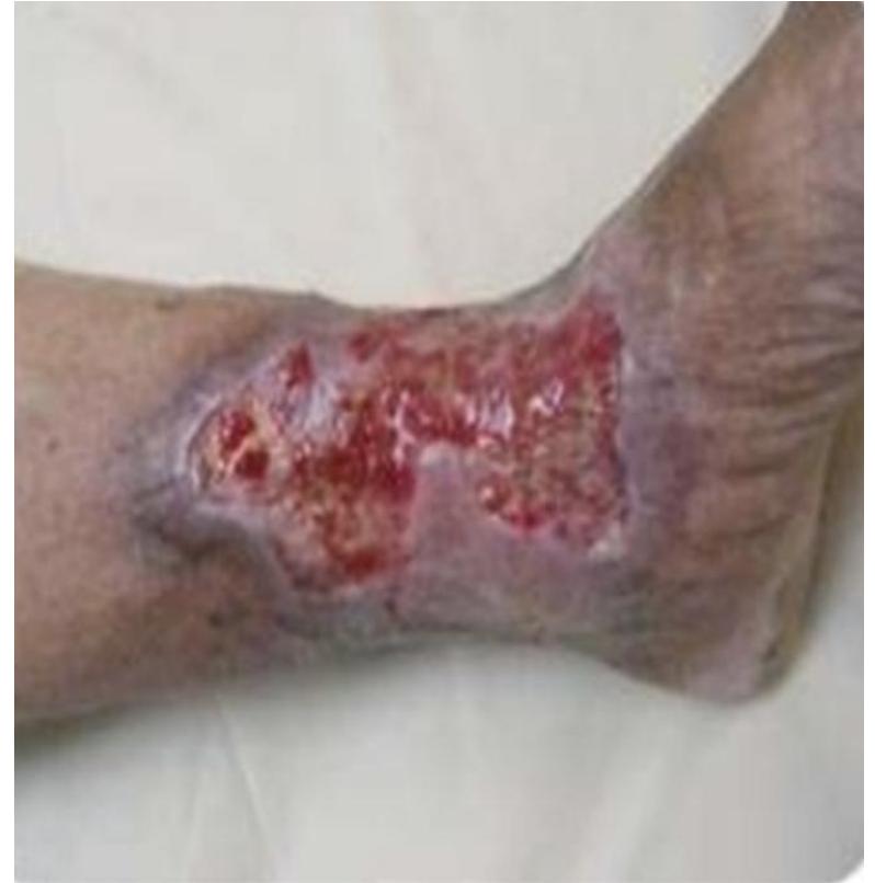
Q1: Mention TWO specific inspection signs visible in this photograph.
- Answer:
- Shallow, irregular ulceration located over the medial lower leg / medial malleolus (Gaiter zone).
- Surrounding stasis hyperpigmentation (hemosiderin staining), lipodermatosclerosis, and chronic venous edema.
Q2: Mention TWO definitive treatment modalities for this condition.
- Answer:
- Multilayer Compression Bandaging (four-layer compression system) after ruling out arterial disease ().
- Endovenous ablation of incompetent superficial veins (to correct underlying reflux and prevent recurrence).
-OSPE Station 3: Lower Extremity Lymphedema
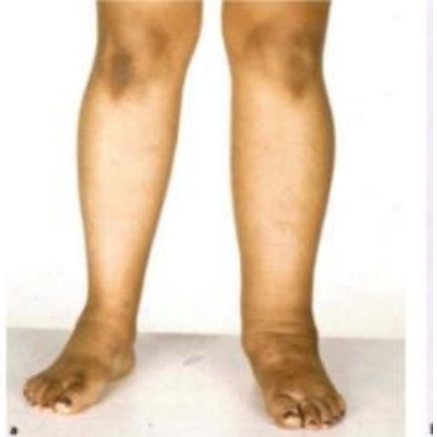
Q1: What is the diagnosis shown in this photograph?
- Answer: Chronic Lymphedema of the lower extremity.
Q2: What is the characteristic physical examination sign for lymphedema?
- Answer: Positive Stemmer’s Sign (inability to pinch and lift a fold of skin at the base of the second toe due to fibrotic skin thickening).
Q3: List TWO primary and TWO secondary causes of lymphedema.
- Answer:
- Primary (Congenital/Hereditary): Milroy’s disease (congenital), Lymphedema praecox (Meige disease), Lymphedema tarda.
- Secondary (Acquired): Surgical lymph node dissection (cancer surgery), Radiotherapy, Filariasis (Wuchereria bancrofti), Malignant lymphatic obstruction.
Q4: What is the cornerstone of non-operative management?
- Answer: Complex Decongestive Therapy (CDT) (manual lymphatic drainage, multilayer compression bandaging, skin hygiene, and exercise).
-OSPE Station 4: Acute Lower Extremity Cellulitis
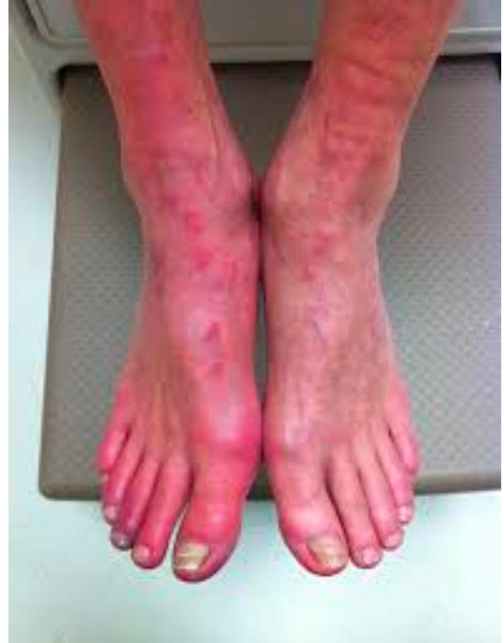
Q1: Describe the clinical findings and state the diagnosis.
- Answer: Diffuse, poorly demarcated erythema, warmth, edema, and tenderness over the lower leg without clear margins; Diagnosis: Acute Lower Extremity Cellulitis.
Q2: What are the most common causative bacterial organisms?
- Answer: Streptococcus pyogenes (Group A Streptococcus) and Staphylococcus aureus.
Q3: Outline the essential management steps.
- Answer:
- Mark the advancing border of erythema with a surgical marker to monitor treatment response.
- Leg elevation to reduce dependent edema.
- Systemic oral or intravenous antibiotics (anti-streptococcal/anti-staphylococcal: Cefazolin, Flucloxacillin, or Vancomycin).
- Identify and treat entry points (tinea pedis, interdigital maceration, minor cuts).
Skin & Soft Tissue
Skin, & Subcutaneous Tumors
Clinical Scenarios (CS)
Case 1: Cutaneous Malignant Melanoma
A 52-year-old fair-skinned male presents with a changing pigmented lesion on his upper back that has grown over 6 months, become darker, and bled intermittently.
Q1: State the “ABCDE” clinical diagnostic criteria used to evaluate pigmented skin lesions.
- Answer:
- A (Asymmetry): One half of the lesion does not match the other half.
- B (Border irregularity): Edges are ragged, notched, blurred, or poorly defined.
- C (Color variation): Non-uniform color (shades of brown, black, red, white, or blue).
- D (Diameter): Diameter (size of a pencil eraser).
- E (Evolving): Any change in size, shape, color, elevation, or new symptom (itching, bleeding).
Q2: What is the correct biopsy technique to confirm the diagnosis?
- Answer: Complete Excisional Biopsy with a 1–2 mm normal skin margin and full subcutaneous fat thickness. (Incisional biopsy or punch biopsy is avoided unless lesion is exceptionally large or on face).
Q3: What is the single most important histological prognostic factor for localized primary melanoma?
- Answer: Breslow Tumor Thickness (vertical depth of invasion from the granular layer of epidermis to the deepest tumor cell in millimeters) presence of ulceration.
Q4: What definitive surgical procedure is indicated once diagnosis is established?
- Answer: Wide Local Excision with surgical margins determined by Breslow thickness:
- Melanoma in situ: margin.
- : margin.
- : margin Sentinel Lymph Node Biopsy (SLNB).
- : margin + SLNB.
Case 2: Acute Perianal Pain - Differential Diagnosis
A 34-year-old male presents with acute severe perianal pain for 2 days, aggravated by defecation.
Q1: Mention FOUR relevant questions to ask in history of present illness.
- Answer:
- Relationship of pain to defecation (sharp “knife-like” tearing pain during and after defecation vs constant throbbing ache).
- Presence of rectal bleeding (bright red blood on toilet paper / streaks on stool vs no bleeding).
- Any palpable tender swelling or lump at the anal verge.
- Systemic symptoms: fever, chills, rigors, or purulent perineal discharge.
Q2: Mention FOUR common differential diagnoses for acute perianal pain.
- Answer:
- Acute Anal Fissure (posterior midline tearing pain with bright red streaks of blood).
- Acute Perianal Abscess (constant throbbing pain, indurated tender mass, fever).
- Thrombosed External Hemorrhoid (acute painful, tense, purplish perianal lump).
- Fistula-in-ano with secondary infection (or Proctalgia fugax / Strangulated internal hemorrhoids).
Q3: Outline initial conservative and medical management for an acute anal fissure.
- Answer:
- High-fiber diet, increased oral fluid intake, and stool softeners (osmotic laxatives) to avoid straining.
- Warm sitz baths after bowel movements.
- Topical pharmacological sphincter-relaxing agents: 0.2% Glyceryl Trinitrate (GTN) ointment OR 2% Diltiazem cream applied BID for 6–8 weeks.
-OSPE Station 1: Cutaneous Malignant Melanoma Evaluation (ABCDE Criteria)
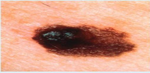
Q1: Apply the ABCDE criteria to the clinical image shown.
- Answer:
- Asymmetry: Asymmetrical in shape.
- Border: Irregular, jagged, notched margins.
- Color: Marked color variegation (black, dark brown, tan).
- Diameter: Exceeds .
- Evolving: Documented recent growth and irregular pigmentation.
Q2: What is the clinical diagnosis?
- Answer: Cutaneous Malignant Melanoma (Superficial Spreading Melanoma).
Q3: Mention TWO microscopic factors that determine the T stage of the primary tumor.
- Answer:
- Breslow thickness (in mm).
- Presence or absence of histological ulceration.
-OSPE Station 2: Pilonidal Sinus Disease
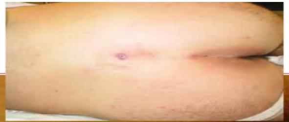
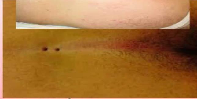
Q1: What is the diagnosis shown in these photographs?
- Answer: Pilonidal Sinus Disease (Sacrococcygeal Pilonidal Sinus).
Q2: Describe the characteristic physical findings.
- Answer: Presence of primary midline epithelialized pits or sinus openings located in the natal cleft overlying the sacrococcygeal region, often with protruding hair tufts, induration, and lateral secondary fistulous tracts.
Q3: What is the accepted pathophysiological etiology?
- Answer: Acquired condition; loose shedding hair fragments collect in the deep natal cleft, friction forces the hair shafts to drill into dilated follicles, causing a foreign body granulomatous reaction and secondary bacterial infection.
Q4: Contrast management of an Acute Pilonidal Abscess versus Chronic Pilonidal Sinus.
- Answer:
- Acute Abscess: Emergency surgical Incision and Drainage (I&D) (off-midline), curettage of hair, and wound packing.
- Chronic Sinus: Elective excision: Off-midline flap reconstruction (e.g., Bascom cleft lift procedure, Karydakis flap, or Limberg flap) to flatten the natal cleft and move the scar away from the midline; permanent laser hair depilation.
-OSPE Station 3: Acute Perianal Abscess
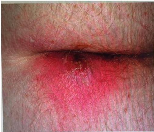
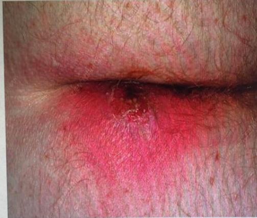
Q1: What is the diagnosis shown in this patient with 2 days of severe perineal pain?
- Answer: Acute Perianal Abscess.
Q2: What is the underlying anatomical etiology (Cryptoglandular Hypothesis)?
- Answer: Obstruction and secondary bacterial infection of the anal crypt glands at the dentate line in the intersphincteric plane, tracking down to the perianal subcutaneous tissue.
Q3: Mention the TWO crucial steps in surgical management.
- Answer:
- Prompt examination under anesthesia (EUA) and Incision and Drainage (I&D): cruciate incision (or de-roofing) close to the anal verge to shorten any potential fistula tract.
- Digital breakdown of all internal loculations, irrigation, and loose packing (antibiotics only if immunocompromised, diabetic, or systemic sepsis).
Q4: What chronic complication develops in up to of patients following drainage?
- Answer: Fistula-in-Ano (Anorectal Fistula).
MIS / Laparoscopy
Introduction to MIS
(No past exam questions recorded in source)
Examples of laparoscopic surgeries
(Systematically cross-referenced across surgical disciplines: Laparoscopic Cholecystectomy for gallstone disease; Laparoscopic Appendectomy for acute appendicitis; Laparoscopic Inguinal Hernia Repair [Transabdominal Preperitoneal - TAPP, or Totally Extraperitoneal - TEP]; Laparoscopic Heller Myotomy for achalasia; Laparoscopic Sleeve Gastrectomy and Roux-en-Y Gastric Bypass for morbid obesity; and Diagnostic Laparoscopy in trauma and acute abdomen).
Metabolic Surgery
Surgical Management of Morbid Obesity
(No past exam questions recorded in source)
Transplant Surgery
organ transplant
(No past exam questions recorded in source)
Pediatric Surgery
Common Pediatric & Neonatal Surgical Emergencies
Clinical Scenarios (CS)
Case 1: Infantile Hypertrophic Pyloric Stenosis
A 4-week-old first-born male infant presents with non-bilious, projectile vomiting immediately after feeds, persistent hunger, and a palpable “olive-shaped” mass in the right epigastrium.
Q1: What classic electrolyte and metabolic derangement is caused by this condition?
- Answer: Hypochloremic, Hypokalemic Metabolic Alkalosis with Paradoxical Aciduria (due to persistent loss of gastric HCl, followed by renal conservation of sodium and hydrogen in exchange for potassium).
Q2: What is the diagnostic imaging modality of choice?
- Answer: Abdominal Ultrasonography (criteria: pyloric muscle thickness and pyloric channel length ; “target sign”).
Q3: What is the crucial initial step in management?
- Answer: Fluid and Electrolyte Resuscitation (pyloric stenosis is a medical emergency, not an immediate surgical emergency): NPO, NG tube decompression, IV rehydration with Normal Saline with Dextrose and Potassium Chloride until serum chloride is and bicarbonate is normal.
Q4: What is the definitive surgical operation?
- Answer: Ramstedt Pyloromyotomy (open or laparoscopic): longitudinal division of hypertrophic pyloric circular muscle down to the bulging, intact submucosa.
Common pediatric surgical problems
Clinical Scenarios (CS)
Case 1: Inguinal and Groin Swellings in Adults vs Children
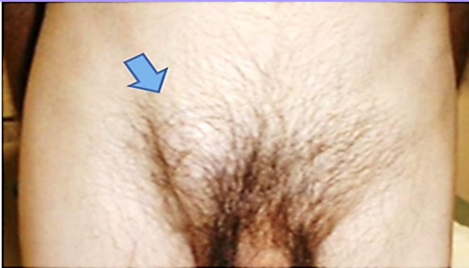
A 55-year-old male presents with a painless bulge in the right groin that appears on standing and straining and disappears when lying flat.
Q1: What THREE relevant questions should you ask regarding the chief complaint?
- Answer:
- Duration and variability (does it reduce spontaneously or require manual reduction?).
- Associated pain, change in color, or sudden irreducibility (signs of incarceration/strangulation).
- Risk factors for raised intra-abdominal pressure: chronic cough, chronic constipation/straining, heavy lifting, or LUTS/urinary straining.
Q2: What THREE physical examination signs should you elicit on palpation?
- Answer:
- Palpable Expansile Cough Impulse.
- “Getting above the swelling” (possible in scrotal hydrocele/cyst, impossible in inguinoscrotal hernia).
- Internal Inguinal Ring Occlusion Test (occluding internal ring above femoral artery midpoint: if hernia is controlled Indirect Inguinal Hernia; if hernia bulges medial to finger Direct Inguinal Hernia).
Q3: Mention THREE differential diagnoses.
- Answer:
- Inguinal hernia (direct vs indirect).
- Femoral hernia (bulge below and lateral to pubic tubercle).
- Hydrocele of the cord or testis (or inguinal lymphadenopathy / saphena varix).
Q4: What is the definitive treatment in adult inguinal hernia?
- Answer: Elective surgical repair: Lichtenstein Tension-Free Mesh Hernioplasty (open polypropylene mesh repair) OR Minimally Invasive Laparoscopic Repair (TEP or TAPP).
-OSPE Station 1: Congenital Pediatric Inguinal Hernia
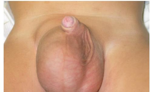
Q1: What is the diagnosis shown in this 1-year-old boy?
- Answer: Congenital Indirect Inguinal Hernia (right-sided inguinoscrotal hernia).
Q2: What is the embryological etiology of this condition?
- Answer: Failure of complete obliteration / closure of the Processus Vaginalis (persistent patent peritoneal pouch).
Q3: Mention TWO recognized complications if left untreated.
- Answer:
- Incarceration / Irreducibility.
- Strangulation with bowel ischemia and secondary testicular infarction.
Q4: What is the surgical procedure of choice in children, and how does it fundamentally differ from adult hernia repair?
- Answer:
- Procedure: High Ligation of the Hernia Sac (Herniotomy).
- Fundamental Difference: Children have normal abdominal wall musculature and internal ring floor; therefore, herniotomy alone (ligating the sac at the internal ring) is curative. Synthetic mesh is NEVER used in pediatric hernia repair.
-OSPE Station 2: Undescended Testis (Cryptorchidism)
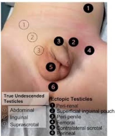
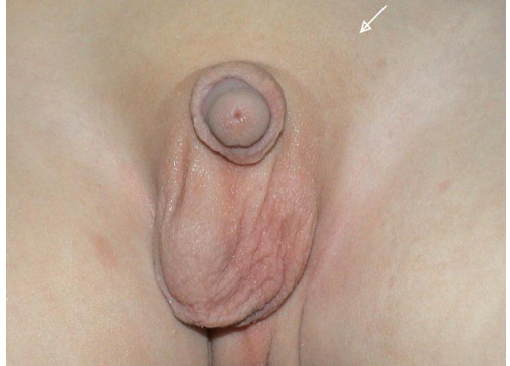
Q1: Describe the physical finding shown in the clinical photograph.
- Answer: Empty Left Hemiscrotum with marked hypoplasia/underdevelopment and loss of rugosity of the left scrotal sac.
Q2: Distinguish between a True Undescended Testis and an Ectopic Testis based on the anatomical diagram.
- Answer:
- True Undescended Testis: Arrested along its normal embryological path of descent (intra-abdominal, deep inguinal ring, inguinal canal, superficial inguinal ring, upper scrotum).
- Ectopic Testis: Deviates away from its normal path into an abnormal anatomical location (superficial inguinal pouch [most common], femoral canal, perineum, pubopenile, or contralateral scrotum).
Q3: Mention TWO major long-term complications of untreated cryptorchidism.
- Answer:
- Subfertility / Infertility (due to thermal degenerative injury to germ cells).
- Testicular Malignancy (primarily Seminoma, risk remains elevated even after orchiopexy). (Also increased risk of testicular torsion and trauma).
Q4: What is the recommended surgical operation and optimal timing?
- Answer:
- Operation: Surgical Orchidopexy (Orchiopexy) (mobilization of spermatic cord and fixation of testis in a subdartos scrotal pouch; laparoscopic Fowler-Stephens for intra-abdominal testis).
- Optimal Timing: Performed between 6 and 12 months of age (and completed before 18 months).
-OSPE Station 3: Pediatric Umbilical Hernia versus Adult Paraumbilical Hernia
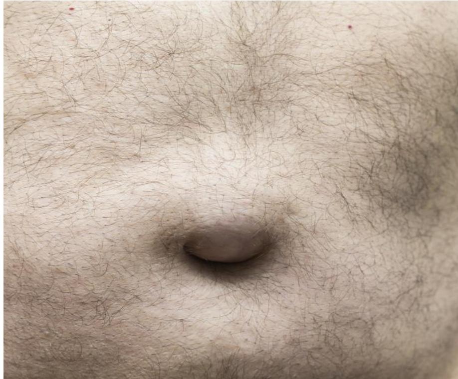
Q1: Contrast the clinical course and management of an Infantile Umbilical Hernia versus an Adult Paraumbilical Hernia.
- Answer:
- Infantile (Pediatric) Umbilical Hernia:
- Anatomical Defect: Congenital defect through the central umbilical fibrous ring.
- Clinical Course: High rate of spontaneous closure ( close spontaneously by age 3–4 years); exceptionally low strangulation risk.
- Management: Watchful waiting; surgical repair (herniotomy) indicated only if defect persists beyond age 4–5 years, is very large (), or becomes symptomatic.
- Adult Paraumbilical Hernia:
- Anatomical Defect: Acquired defect in the linea alba immediately adjacent to the umbilical cicatrix.
- Clinical Course: Does NOT close spontaneously; narrow rigid fibrous neck causes very high risk of incarceration and strangulation.
- Management: Mandatory elective surgical repair: Primary suture repair or Mesh Hernioplasty (open or laparoscopic).
- Infantile (Pediatric) Umbilical Hernia: