Collected from Haneen H. Alsultan, Faisal Alkharji, Faisal Alturki, Talal M. Ak., Toqa Alsulaim, Rawan Althaqib, Abdul Rhman Abou Al-Shaar, Mohammed Al Khaldi, Basim Alhomida, , Muath Bin Abdullah Muath bin abdullah, Khalid Alanazi.
اللهم يا معلّم موسى علّمني، ويا مفهم سليمان فهّمني، ويا مؤتي لقمان الحكمة وفصل الخطاب آتني الحكمة وفصل الخطاب اللهم اجعل ألستنا عامرة بذكرك، وقلوبنا بخشيتك، وأسرارنا بطاعتك، إنك على كل شيء قدير، حسبنا الله ونعم الوكيل
Table of Contents
- 1. General Principles & Perioperative Care
- 2. Trauma & Emergency Surgery
- 3. Head & Neck
- 4. Neurosurgery
- 5. Breast
- 6. Thoracic
- 7. Upper GI Surgery
- 8. Abdominal Wall, Hernia & Stoma
- 9. Hepatobiliary & Pancreatic Surgery
- 10. Colorectal & Anorectal Surgery
- 11. Urology
- 12. Vascular Surgery
- 13. Skin & Soft Tissue
- 14. Minimally Invasive Surgery (MIS) / Laparoscopy
- 15. Pediatric Surgery
1. General Principles & Perioperative Care
Station 1: Nasogastric Tube (NGT) Insertion

A. Indications & Contraindications (Past Exam Recalls)
- Indications:
- Gastric Decompression (Suction): Relieve bowel obstruction, paralytic ileus, or acute gastric dilatation.
- Enteral Feeding / Nutrition: Administer nutrition and oral medications in patients unable to swallow (e.g., CVA, impaired consciousness).
- Diagnostic Aspiration: Aspirate gastric fluid to assess upper GI bleeding (fresh blood vs coffee-ground) or laboratory analysis.
- Contraindications:
- Absolute: Basal skull fracture (Battle’s sign, raccoon eyes, CSF leak) and severe midface/facial bone fractures (risk of intracranial entry through fractured cribriform plate).
- Relative: Nasal deformity or recent nasal/ENT surgery; severe coagulopathy / anticoagulant therapy (if INR > 3, high epistaxis risk; require extreme caution and lubrication).
B. Tube Types & Sizing
- Tube Types:
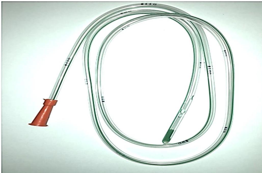
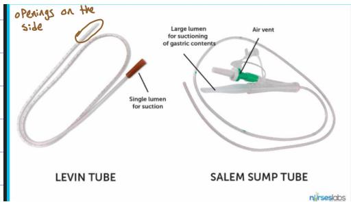
- Levin Tube: Single-lumen tube, primarily used for feeding.
- Salem Sump Tube: Double-lumen tube with an air vent lumen to prevent mucosal suction damage, preferred for bowel obstruction decompression.
- Sizing & Equipment:
- 12 Fr: Softer, bends easily, harder to insert, but more comfortable for the patient.
- 14–16 Fr: Stiffer, easier to pass, standard adult size.
- Lubricant: 2% lidocaine gel (preferred because it numbs the mucosa, applied to the distal 10–15 cm), water-soluble, or silicone gel.
- Tray: Catheter-tip syringe, kidney dish, cup of water with straw, gloves, litmus paper, hypoallergenic tape.
C. Six-Step Clinical Insertion Procedure ()
- WIPES & Patient Preparation:
- Confirm patient identity and review file/nurse report (indications, consciousness, stability, and coagulation status; note if INR > 3).
- Wash hands, introduce yourself, explain procedure, obtain consent.
- Warn patient that the procedure is uncomfortable. Agree on a stop signal: “If you want me to pause, simply raise your hand and I will stop.”
- Position: 45° sitting position (High Fowler’s). Expose abdomen to head; cover chest. Ask nurse to assist.
- Nostril Patency & NEX Measurement:
- Inspect nose for polyps or severe septal deviation.
- Patency Test: Ask patient to occlude one nostril, inhale, repeat on the other side; select the more patent nostril. (Patient can breathe through mouth during insertion).
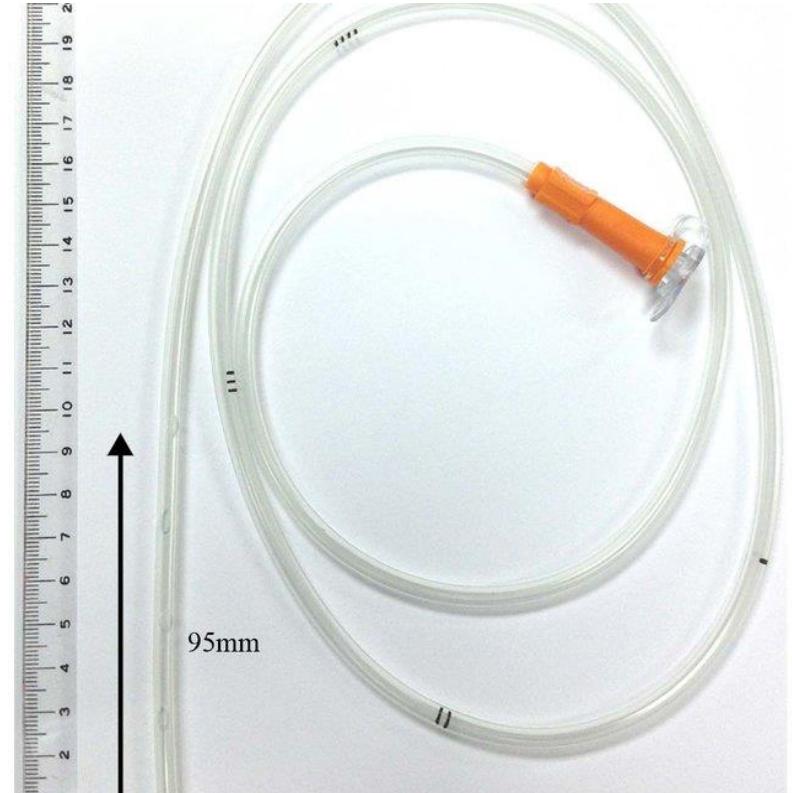
- NEX Measurement: Measure from Nose tip to Earlobe, and from earlobe to Xiphoid process (N-E-X). Observe the printed centimeter depth markings (tube also features a blue radiopaque line visible on X-ray).
- Insertion:
- Lubricate first 10–15 cm with lidocaine gel.
- Insert tube horizontally along the floor of the nose (NOT upwards) until it reaches the posterior pharynx.
- Head Flexion & Swallowing:
- Ask patient to flex their head forward (chin to chest) and take repeated sips of water through the straw.
- Advance the tube smoothly with each swallow until the measured mark is reached.
- If severe coughing, gagging, or cyanosis occurs, withdraw immediately into pharynx (tracheal entry).
- Confirmation of Position (5 Methods from Backup):
- Aspiration by Syringe: Remove caps and aspirate. Normal gastric juice is yellow, green, or brown. If empty, tube may be in lung, has not reached stomach, or patient is fasting.
- pH Paper: Test aspirate on litmus paper; pH < 5 confirms gastric acid placement (normal stomach pH is 2–3).
- Air Insufflation (Whoosh Test): Inject 30 cc of air while auscultating epigastrium with stethoscope to hear a bubble sound.
- Erect Chest X-Ray (Must see all 3 criteria):
- Tube runs in the midline/center of the chest.
- Crosses the diaphragm.
- Tip ends below the left dome of the diaphragm in the gastric bubble.
- Capnogram: High CO2 indicates incorrect placement in the airways/lung.
- Securing & Maintenance:
- Tape securely to nose without alar pressure. Change or remove every 2 weeks, or convert to a gastrostomy (PEG) or jejunostomy tube if prolonged enteral feeding is required.
- Complications: Patient discomfort, irritation, nasopharyngeal injury, infection, esophagitis.
Station 2: Surgical Drains — Closed Suction (Jackson-Pratt) Drain
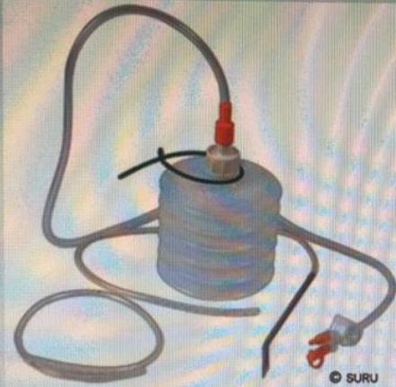
Q&A & Clinical Principles (Past Exam Recalls)
- Q1: Name this device.
- Answer: Closed suction drain (Jackson-Pratt / Blake / Suru drain).
- Q2: What is this used for?
- Answer:
- To drain potential fluid, seroma, hematoma, or pus collections postoperatively.
- Obliteration of surgical dead space (e.g., after mastectomy, hernia repair, or major abdominal/pelvic resections).
- Evacuation of bile, pancreatic fluid, or lymph.
- Answer:
- Q3: How does it work?
- Answer: Negative pressure (closed suction); manual compression of the flexible silicone reservoir bulb creates a continuous vacuum that draws fluid through the perforated tubing.
- Q4: Mention two problems or complications of its use.
- Answer:
- Infection (retrograde bacterial contamination).
- Tube blockage (clot or tissue plugging the holes).
- Visceral/vascular erosion or pain during removal.
- Answer:
- Q5: Criteria for removal:
- Answer: When daily output decreases to of serous fluid with no signs of bile leak or hemorrhage.
Station 3: Foley’s Catheter Insertion & Management
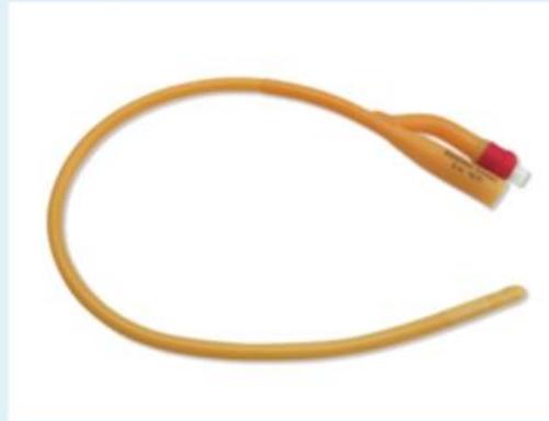
Q&A & Clinical Principles (Past Exam Recalls)
- Q1: What is this instrument?
- Answer: Foley’s (urinary) catheter (indwelling balloon retention catheter).
- Q2: Mention three common indications to use it.
- Answer:
- Accurate monitoring of input and urine output in critically ill, shock, or trauma resuscitation patients ().
- Intraoperative bladder decompression to prevent accidental surgical bladder injury and maintain clear pelvic visualization during major surgery.
- Relief of acute or chronic urinary obstruction / retention (e.g., BPH, urethral stricture, clot retention).
- Answer:
- Q3: What fluid is used to inflate the retention balloon?
- Answer: 10 mL of sterile water (normal saline is avoided to prevent crystal precipitation obstructing the inflation channel). Standard adult size is 14–16 Fr.
- Q4: What is the primary contraindication in trauma?
- Answer: Suspected urethral disruption (suggested by blood at the urethral meatus, perineal hematoma, or high-riding prostate in pelvic fractures); perform retrograde urethrogram before catheterization.
Station 4: Volodyne Incentive Spirometer
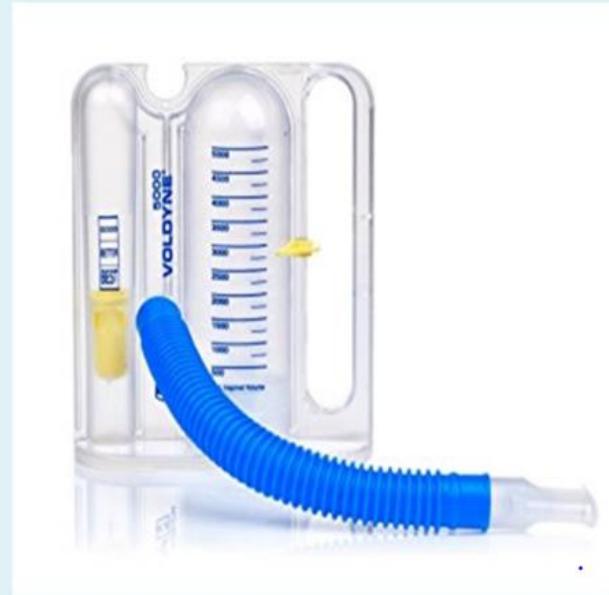
Q&A & Clinical Principles (Past Exam Recalls)
- Q1: Name this tool.
- Answer: Spirometer (Volodyne Incentive Spirometer).
- Q2: Mention TWO of its clinical uses.
- Answer:
- Postoperative care: To help get rid of secretions, expand the lungs, and prevent postoperative pulmonary atelectasis and pneumonia (especially after upper abdominal or thoracic surgery).
- Asthma monitoring: For asthmatic patients to monitor their respiratory condition and lung function (can be used at home).
- Answer:
Station 5: Wound Assessment & Laceration Management

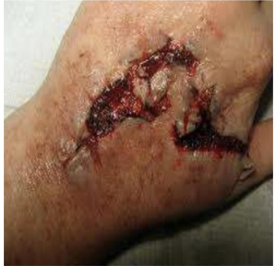
Q&A & Clinical Principles (Past Exam Recalls)
- Q1: What is the type of wound shown?
- Answer:
- Lacerated wound (jagged, irregular edges on hand or knee).
- Clean wound / surgical incision (sharply incised wound edges).
- Answer:
- Q2: What is the mechanism of injury?
- Answer: Blunt trauma / mechanical injury due to friction (for lacerations) or sharp cutting injury (for incised wounds).
- Q3: Give 5 steps for treatment of a contaminated or traumatic wound:
- Answer:
- Debridement of non-viable tissue.
- Wash out / copious irrigation and disinfection.
- Antibiotics (prophylactic or therapeutic).
- Let it open (delayed primary or secondary intention healing; or primary suturing and daily dressing if clean).
- Tetanus toxoid prophylaxis.
- Answer:
- Q4: Clinical classification pearl ():
- Answer: All uncomplicated abdominal wall hernias are classified as clean wounds; prosthetic mesh should only be placed in clean wounds to avoid mesh infection.
Station 6: Blood Transfusion in Surgery
Q&A & Clinical Principles (Past Exam Recalls)
- Scenario: A patient presents with episodes of melena. Lab investigations reveal Hemoglobin: 8.1 g/dL (normal: 13–18 g/dL).
- Q1: How many units of blood is needed to raise hemoglobin to 10 g/dL?
- Answer: Equal or more than 2 units of Packed Red Blood Cells (PRBC). (Dosage rule: 1 unit of PRBC typically raises adult hemoglobin by approximately ).
- Q2: Mention FOUR symptoms of a transfusion reaction.
- Answer:
- Fever
- Chills
- Respiratory distress (dyspnea/wheezing)
- Itching / urticaria
- Answer:
- Q3: Immediate management upon suspecting a reaction:
- Answer:
- Stop the transfusion immediately.
- Keep IV line patent with normal saline.
- Verify patient ID against blood product label.
- Return blood unit and tubing to blood bank with fresh patient samples for repeat crossmatch.
- Supportive care (oxygen, fluids, antihistamines).
- Answer:
2. Trauma & Emergency Surgery
Station 7: Polytrauma Initial Assessment (Collapsing Trauma Patient)
Clinical Scenario & Exam Steps
A trauma victim arrives at the Emergency Department collapsing and in severe respiratory distress.
- Q1: Outline your immediate stepwise management.
- Answer:
- ATLS Primary Survey (ABCDE):
- A (Airway): Assess airway patency with strict in-line cervical spine protection / rigid collar.
- B (Breathing): Inspect chest expansion, auscultate breath sounds bilaterally, percuss for hyperresonance.
- C (Circulation): Two large-bore IV cannulas (14–16G), IV fluid resuscitation, control any external bleeding.
- D (Disability): Rapid GCS and pupillary reflexes.
- E (Exposure): Completely expose patient to check for hidden injuries; prevent hypothermia.
- Crucial Exam Step: Explicitly state to the examiner that an urgent portable Chest X-Ray is required.
- ATLS Primary Survey (ABCDE):
- Answer:
- Q2: The examiner provides the Chest X-Ray showing a massive pneumothorax. What is the immediate life-saving intervention?
- Answer:
- Immediate needle decompression (2nd ICS midclavicular line or 4th/5th ICS anterior axillary line) followed immediately by tube thoracostomy (chest tube insertion) in the Triangle of Safety (5th ICS, anterior to midaxillary line).
- Transition to secondary survey once hemodynamically stabilized.
- Answer:
Station 8: Splenic Trauma / Splenic Rupture
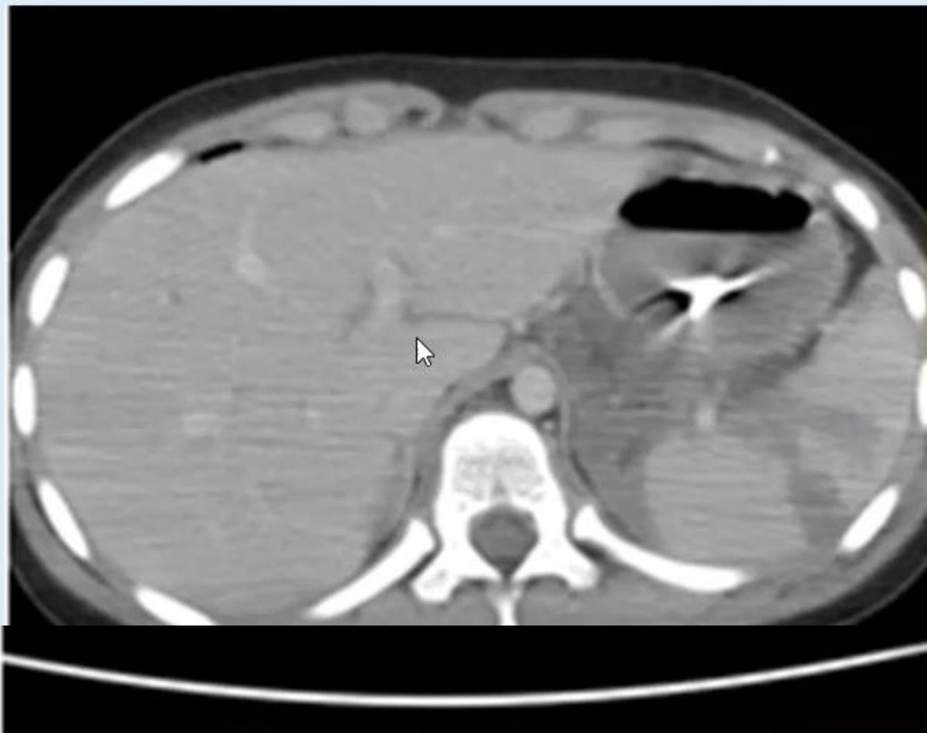
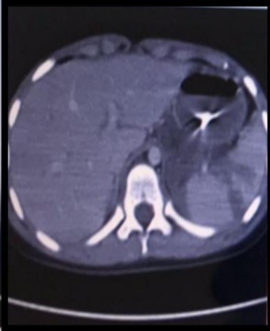
Q&A & Clinical Management (Past Exam Recalls)
- Scenario: A car accident victim is brought to the ER with abdominal pain. Vital signs: pulse 96/min, BP 118/74 mmHg. IV fluid infusion was started and NGT was placed. Abdominal CT is shown.
- Q1: What is the main finding in this CT scan?
- Answer: Splenic rupture / splenic injury (large hypodense intrasplenic hematoma / parenchymal laceration).
- Q2: Mention steps in the initial management of this patient:
- Answer (Past Exam Key Points):
- ABC (Airway, Breathing, Circulation with cervical spine control).
- IV fluid resuscitation & blood transfusion.
- Cross matching of blood.
- Anti-tetanus toxoid.
- Foley’s catheter insertion (to monitor urine output).
- Answer (Past Exam Key Points):
- Q3: What is your definitive management plan?
- Answer:
- If patient is unstable or develops peritonitis: Emergency laparotomy and splenectomy.
- If patient is hemodynamically stable: Conservative non-operative management (ICU monitoring) and angioembolization.
- Answer:
Station 9: Burns Assessment & Split-Thickness Skin Graft (STSG)
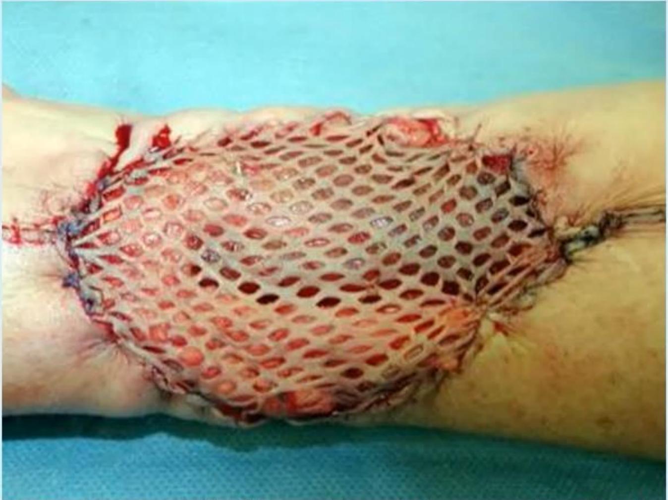
Q&A & Clinical Principles (Past Exam Recalls)
- Scenario: A patient suffered a deep burn. The photograph shows a split-thickness skin graft used to cover the burn wound.
- Q1: Split skin graft is made up of which part of the skin?
- Answer: Epidermis + portion of dermis (papillary dermis).
- Q2: Mention TWO disadvantages of split skin graft:
- Answer (Past Exam Key Points):
- Color mismatch.
- Poor sensation at the grafted area.
- Need for general anesthesia (for large donor harvesting).
- Less cosmetic.
- More susceptible to trauma.
- Answer (Past Exam Key Points):
Station 10: Acute Abdomen — Pneumoperitoneum / Perforated Peptic Ulcer
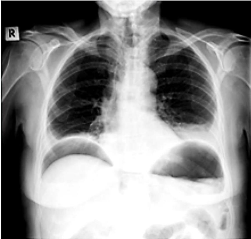
Q&A & Clinical Management (Past Exam Recalls)
- Q1: Name (A) the imaging study and (B) the main abnormality visible in the image.
- Answer:
- A) Imaging study: Erect Chest X-Ray.
- B) Main abnormality: Air under the diaphragm (pneumoperitoneum / subdiaphragmatic crescentic free gas).
- Answer:
- Q2: Name TWO clinical conditions where you can find this abnormality:
- Answer (Past Exam Key Points):
- Perforated hollow viscus (perforated peptic ulcer, perforated diverticulum).
- Postoperative state (residual gas following laparotomy or laparoscopy).
- Penetrating abdominal injury (stab/knife wound).
- Answer (Past Exam Key Points):
- Q3: What clinical history and examination signs identify a perforated peptic ulcer?
- Answer:
- History: Sudden-onset, severe epigastric pain rapidly becoming generalized; past history of dyspepsia.
- Examination: Patient lies still with shallow breathing, board-like abdominal rigidity, severe guarding/rebound tenderness, loss of liver dullness on percussion, and absent bowel sounds (silent abdomen).
- Answer:
- Q4: Mention 4 important steps in the management (including the most important if the patient is unstable):
- Answer (Past Exam Key Points):
- ABC Resuscitation (Airway, breathing, oxygen, large-bore IV access).
- IV fluid resuscitation with isotonic crystalloids (and Foley catheter to monitor output).
- Analgesia and broad-spectrum IV antibiotics + IV proton pump inhibitor (PPI).
- Emergency Surgery (exploratory laparotomy and omental patch repair / Graham patch) — the most important definitive step.
- Answer (Past Exam Key Points):
Station 11: Ruptured Appendicitis with Intra-abdominal Abscess
Q&A & Clinical Principles (Past Exam Recalls)
- Scenario: CT of a patient who had ruptured appendicitis 2 weeks ago and presents with persistent lower abdominal pain and fever.
- Q1: Give 4 clinical examination signs you will elicit for this case:
- Answer (Past Exam Key Points):
- Rebound tenderness in RIF (Blumberg’s sign — indicates localized peritoneal irritation).
- Rovsing’s sign (deep pressure in LIF elicits pain in RIF).
- Psoas sign (pain on passive right hip extension, indicates retrocecal appendix irritating iliopsoas).
- Obturator sign (pain on passive internal rotation of flexed right hip, indicates pelvic appendix irritating obturator internus).
- Answer (Past Exam Key Points):
- Q2: Give (2) options of treatment:
- Answer (Past Exam Key Points):
- A) Non-surgical (Conservative):
- Percutaneous image-guided drainage of the abscess.
- IV broad-spectrum antibiotics.
- B) Surgical:
- Appendectomy (interval appendectomy 6–8 weeks after resolution).
- A) Non-surgical (Conservative):
- Answer (Past Exam Key Points):
Station 12: Upper GI Bleeding — Duodenal Ulcer & Gastric Lesions
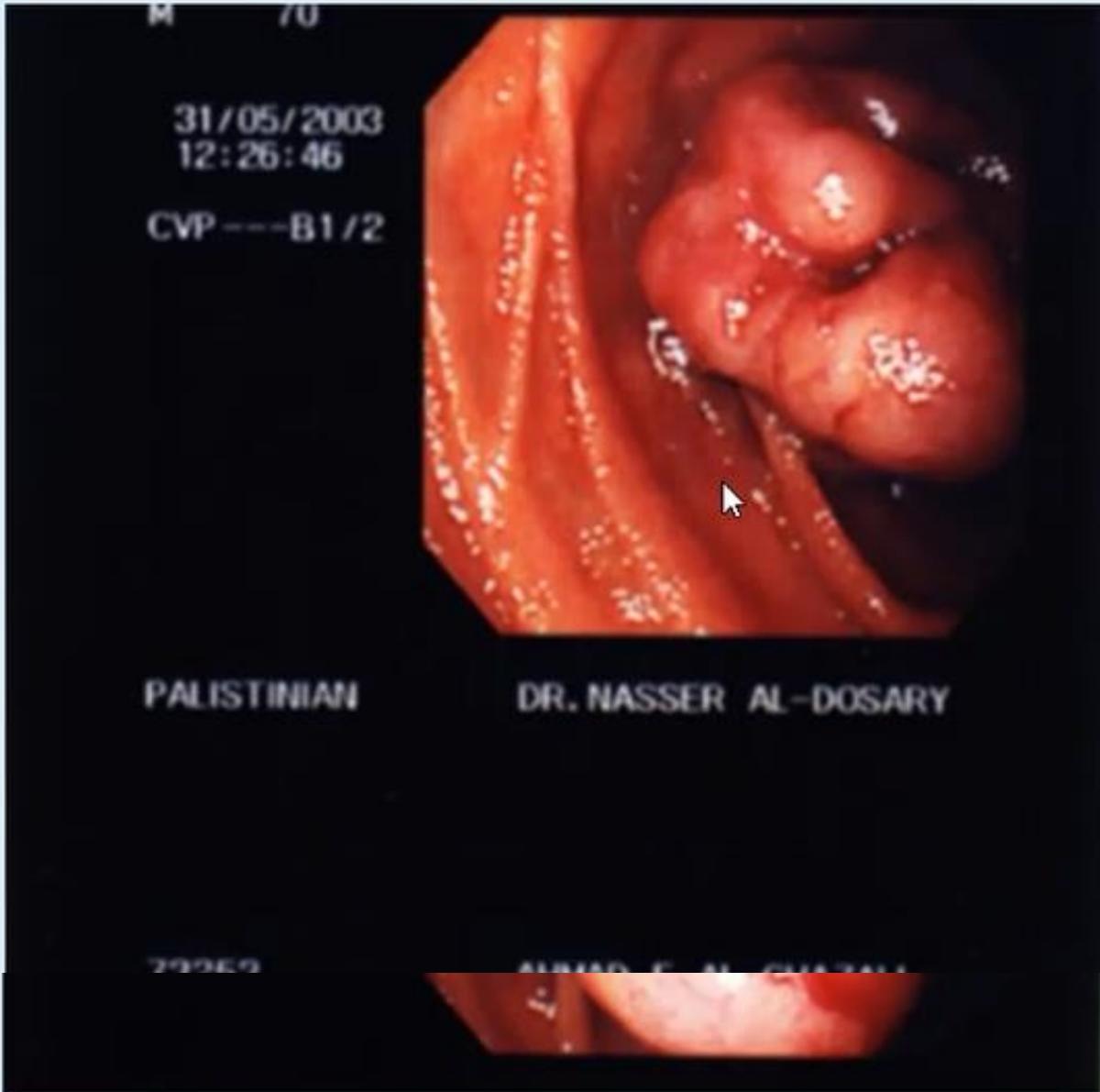
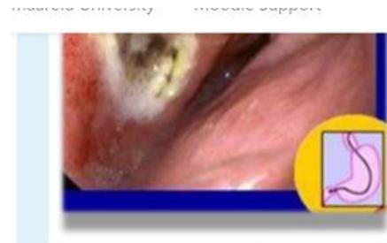
Q&A & Clinical Principles (Past Exam Recalls)
- Part 1: Duodenal Ulcer
- Scenario: Endoscopic view of the duodenum in a patient with recurrent abdominal pain for 3 weeks.
- Q1: What is the most likely abnormality?
- Answer: Duodenal ulcer.
- Q2: Mention THREE usual criteria of the abdominal pain related to this condition:
- Answer (Past Exam Key Points):
- Epigastric pain.
- Night pain (awakens patient from sleep).
- Hunger pain (relieved by eating food or antacids).
- Sharp / burning pain.
- Answer (Past Exam Key Points):
- Q3: Mention THREE common complications related to this condition:
- Answer (Past Exam Key Points):
- Perforation (acute peritonitis).
- Bleeding / hemorrhage (erosion into gastroduodenal artery).
- Penetration (into adjacent pancreas or liver).
- Obstruction (gastric outlet obstruction due to pyloric scarring).
- Answer (Past Exam Key Points):
- Part 2: Gastric Lesion
- Scenario: Patient presented with melena; vital signs were stable. Upper GI endoscopy shows a large polypoid lesion.
- Q1: Mention TWO differential diagnoses:
- Answer (Past Exam Key Points):
- Gastric carcinoma.
- GIST (Gastrointestinal Stromal Tumor).
- Gastrinoma.
- Answer (Past Exam Key Points):
- Q2: What TWO further specialized investigations (in order) would you perform?
- Answer (Past Exam Key Points):
- Endoscopy with biopsy.
- Endoscopic ultrasound (EUS).
- CT scan.
- Serum gastrin level (if gastrinoma suspected).
- Answer (Past Exam Key Points):
Station 13: Small Bowel Obstruction (SBO)
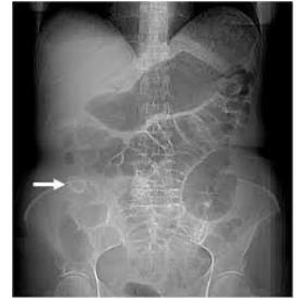
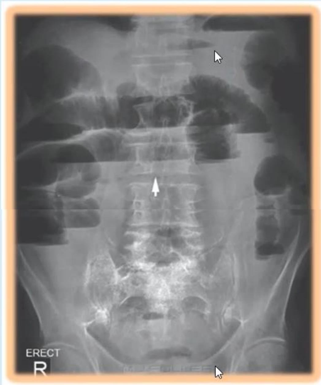
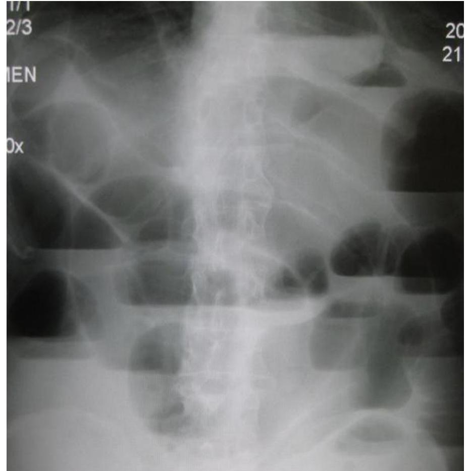
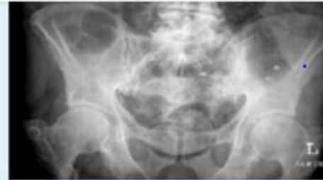
A. Framework for Reading Plain Abdominal X-Rays (‘s ABCDE)
- A (Abnormal Air): Under diaphragm (pneumoperitoneum) or multiple air-fluid levels in bowel.
- B (Bowel): Dilated vs normal:
- Small Bowel: Features valvulae conniventes (“coin-stack” appearance) extending completely across the lumen width (characteristic of jejunum; ileum is featureless).
- Large Bowel: Features haustrations that extend only partially across the lumen.
- C (Calcification): Stones (gallstones, urinary calculi), calcified prostate.
- D (Deformity): Spine fractures.
- E (Everything Else): Foreign bodies, lines, and tubes.
- Look at: Gallstone ileus (pneumobilia + SBO + ectopic gallstone), SBO, Foreign body, Volvulus.
B. Q&A & Clinical Management (Past Exam Recalls)
- Scenario 1: A patient presents with colicky abdominal pain, vomiting, and constipation for 2 days. Abdominal examination shows an RLQ surgical scar, mild distension, and hyperactive bowel sounds.
- Q1: What is the most likely diagnosis?
- Answer: Small bowel obstruction.
- Q2: What is the commonest cause of this condition?
- Answer: Adhesions (postoperative intra-abdominal adhesions); second cause: Hernia.
- Q3: Give 2 Differential Diagnoses on X-Ray:
- Answer: Volvulus, Small bowel tumor.
- Q4: Mention 4 important steps in the initial treatment:
- Answer (Past Exam Key Points):
- NPO (nil per os).
- IV fluids resuscitation.
- Nasogastric (NG) tube decompression.
- Pain killers (analgesia).
- Answer (Past Exam Key Points):
- Q5: Give 2 complications of bowel obstruction:
- Answer: Perforation, Strangulation / bowel ischemia.
- Q1: What is the most likely diagnosis?
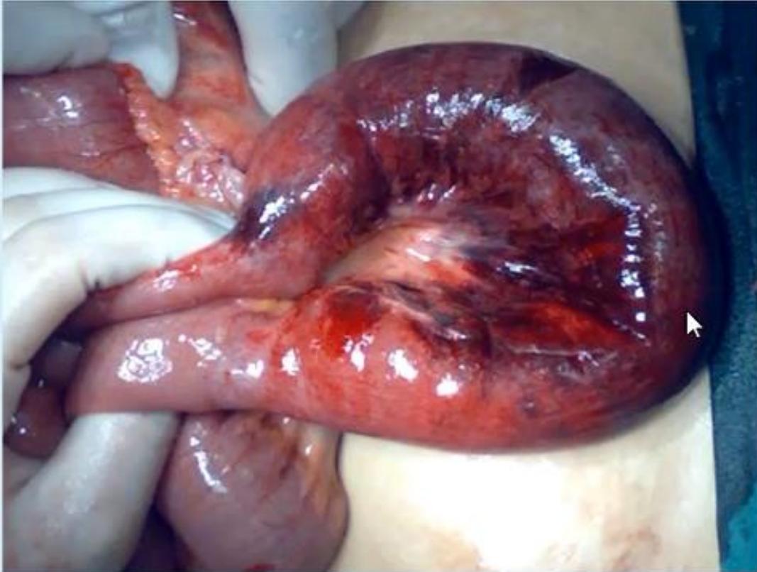
- Scenario 2: Laparotomy finding in a patient undergoing emergency surgery for acute abdomen showing a dark, dilated, congested loop of bowel.
- Q1: What is your diagnosis?
- Answer: Bowel strangulation.
- Q2: Mention TWO causes for this finding in an acute abdomen:
- Answer:
- Fibrous adhesion (Ladd’s band).
- Volvulus.
- Answer:
- Q1: What is your diagnosis?
Station 14: Large Bowel Obstruction (LBO)
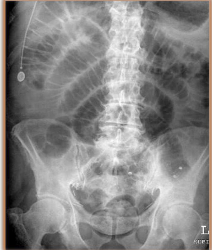
Q&A & Clinical Principles (Past Exam Recalls)
- Q1: What is the diagnosis on plain abdominal X-ray?
- Answer: Bowel obstruction (Large bowel obstruction / small bowel obstruction).
- Q2: Mention 4 physical examination findings to elicit on abdominal palpation:
- Answer:
- Tenderness (localized or generalized).
- Guarding (voluntary or involuntary).
- Rigidity (board-like rigidity indicating peritonitis).
- Palpable abdominal mass (e.g., obstructing neoplasm or volvulus loop).
- Answer:
- Q3: List 4 common causes of bowel obstruction:
- Answer:
- Postoperative adhesions.
- Incarcerated/strangulated hernia.
- Colorectal carcinoma / tumor.
- Volvulus (sigmoid or cecal).
- Answer:
- Q4: List 2 complications of bowel obstruction:
- Answer:
- Bowel ischemia / strangulation.
- Bowel perforation.
- Answer:
Station 15: Surgical Lab Interpretation — Coagulation Profile & DIC
Q&A & Clinical Principles (Past Exam Recalls)
- Q1: What are the laboratory abnormalities and definitive diagnosis in this coagulation panel?
- Laboratory Findings:
- PT: Prolonged / High
- aPTT: Prolonged / High
- INR: Elevated / High
- Pathophysiological Mechanisms:
- Depletion of circulating clotting factors.
- Depletion / malabsorption of Vitamin K.
- Definitive Diagnosis: Disseminated Intravascular Coagulation (DIC).
- Laboratory Findings:
- Q2: Outline the management of this patient:
- Answer (Past Exam Key Points):
- Treat the underlying cause (surgical hemostasis, source control of sepsis).
- Vitamin K administration.
- Blood component replacement therapy:
- Fresh Frozen Plasma (FFP) to restore clotting factors.
- Cryoprecipitate to replenish fibrinogen.
- Platelet transfusion.
- Packed Red Blood Cell (PRBC) transfusion for anemia.
- Heparin in selected non-bleeding thrombotic cases.
- Answer (Past Exam Key Points):
- Q3: A patient presents with hematemesis due to liver disease / ruptured esophageal varices. Coagulation profile shows high PT, high aPTT, and high INR. What is the management?
- Answer:
- Treat the cause: Emergency resuscitation and endoscopic variceal band ligation (EVBL).
- Administer IV Vitamin K.
- Transfuse blood products: FFP, Cryoprecipitate, Platelets, and PRBCs.
- Answer:
Station 16: Surgical Lab Interpretation — Arterial Blood Gas (ABG)
Q&A & Clinical Principles (Past Exam Recalls)
- Scenario: An Arterial Blood Gas (ABG) result from a diabetic patient presenting with acute abdominal distress shows:
- Low pH
- Low
- Low
- Q1: What is your diagnosis?
- Answer: Metabolic Acidosis (Diabetic Ketoacidosis / DKA with compensatory respiratory alkalosis).
- Q2: Give TWO laboratory tests you will immediately order:
- Answer (Past Exam Key Points):
- Blood glucose level.
- Urinalysis for ketones.
- Answer (Past Exam Key Points):
3. Head & Neck
Station 17: Clinical Neck & Thyroid Examination Guide
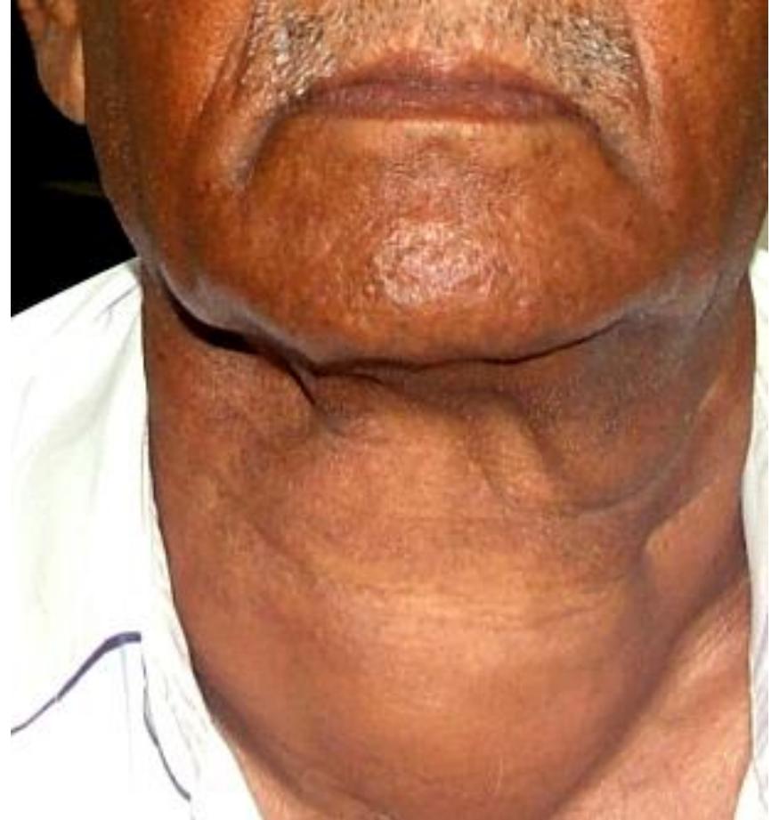
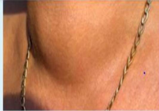
Complete Examination Checklist
- Introduction & WIPES:
- Wash hands, introduce yourself, identify patient, explain examination, obtain consent.
- Position & Exposure: Patient sitting comfortably upright on an exam chair. Exposure is from the chin down to the nipple line (essential to inspect for dilated veins, thoracic inlet obstruction, and retrosternal extension).
- Clinical pearl (The 3 Hidden Areas in General Surgery): Inspection is normally strictly visual, but there are 3 specific “hidden areas” where gentle touching/retraction is permitted during inspection: (1) In between the toes and behind the heels; (2) Underside of the breast (inframammary fold); (3) The abdominal pannus in obese patients.
- The 3 Lumps & Mandatory Pre-Palpation Maneuvers:
- Thyroid Lump: Ask patient to swallow a sip of water.
- Breast Lump: Ask patient to flex pectoralis major (hands pressed firmly on hips).
- Hernia Lump: Ask patient to cough (expansile impulse).
- General & Peripheral Inspection:
- Hands: Thyroid acropachy, onycholysis (Plummer’s nails), fine postural tremor (test with sheet of paper over outstretched fingers), palmar erythema, pulse rate (tachycardia in thyrotoxicosis, bradycardia in hypothyroidism).
- Eyes: Exophthalmos (inspect from above/behind the patient’s forehead), lid retraction (Dalrymple’s sign — sclera visible above superior limbus), lid lag (von Graefe’s sign), chemosis, ophthalmoplegia.
- Local Inspection of the Neck:
- Inspect anterior triangle for swelling, scars, visible skin redness, dilated superficial veins, or visible pulsations.
- Swallowing Test: Hand the patient a glass of water. Ask them to take a sip and hold it, then swallow while observing the swelling.
- Clinical significance: The thyroid gland and thyroglossal duct cysts are enveloped by the pretracheal fascia, which attaches to the thyroid cartilage and hyoid bone; therefore, they rise during swallowing. Cervical lymph nodes, lipomas, and branchial cysts do NOT move with swallowing.
- Tongue Protrusion Test: Ask the patient to stick their tongue out straight.
- Clinical significance: A thyroglossal duct cyst rises with tongue protrusion (due to attachment to the foramen cecum via the thyroglossal tract); a thyroid goiter or nodule does NOT move with tongue protrusion.
- Palpation of the Thyroid Gland:
- Stand BEHIND the patient. Ask patient to flex neck slightly forward to relax the sternocleidomastoid (SCM) muscles.
- Rest thumbs on the nape of the neck with finger pads pointing upwards to support and steady the head.
- Bimanual Technique Pearl: When palpating the right thyroid lobe, apply gentle counter-pressure on the left side of the larynx/thyroid to make the right lobe protrude laterally; repeat for the opposite side. NEVER move both hands at the same time while palpating the thyroid gland (simultaneous bilateral palpation is permitted only when examining lymph nodes).
- Systematically evaluate the mass using The 4 S’s and C:
- Site: Left lobe, right lobe, isthmus (over 2nd/3rd tracheal rings), or diffuse.
- Size: Dimensions measured in centimeters.
- Shape: Symmetrical, asymmetrical, lobulated.
- Surface: Smooth (diffuse goiter) vs nodular/bosselated (multinodular goiter).
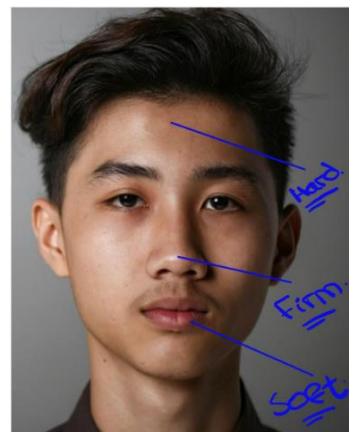
- Consistency: Reference standard: Hard = forehead; Firm = bridge of nose; Soft = lips/chin. (99% of goiters in clinical OSCE exams present as Firm; stony hard suggests carcinoma or Riedel’s thyroiditis; soft/fluctuant suggests a thyroid cyst).
- Tenderness: Suggests subacute thyroiditis (de Quervain’s) or hemorrhage into a cyst.
- Lower Border: Always palpate for the lower poles while patient swallows; if fingers can get below the lower border, retrosternal goiter is ruled out. If lower border is not palpable, suspect retrosternal goiter.
- Pemberton’s Sign: Instruct patient to raise both arms straight above their head for 1 minute.
- Positive sign: Facial congestion, plethora, cyanosis, and inspiratory stridor due to mass compression at the thoracic inlet.
- Trachea & Carotid Assessment:
- Tracheal Deviation: Stand in front of patient. Place index and ring fingers on sternoclavicular joints and middle finger gently over tracheal rings above suprasternal notch to evaluate lateral displacement.
- Carotid Pulses & Berry’s Sign: Palpate carotid pulsation bilaterally against the transverse process of the C6 vertebra (Chassaignac’s tubercle).
- Deviation vs Encasement: If the carotid pulse is displaced laterally but the superficial temporal pulse is felt, the carotid is displaced by a benign goiter. If BOTH the carotid and superficial temporal pulsations are absent, this confirms Berry’s sign (invasion and encasement of the carotid sheath by anaplastic or advanced thyroid carcinoma).
- Systematic Palpation of Cervical Lymph Nodes:
- Stand behind the patient and palpate all cervical lymph node stations in sequence (both hands may move simultaneously):
- Transverse Group: Submental (Level IA), Submandibular (Level IB), Preauricular, Postauricular, Suboccipital.
- Longitudinal Group: Upper deep cervical (Level II), Mid-jugular (Level III), Lower jugular (Level IV), Posterior triangle (Level V — test trapezius by shoulder shrugging, SCM by head rotation against resistance), Anterior central compartment (Level VI), and Supraclavicular (Virchow’s node / Troisier’s sign — ask patient to shrug shoulders slightly).
- Stand behind the patient and palpate all cervical lymph node stations in sequence (both hands may move simultaneously):
- Percussion & Auscultation:
- Percuss directly over the manubrium sterni: Retrosternal dullness indicates thoracic inlet extension.
- Auscultate over the superior pole of the thyroid lobes (where the superior thyroid artery enters): Continuous machinery murmur / systolic bruit confirms Graves’ thyrotoxicosis (distinguish from carotid bruit by light pressure or breath-holding).
Station 18: History Taking for Neck Swelling / Goiter
Structured Clinical History Checklist
- Analysis of Presenting Complaint (The Lump):
- Onset (sudden painful enlargement suggests hemorrhage into a nodule; rapid growth over weeks/months in elderly suggests anaplastic cancer or lymphoma; gradual growth over years suggests colloid goiter).
- Duration, rate of enlargement, changes in size, presence of pain.
- Communication pearl: Never touch the patient while taking history. Always use the word “swelling” rather than “mass” when speaking with the patient.
- Pulsation distinction:
- Expansile pulsation: Place fingers on opposite sides of the swelling; if fingers are pushed apart, this indicates an arterial aneurysm.
- Transmitted pulsation: Place fingers on the swelling; if fingers move up and down without separating, this indicates a solid mass overlying an artery (classic DDx: Carotid Body Tumor / Chemodectoma, located anterior to the middle third of the sternocleidomastoid; palpated one finger anterior to the SCM).
- Lymph node character: Lymphoma nodes are classically rubbery, soft, and discrete; tuberculous lymph nodes are matted and firm.
- Local Compressive Symptoms (Red Flag Symptoms):
- Dyspnea or stridor: Tracheal compression (especially on lying flat).
- Dysphagia: Esophageal compression (solids first).
- Hoarseness of voice: Unilateral recurrent laryngeal nerve infiltration (highly suspicious for thyroid malignancy until proven otherwise).
- Pain or sensation of local fullness/pressure.
- Functional Thyroid Status (Thyrotoxic vs Hypothyroid):
- Hyperthyroidism: Heat intolerance, excessive sweating, palpitation, weight loss despite increased appetite, diarrhea/hyperdefecation, anxiety, tremor, oligomenorrhea.
- Hypothyroidism: Cold intolerance, weight gain, fatigue, constipation, dry coarse skin, hair loss, menorrhagia, hoarseness, depressed mood.
- Risk Factors & Background:
- History of childhood or external head/neck ionizing radiation exposure (major risk factor for papillary thyroid cancer).
- Family history of goiter, thyroid cancer, or Multiple Endocrine Neoplasia type 2 (MEN 2A/2B: medullary thyroid cancer, pheochromocytoma, hyperparathyroidism).
- Medication history: Amiodarone, lithium, antithyroid medications. Dietary iodine exposure.
Station 19: Solitary Thyroid Nodule & Multinodular Goiter — Diagnostic Workup & DDx
Q&A & Clinical Principles
- Q1: Mention clinical differential diagnoses for an anterior/lateral neck swelling.
- Answer:
- Thyroid Mass: Colloid nodule, follicular adenoma, multinodular goiter, or thyroid carcinoma (papillary, follicular, medullary, anaplastic, lymphoma).
- Thyroglossal Duct Cyst: Midline cystic mass that moves upwards on swallowing AND on tongue protrusion.
- Cervical Lymphadenopathy: Reactive lymphadenitis, tuberculous lymphadenitis (matted nodes), lymphoma, or metastatic carcinoma.
- Branchial Cleft Cyst: Lateral neck mass along the anterior border of the upper third of SCM.
- Carotid Body Tumor: Transmitted pulsation, splaying carotid bifurcation.
- Submandibular Salivary Gland Swelling / Plunging Ranula.
- Soft Tissue Neoplasms: Lipoma, cystic hygroma (transilluminates brilliantly).
- Answer:
- Q2: Outline the sequential three-step diagnostic evaluation of a solitary thyroid nodule.
- Answer:
- Serum TSH (First-line functional test):
- If TSH is suppressed/low Perform Radionuclide Thyroid Uptake Scan (I-123 or Tc-99m): “Hot” (hyperfunctioning) nodules are almost always benign; “cold” (non-functioning) nodules carry a 15–20% risk of malignancy and require ultrasound/FNA.
- If TSH is normal or elevated Proceed directly to high-resolution neck ultrasound.
- High-Resolution Neck Ultrasound (First-line anatomical test):
- Evaluates nodule size, composition (solid vs cystic), echogenicity (hypoechoic), margins (microlobulated/spiculated), calcifications (microcalcifications), shape (taller-than-wide), and abnormal cervical lymphadenopathy (TIRADS classification).
- Ultrasound-Guided Fine Needle Aspiration (US-FNA):
- Indicated for nodules with suspicious ultrasound features or with low-risk features. Results reported via the Bethesda System for Reporting Thyroid Cytopathology (Categories I to VI).
- Serum TSH (First-line functional test):
- Answer:
- Q3: What are the surgical indications for a multinodular goiter?
- Answer:
- Compressive symptoms: Tracheal compression with stridor/airway compromise, dysphagia, or superior vena cava syndrome.
- Suspicion of malignancy or documented indeterminate/malignant cytology on FNA (Bethesda IV, V, VI).
- Secondary hyperthyroidism (toxic multinodular goiter / Plummer’s disease).
- Retrosternal / intrathoracic extension.
- Cosmetically unacceptable anterior neck mass or nodule .
- Answer:
Station 20: Graves’ Disease & Thyroid Eye Disease
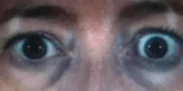
Q&A & Clinical Principles
- Q1: Describe the ocular abnormalities shown and name the primary diagnosis.
- Answer:
- Ocular Abnormalities: Bilateral upper eyelid retraction (Dalrymple’s sign with visible sclera between upper limbus and eyelid margin), bilateral proptosis/exophthalmos, and a wide-eyed staring appearance.
- Primary Diagnosis: Graves’ Disease with Graves’ Ophthalmopathy (Thyroid Eye Disease).
- Answer:
- Q2: Explain the underlying pathogenesis of ophthalmopathy in Graves’ disease.
- Answer: Autoimmune process driven by TSH-receptor antibodies (TRAb / TSI) that cross-react with TSH receptors located on retro-orbital fibroblasts and preadipocytes. This activates T-cell infiltration, releasing cytokines (TNF-, IFN-) that stimulate massive production of hydrophilic glycosaminoglycans (hyaluronic acid). The resulting severe osmotic retro-orbital fat expansion, muscle edema, and fibrosis pushes the globes forward.
- Q3: What three treatment options exist for Graves’ hyperthyroidism, and what is the definitive surgical procedure?
- Answer:
- Medical Therapy: Antithyroid drugs (Thionamides: Methimazole or Propylthiouracil / PTU in 1st trimester pregnancy) for 12–18 months + Beta-blockers (Propranolol) for immediate symptom control.
- Radioactive Iodine Ablation (RAI, I-131): Destroys hyperfunctioning thyroid follicular tissue (contraindicated in pregnancy, lactation, and severe active eye disease).
- Definitive Surgical Procedure: Total (or Near-Total) Thyroidectomy. Indicated for large goiters with compression, moderate-to-severe ophthalmopathy, suspicion of cancer, women planning pregnancy within 6 months, or patient preference. Patient must be rendered euthyroid preoperatively with thionamides and Lugol’s iodine (to decrease thyroid vascularity).
- Answer:
Station 21: Surgical Calcium Disorders & Basic Metabolic Panel (BMP)
Q&A & Clinical Interpretation (Past Exam Recalls)
-
Part 1: Basic Metabolic Panel (BMP) Lab Interpretation
Component Patient Value Reference Range Units Sodium 142 136–144 mmol/L Potassium 3.9 3.3–5.1 mmol/L Chloride 107 98–108 mmol/L 27 20–32 mmol/L BUN 10 7–22 mg/dL Creatinine 0.80 0.7–1.5 mg/dL Glucose 100 70–100 mg/dL Calcium 8.5 (Low) 8.9–10.3 mg/dL -
Q1: What is your interpretation of this laboratory panel?
- Answer: This patient has mild hypocalcemia.
-
Q2: What other test would you like to order?
- Answer: Serum albumin. (Clinical rationale: If the serum albumin level is low, this lowers the total serum calcium level [pseudohypocalcemia]; corrected calcium must be calculated).
-
Q3: What is the treatment of hypocalcemia?
- Answer (Past Exam Key Points):
- IV Calcium: Calcium gluconate or calcium chloride at () for symptomatic patients.
- Oral Calcium: Calcium citrate or calcium carbonate.
- Vitamin (Calcitriol): Increases intestinal calcium absorption.
- Answer (Past Exam Key Points):
-
Part 2: Treatment of Hypercalcemia
- Management Steps (Past Exam Key Points):
- Promote diuresis by infusion of 0.9% normal saline.
- Add KCl 20–30 mEq/L to IV fluids.
- Furosemide to enhance renal calcium excretion.
- Treat the underlying cause:
- Primary Hyperparathyroidism: Parathyroidectomy / Surgery.
- Bone metastasis / malignancy: Bisphosphonates, calcitonin, steroids.
- Management Steps (Past Exam Key Points):
4. Neurosurgery
Station 22: Epidural Hematoma (EDH)
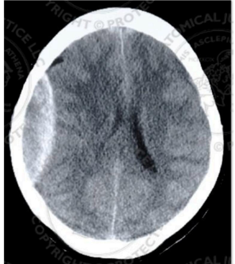
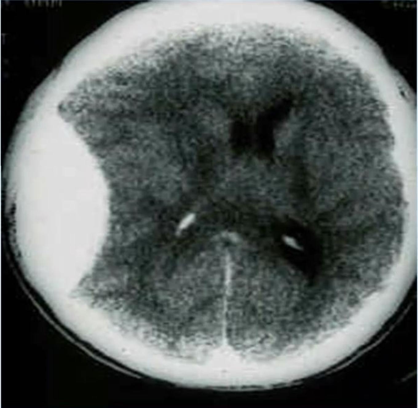
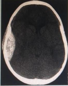
Q&A & Emergency Protocol (Past Exam Recalls)
- Scenario: A trauma victim is brought to the ER following a head injury and undergoes a non-contrast brain CT scan.
- Q1: What is the main finding / diagnosis on CT?
- Answer: Epidural hematoma (biconvex / lenticular-shaped hyperdense extra-axial collection bounded by cranial suture lines).
- Q2: What is the main cause of this condition?
- Answer: Trauma with injury to the middle meningeal artery (often associated with skull fracture at the pterion).
- Q3: What are the THREE characteristic sequences of presentation in this condition?
- Answer (Past Exam Key Points):
- Loss of consciousness (initial post-traumatic concussion).
- Regaining consciousness (the “lucid interval”).
- Loss of consciousness again (secondary rapid deterioration from expanding arterial hematoma and rising ICP).
- Answer (Past Exam Key Points):
- Q4: What is the treatment for epidural hematoma?
- Answer (Past Exam Key Points):
- Surgical Treatment (Definitive / Main Treatment):
- Emergency surgical evacuation (Craniotomy) with clot evacuation and ligation of the bleeding middle meningeal vessel.
- Minimally invasive / temporizing: Burr holes decompression, drain insertion, or endovascular embolization.
- Conservative Treatment (in stable patient with small hematoma):
- Head of bed elevation to 30°.
- Diuretics and hyperventilation (to reduce ICP).
- IV fluid support.
- Reversal of coagulopathy (Vitamin K, FFP, cryoprecipitate, protamine sulfate).
- Surgical Treatment (Definitive / Main Treatment):
- Answer (Past Exam Key Points):
Station 23: Basal Skull Fracture
Q&A & Clinical Management (Past Exam Recalls)
- Scenario: A trauma patient presents with bilateral periorbital ecchymosis (“raccoon eyes”).
- Q1: What is the diagnosis?
- Answer: Basal skull fracture (anterior cranial fossa fracture).
- Q2: Mention characteristic physical examination signs of basal skull fracture:
- Answer (Past Exam Key Points):
- Raccoon eyes (bilateral periorbital ecchymosis without direct eye trauma).
- Battle’s sign (postauricular / mastoid ecchymosis indicating petrous temporal fracture).
- CSF leak (CSF rhinorrhea from nose or CSF otorrhea from ear canal).
- Hemotympanum (blood behind the tympanic membrane).
- Answer (Past Exam Key Points):
- Q3: How do you manage this condition and what is the strict contraindication?
- Answer:
- Strict Contraindication: DO NOT insert a Nasogastric Tube (NGT) or nasotracheal tube (risk of intracranial entry through fractured cribriform plate).
- Management: Conservative management in most cases (bed rest with head elevation 30°, neurological observation for GCS deterioration and signs of meningitis). Surgery is reserved for persistent CSF leaks.
- Answer:
5. Breast
Station 24: Clinical Breast & Axillary Examination Guide
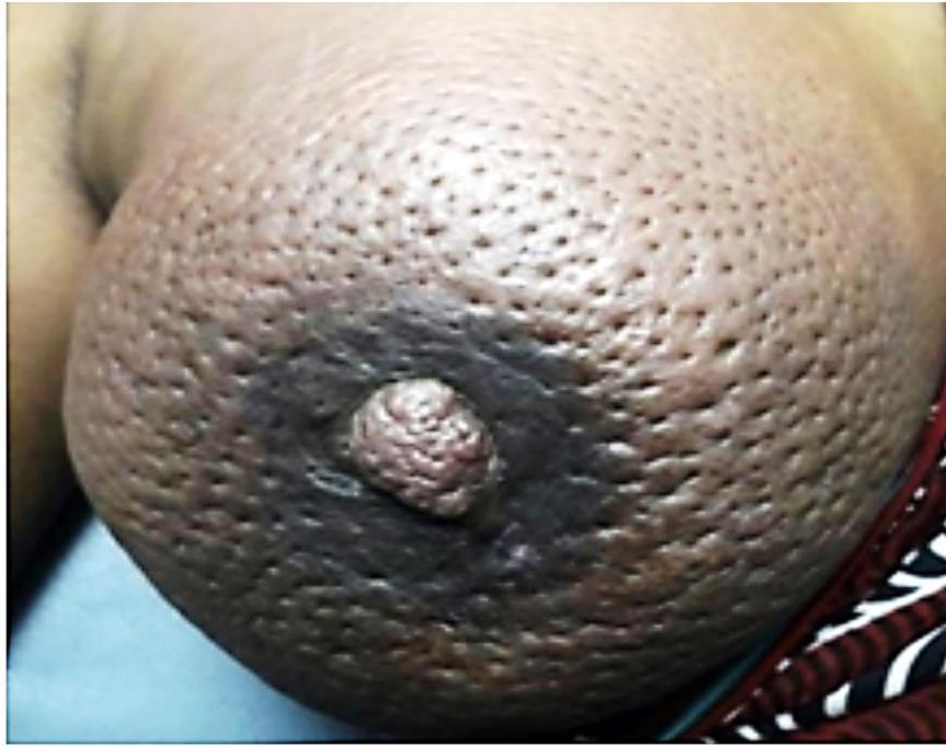
Past Exam Scenario
- Scenario: Photograph from a 55-year-old female complaining of right breast pain.
- Q1: Mention 2 clinical findings on inspection:
- Answer: Breast enlargement, Peau d’orange skin, change in skin color (or visible mass).
- Q2: Describe STEPWISE how to clinically perform palpation of the right breast:
- Answer:
- Patient lying in 45° semi-sitting position (prevents breast falling flat/laterally).
- Take permission, ensure privacy with female chaperone, and expose waist and above.
- Start palpation by the palmar surface of the fingers (not tips) in a circular manner from outside to inside (or quadrant by quadrant) covering all 4 quadrants including the nipple-areolar complex.
- Palpate the axillary lymph nodes.
- Answer:
’s Complete Clinical Checklist
- Preparation & Position:
- WIPES: Wash hands, introduce yourself, seek permission/consent, ensure strict privacy, and confirm female chaperone.
- Position: 45° semi-sitting position (if sitting upright, the breast drops down; if fully supine, the breast falls laterally). Fowler’s position: If patient states lump is felt only in a specific position, examine at 45° first, then in that position.
- Exposure: From waist and above.
- Inspection (Stand in front of patient):
- Inspect normal breast first, then diseased breast.
- Size & Symmetry: Normal/large/small for age; bilateral symmetry.
- Skin Changes: Discoloration (erythema), ulceration, visible masses, dilated veins, scars, peau d’orange, and signs of inflammation.
- Tethering: Pulling of skin inward due to tumor invading Cooper’s ligament. Dynamic maneuvers:
- Arms resting at sides.
- Hands pressed firmly against hips (contracts pectoralis major; if mass is attached to muscle, it becomes fixed/less mobile).
- Arms raised above head (accentuates inframammary fold lesions and subtle tethering).
- Nipple-Areola Complex: Retraction/inversion (congenital vs acquired), displacement along the mammary line (mid-clavicular to inguinal), deviation direction, areolar darkening (darker in malignancy/pregnancy), and discharge.
- Palpation (Stand on patient’s right side):
- Palpate normal breast first, then the affected breast.
- Technique: Use palmar surface of fingers (not tips), moving circularly from outside to inside across all 4 quadrants and the axillary tail.
- If a mass is found, describe:
- Site: Quadrant and “o’clock” position.
- Size: Measurements in cm.
- Shape & Surface: Smooth (cyst/fibroadenoma) vs irregular/nodular.
- Edge: Well-defined vs ill-defined.
- Consistency: Soft (lipoma), firm/rubbery (fibroadenoma), hard (carcinoma).
- Temperature & Tenderness: Check temperature using dorsum/nail side of fingers; observe patient’s face/eyes for tenderness.
- Mobility & Attachment: Hold mass with one hand, move with the other. Ask patient to press hands on hips: if mass becomes fixed, it is attached to pectoralis muscle/fascia. “Slip sign” is characteristic of fibroadenoma or lipoma.
- Nipple Compression: Palpate subareolar region. Squeeze nipple-areola complex with thumb and index finger (or ask patient to squeeze) to check for discharge from the 15 lactiferous ducts (pus, milky, or bloody).
- Axillary Lymph Node Palpation:
- To examine the patient’s LEFT axilla, hold and support the patient’s left elbow/forearm with your RIGHT hand (keeping arm slightly flexed and relaxed), and palpate with your other hand.
- Palpate by the bulb/pads of fingers systematically against the chest wall:
- Anterior (Pectoral) group: Posterior border of pectoralis major.
- Posterior (Subscapular) group: Anterior border of latissimus dorsi / subscapularis.
- Medial group: On serratus anterior / chest wall.
- Lateral group: Upper medial surface of arm/humerus (biceps).
- Central group: Deep in center of axilla.
- Apical group: High in apex beneath clavicle (warn patient this may cause discomfort).
- Examine supraclavicular and infraclavicular nodes.
- Past Exam Pearl (Model Station): Diffuse, matted axillary lymphadenopathy classically suggests Tuberculous (TB) lymphadenitis.
- Completion of Examination (Metastatic Survey):
- Abdominal exam: Palpate for hepatomegaly and percuss for ascites.
- Spine: Palpate/percuss lower back for low back tenderness (bone metastasis).
Station 25: History Taking for Breast Lump & Nipple Discharge
Structured History Points (Past Exam Recalls)
- Lump Analysis:
- Duration, onset, progression (rapidly enlarging vs stable).
- Pain: Painful vs painless (most carcinomas are painless).
- Menstrual relation: Changes in size or tenderness with menstrual cycle (cyclical pain/swelling suggests fibrocystic disease).
- Nipple Discharge & Retraction:
- Color: Bloody/serosanguinous (duct papilloma or carcinoma), green/brown (duct ectasia), purulent (abscess), milky (galactorrhea).
- Orifices: Unilateral vs bilateral; single duct vs multiple ducts.
- Retraction: Congenital (longstanding, bilateral, can be pulled out) vs recent acquired retraction.
- Transverse slit-like retraction: Classic for mammary duct ectasia (strongly associated with cigarette smoking).
- Risk Factors & Background:
- Menstrual & Obstetric: Age at menarche, age at menopause, age at first pregnancy, number of children, history of breastfeeding.
- Family history: Breast, ovarian, colon, or other cancers in first-degree relatives and age at diagnosis.
- Past medical/surgical: Prior breast lumps, biopsies, or chest radiation exposure.
- Smoking history.
Station 26: Breast Carcinoma
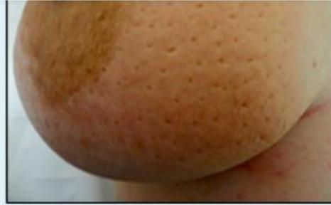
Past Exam Scenario
- Scenario: Finding during clinical examination of a 58-year-old female complaining of a painless lump in the right breast for 1 year.
- Q1: Mention TWO clinical findings observed on inspection:
- Answer:
- Peau d’orange (orange peel skin appearance).
- Nipple retraction (and elevation/inversion).
- Answer:
- Q2: What is the most likely diagnosis?
- Answer: Breast Carcinoma (Breast cancer).
- Q3: How do you confirm the definitive diagnosis?
- Answer: Core needle biopsy (Tru-cut biopsy) for histopathological examination.
- Q4: What is the pathophysiology of peau d’orange?
- Answer: Obstruction of subdermal lymphatic drainage by malignant tumor cells, leading to localized cutaneous edema with dimpling at hair follicles.
- Q5: What is the difference between multifocal and multicentric breast cancer?
- Answer:
- Multifocal: More than one tumor focus within the same quadrant.
- Multicentric: Multiple tumor foci in different quadrants of the breast (mandates mastectomy).
- Answer:
Station 27: Acute Breast Abscess & Lactational Mastitis
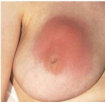
Past Exam Scenarios
- Clinical Scenario: A lactating female presents with a painful, erythematous, fluctuant swelling in her right breast for 2 days.
- Q1: What is the diagnosis?
- Answer: Acute breast abscess (complicating acute lactational mastitis).
- Q2: What is the commonest microorganism causing this condition?
- Answer: Staphylococcus aureus (including MRSA strains).
- Q3: Mention TWO key steps in the management:
- Answer:
- Incision and drainage (or needle aspiration / catheter drainage under ultrasound guidance for small, loculated abscesses).
- Systemic antibiotics (anti-staphylococcal coverage, e.g., flucloxacillin or co-amoxiclav).
- Encourage continued regular breast emptying / breastfeeding or pumping from the affected breast.
- Answer:
6. Thoracic
Station 28: Lung Tumor / Solitary Pulmonary Nodule on Chest X-Ray
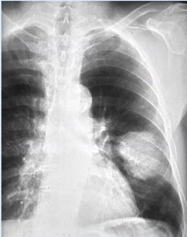
Past Exam Scenario
- Scenario: Chest X-ray from a 60-year-old man presenting with cough and hemoptysis.
- Q1: What is the main abnormality you note in this x-ray?
- Answer: Round consolidation / opacity in the middle of the left lung.
- Q2: What are the TWO most likely differential diagnoses?
- Answer:
- Small cell lung carcinoma.
- Non-small cell lung carcinoma.
- Answer:
- Q3: What further THREE specific investigations will you initially perform for a diagnosis?
- Answer:
- CT chest.
- Fiberoptic bronchoscopy with biopsy.
- MRI chest.
- Answer:
Station 29: Tube Thoracostomy (Chest Tube Insertion)
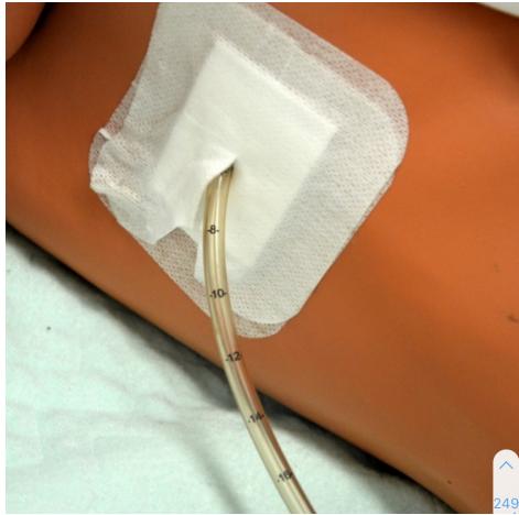
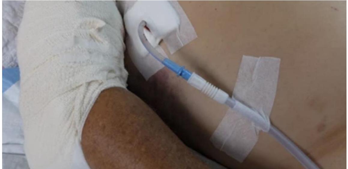
Past Exam Scenario
- Q1: Name the procedure shown in the photograph:
- Answer: Tube thoracocentesis (chest tube insertion).
- Q2: Mention THREE indications for this procedure:
- Answer:
- Pneumothorax (or tension pneumothorax after decompression).
- Hemothorax.
- Chylothorax (or empyema/pleural effusion).
- Answer:
- Q3: Mention FOUR complications associated with this procedure:
- Answer:
- Infection (empyema, cellulitis).
- Bleeding / vascular injury (intercostal vessel laceration).
- Organ penetration (lung laceration, diaphragm, liver, or spleen perforation).
- Tube blockage / kinking.
- Tube displacement / movement causing severe pain and discomfort.
- Answer:
- Key Anatomical Principles (Triangle of Safety):
- Borders: Anterior border: lateral edge of Pectoralis major; Posterior border: anterior edge of Latissimus dorsi; Base: 5th intercostal space (nipple level in males).
- Insertion Technique: Always dissect and introduce the tube over the superior border of the rib to avoid injury to the intercostal neurovascular bundle (V-A-N: Vein, Artery, Nerve) located in the subcostal groove along the inferior border of the rib. Confirm pleural entry by digital exploration. Connect to underwater seal drain.
7. Upper GI Surgery
Station 30: Achalasia Cardia
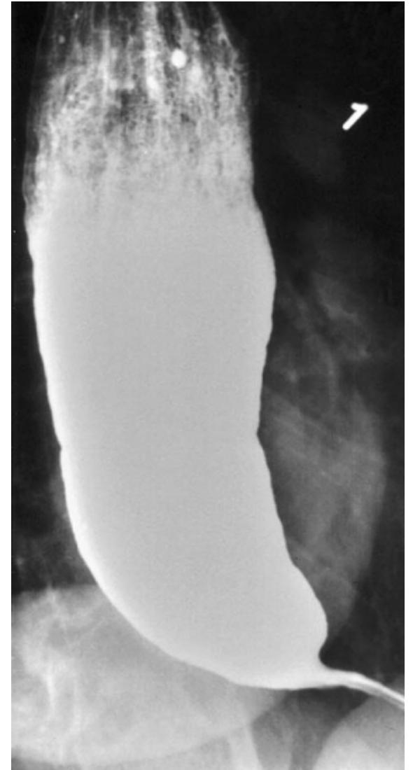
- Clinical Scenario: A 40-year-old female presents with progressive dysphagia to both solids and liquids. A contrast study is performed
- Q1: What is the most likely diagnosis?
- Answer: Achalasia Cardia.
- Q2: What imaging modality is shown and what is the characteristic finding?
- Answer:
- Modality: Barium swallow / Barium meal study.
- Characteristic Sign: Dilated esophagus with smooth, symmetrical, tapered narrowing at the gastroesophageal junction (pencil-shape, rat-tail shape, or bird’s-beak sign).
- Answer:
- Q3: What is the single best / gold standard investigation to confirm the diagnosis?
- Answer: High-resolution esophageal manometry (demonstrates incomplete/absent lower esophageal sphincter [LES] relaxation and aperistalsis in the esophageal body).
- Q4: Mention treatment modalities for this condition:
- Answer (Past Exam Key Points):
- Surgical (Definitive): Laparoscopic Heller’s myotomy (LES myotomy, typically with partial fundoplication e.g., Dor or Toupet).
- Endoscopic Interventions:
- Pneumatic balloon dilatation of the LES.
- Botulinum toxin (Botox) injection into the LES (preferred for elderly or poor surgical candidates).
- Medical / Pharmacological: Oral smooth muscle relaxants (Nitrates and Calcium Channel Blockers, e.g., nifedipine).
- Answer (Past Exam Key Points):
Station 31: Esophageal Carcinoma
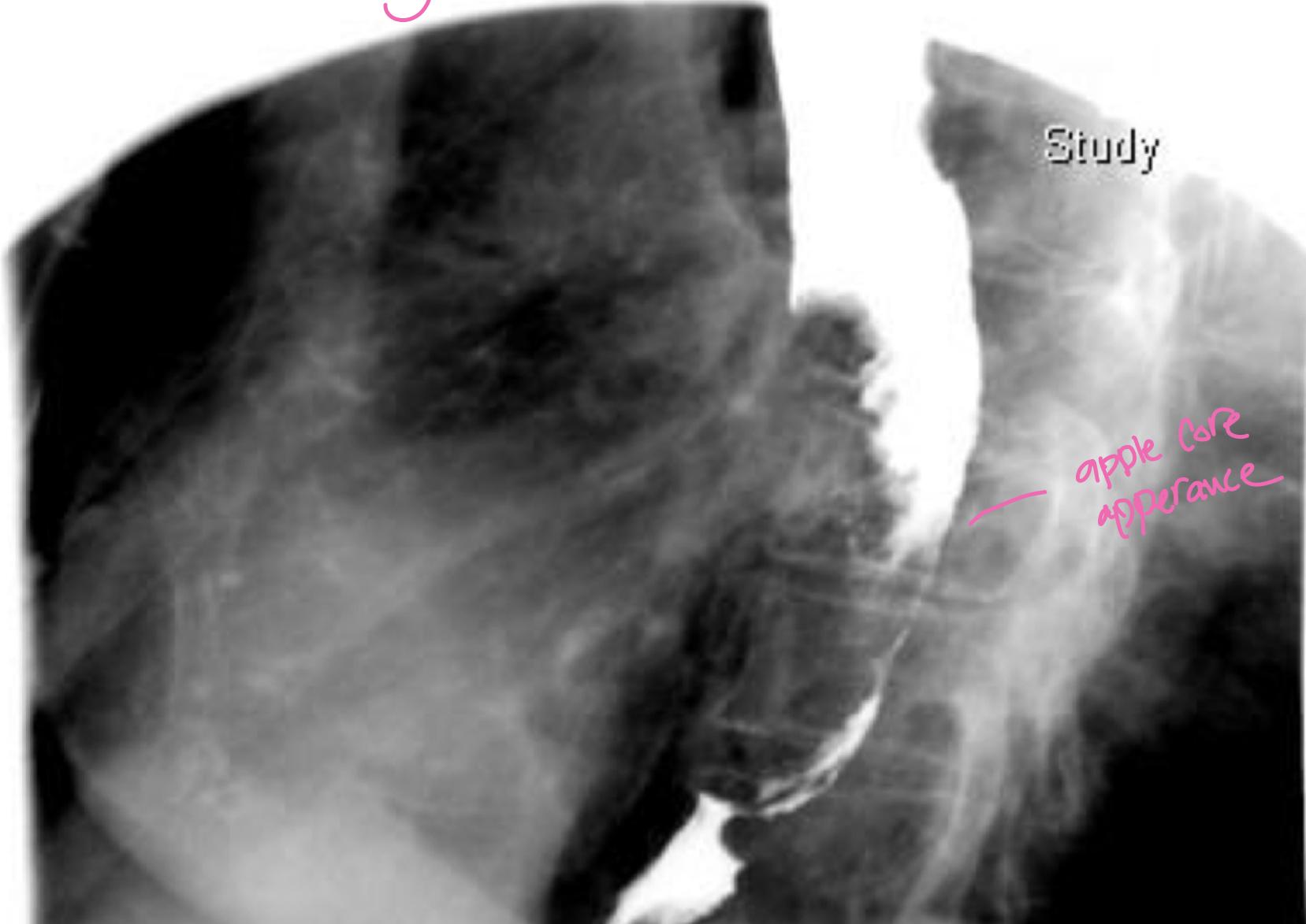
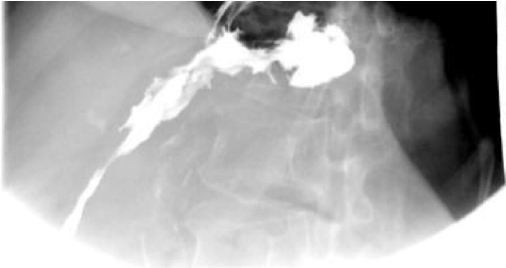
Past Exam Scenario
- Diagnostic Finding on Study: Apple core appearance on esophagogram / barium swallow (annular, irregular narrowing with shouldered margins).
- Diagnosis: Esophageal cancer (carcinoma of the esophagus).
- Clinical Presentation: Progressive dysphagia (first to solids, then progressing to liquids) and significant involuntary weight loss.
- Key Diagnostic Investigations:
- Upper GI Endoscopy with biopsy (confirms tissue diagnosis).
- CT chest and abdomen (metastatic staging).
- Endoscopic Ultrasound (EUS) (assesses depth of invasion T stage and nodal status N stage).
8. Abdominal Wall, Hernia & Stoma
Station 32: Stomas — End Colostomy vs End Loop Ileostomy
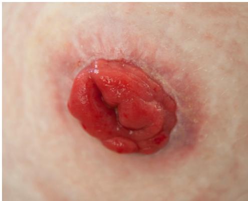
Past Exam Scenarios
- Scenario 1: Patient recently had abdominal surgery.
- Q1: What is this?
- Answer: Colostomy.
- Q2: Mention TWO indications:
- Answer:
- Bowel obstruction.
- Tumor (colorectal cancer).
- Answer:
- Q3: Mention FOUR complications:
- Answer:
- Prolapse.
- Retraction.
- Ischemia / necrosis.
- Infection.
- Answer:
- Q1: What is this?
- Scenario 2: Photograph of the abdomen from a patient who recently had abdominal surgery.
- Q1: What do you see?
- Answer: End loop ileostomy.
- Q2: Name TWO indications for this procedure:
- Answer:
- Fistula.
- Intestinal obstruction.
- Answer:
- Q3: Mention THREE complications associated with this procedure:
- Answer:
- Bleeding.
- Infection.
- Parastomal hernia.
- Answer:
- Q1: What do you see?
Core Anatomical & Clinical Comparisons
| Feature | Colostomy | Ileostomy |
|---|---|---|
| Location | Typically in Left Lower Quadrant (LLQ) | Typically in Right Lower Quadrant (RLQ) |
| Height | Flush with skin | Spouted ( protrusion to keep caustic effluent off skin) |
| Effluent | Formed, solid brown stool | Liquid to semi-solid, green-yellow fluid rich in enzymes |
Station 33: Para-umbilical Hernia
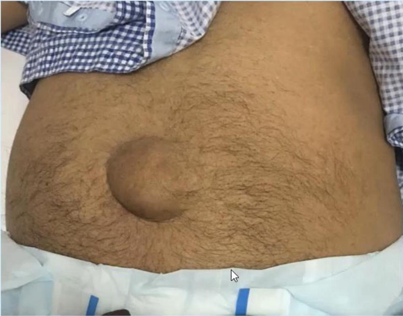
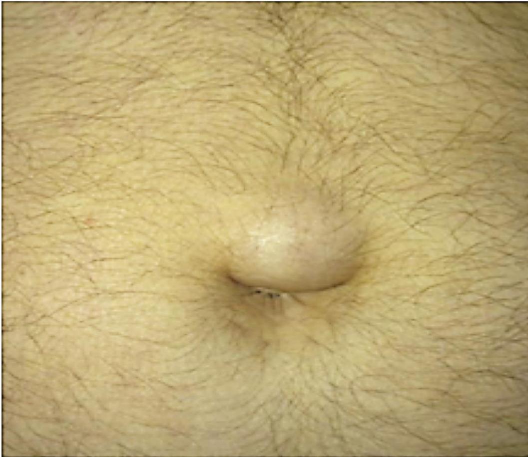
Past Exam Scenarios
- Scenario: A 40-year-old male presents with this painless abdominal wall swelling which increases in size after a day’s activity/straining and becomes smaller after night rest.
- Q1: What is the most likely diagnosis?
- Answer: Para-umbilical hernia.
- Q2: Mention FOUR possible complications of this condition:
- Answer (Past Exam Key Points):
- Incarceration (irreducibility; high risk due to the rigid, narrow fibrous defect).
- Strangulation (vascular compromise leading to bowel necrosis).
- Mechanical bowel obstruction.
- Overlying skin ulceration / infection.
- Answer (Past Exam Key Points):
- Q3: What is the definitive treatment?
- Answer: Surgical repair with prosthetic mesh (tension-free mesh hernioplasty).
Station 34: Incisional Hernia
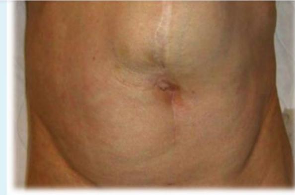
Past Exam Scenario
- Scenario: An old male complained of abdominal pain & repeated vomiting. Photograph of the abdomen is shown.
- Q1: What is the abnormality?
- Answer: Incisional hernia.
- Q2: What is the definitive treatment?
- Answer: Surgical repair for the hernia and mesh.
- Key Surgical Principles:
- Commonest cause/risk factor: Surgical site infection (SSI), followed by obesity, chronic cough, and increased intra-abdominal pressure.
- Mesh repair is mandatory to minimize high recurrence rates of primary suture repair.
Station 35: Inguinal Hernia
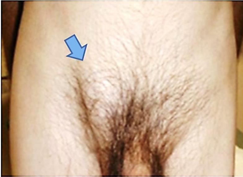
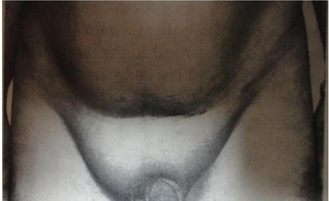
Past Exam Scenarios
- Scenario: A patient presents with a visible groin swelling.
- Q1: What is the most likely diagnosis?
- Answer: Inguinal hernia (indirect or direct inguinal hernia).
- Q2: Mention THREE differential diagnoses for an inguinal swelling:
- Answer:
- Direct or Indirect Inguinal Hernia.
- Femoral Hernia (emerges below and lateral to the pubic tubercle).
- Inguinal Lymphadenopathy (reactive or neoplastic groin lymph node enlargement).
- (Other DDx: saphena varix, hydrocele of the cord, lipoma of the spermatic cord).
- Answer:
- Q3: Mention THREE things to perform / elicit on palpation:
- Answer:
- Expansile cough impulse.
- Reducibility (check if reducible manually or on lying flat; note direction of reduction).
- Tenderness (or signs of irreducibility/strangulation).
- Answer:
- Q4: What is the definitive treatment?
- Answer: Surgical repair with prosthetic mesh (Lichtenstein tension-free open mesh hernioplasty or laparoscopic TAPP/TEP repair).
Station 36: Abdominal Wall & Inguinal Hernia Physical Examination Guide
’s Clinical Checklist & Pearls
- Preparation & Exposure:
- WIPES: Permission, privacy, chaperone.
- Exposure: From waist down to mid-thighs.
- Examine in standing position first (hernia becomes more prominent), then re-examine supine.
- Inspection:
- Look for visible bulge, symmetry, skin changes, previous scars.
- Prior Appendectomy Scar: Always note/ask about previous appendectomy (injury to the ilioinguinal nerve denervates conjoint tendon fibers, predisposing to direct/indirect right inguinal hernia).
- Ask patient to turn head and cough: Look for an expansile cough impulse.
- Inguinal vs Femoral Landmark:
- Locate the pubic tubercle:
- Inguinal Hernia: Emerges above and medial to the pubic tubercle.
- Femoral Hernia: Emerges below and lateral to the pubic tubercle.
- Locate the pubic tubercle:
- Scrotal Swelling vs Inguinoscrotal Hernia (“Getting Above the Swelling”):
- Gently palpate the neck of the scrotum:
- If you can get above the swelling, it is purely scrotal (e.g., hydrocele, epididymal cyst, varicocele).
- If you cannot get above the swelling, it is an inguinoscrotal hernia descending from the groin.
- Gently palpate the neck of the scrotum:
- Tactile Sensation on Reduction:
- Gurgling / Gargling: Characteristic of reducing hollow bowel (enterocele).
- Doughy sensation: Characteristic of reducing omentum (omentocele).
- Direction of Reduction:
- Inguinal Hernia: Reduces backward, upward, and laterally toward the deep internal ring.
- Umbilical / Para-umbilical Hernia: Reduces straight backward.
- Deep (Internal) Ring Occlusion Test:
- Reduce the hernia completely while patient is supine.
- Occlude the deep inguinal ring with your thumb ( above the midinguinal point / femoral pulse).
- Ask patient to stand and cough:
- Indirect Inguinal Hernia: The hernia remains controlled / does not appear while the ring is occluded; emerges once the thumb is released.
- Direct Inguinal Hernia: The hernia escapes / bulges forward medial to the thumb through Hesselbach’s triangle despite occlusion of the deep ring.
- Invagination Test (External Ring Impulse):
- Invaginate scrotal skin along spermatic cord toward the external ring at pubic tubercle; ask patient to cough:
- Impulse on the tip of the finger Indirect hernia.
- Impulse on the pulp / pad of the finger Direct hernia.
- Invaginate scrotal skin along spermatic cord toward the external ring at pubic tubercle; ask patient to cough:
- Wound Classification & Mesh Rules:
- Elective, uncomplicated hernia repair = Clean wound Mesh repair is safe and standard.
- Strangulated hernia with necrotic/gangrenous bowel = Contaminated / dirty wound Synthetic mesh is contraindicated; perform resection and tissue suture repair.
9. Hepatobiliary & Pancreatic Surgery
Station 37: History Taking for Jaundice & Pruritus
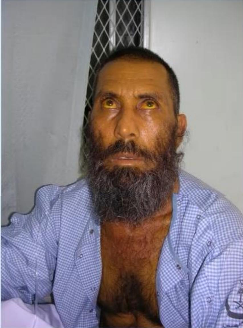
Past Exam Scenario
- Scenario: A patient presents with yellow discoloration of the sclera (jaundice) and dark urine.
- Q1: What TWO important questions will you ask regarding the presenting complaint?
- Answer:
- Pruritus (generalized itching due to cutaneous bile salt deposition).
- Pain (severe colicky RUQ pain suggestive of gallstones/choledocholithiasis vs dull/painless jaundice suggestive of periampullary malignancy). (Also ask about: Stool color [pale/clay-colored], history of travel, and history of blood transfusion).
- Answer:
- Q2: What initial imaging study is indicated?
- Answer: Ultrasound (US) of the abdomen.
Structured History Points
- Character of Jaundice: Onset, duration, progression (progressive in cancer vs fluctuating in choledocholithiasis).
- Urine & Stool: Dark, tea-colored urine (conjugated bilirubin excreted by kidneys); pale, acholic, clay-colored stool (absence of intestinal stercobilin).
- Risk Factors: Blood transfusion, dental procedures, tattoos, IV drug use, alcohol intake, travel history, family history of hemolytic diseases, drug history.
Consultant Pearls ( & Exam Recalls)
- Painless vs Painful Jaundice: Carcinoma of the head of the pancreas causes painless, progressive jaundice; choledocholithiasis or ascending cholangitis causes painful, intermittent/fluctuating jaundice.
- Laboratory Pattern: Obstructive jaundice shows markedly elevated Direct (conjugated) Bilirubin, ALP, and GGT. Initial imaging investigation is always Ultrasound (US).
- Cystic Duct Obstruction Sequence: Persistent obstruction leads to a distended gallbladder filled with clear sterile mucus (Mucocele); secondary bacterial infection progresses to Pyocele (Empyema of the gallbladder).
- Biliary Imaging Modalities:
- MRCP: Non-invasive; visualizes biliary ductal tree both above and below the obstruction.
- ERCP: Invasive; visualizes above obstruction and allows therapeutic interventions (sphincterotomy, stone extraction, stenting). All patients whose stones are cleared by ERCP should undergo elective cholecystectomy afterwards.
- PTC: Visualizes below the obstruction when ERCP is unsuccessful or anatomically impossible.
Station 38: The Gallstone Disease Spectrum & Complications
Core Exam Questions & Clinical Pearls
- Biliary Colic vs Acute Cholecystitis:
- Biliary Colic: Transient cystic duct obstruction; episodic RUQ pain lasting hours, resolves completely, no fever or leukocytosis.
- Acute Cholecystitis: Persistent cystic duct obstruction; continuous RUQ pain hours, fever, leukocytosis, positive Murphy’s sign.
- Gallstone Complications: Detailed under Station 39 below (Pancreatitis, Obstructive jaundice, Gallbladder cancer, Cholangitis).
- Gallstone Ileus:
- Small bowel mechanical obstruction caused by a large gallstone eroding into the duodenum via a cholecystoduodenal fistula and impacting at the ileocecal valve.
- Rigler’s Triad on AXR/CT: Pneumobilia (gas in biliary tree), small bowel obstruction, and ectopic radiopaque gallstone in the right iliac fossa.
Past Exam Scenario: Acute Cholangitis (Batch 16 Recall)
- Scenario: A 30-year-old female was admitted with abdominal pain, jaundice, and fever (). Labs:
- WBC: (normal 4–11)
- Total Bilirubin: (normal )
- Direct Bilirubin: (normal )
- ALT: (normal 5–30)
- AST: (normal 10–40)
- Alkaline Phosphatase (ALP): (normal 25–120)
- Q1: What is the most likely diagnosis?
- Answer: Acute Cholangitis (Ascending cholangitis presenting with Charcot’s triad of fever, jaundice, and abdominal pain).
- Q2: Mention FOUR initial management steps:
- Answer:
- Keep strictly NPO.
- IV fluid resuscitation and fluid balance/urine output monitoring.
- Broad-spectrum IV antibiotics (covering enteric gram-negative bacilli and anaerobes).
- Analgesia and close hemodynamic vital sign monitoring.
- Answer:
- Q3: Mention TWO specific diagnostic modalities:
- Answer:
- Ultrasound (US) of abdomen (initial imaging).
- ERCP (definitive diagnostic and therapeutic biliary decompression).
- Answer:
Station 39: Chronic Calculous Cholecystitis on Ultrasound
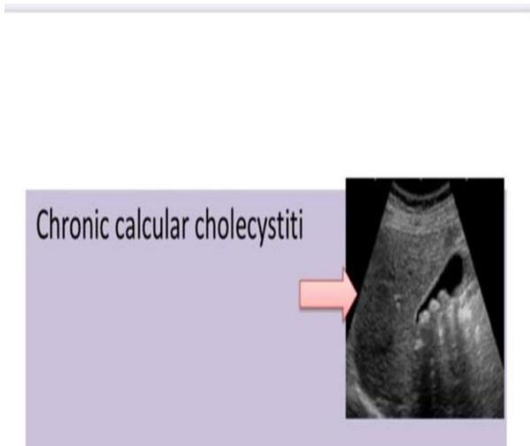
Past Exam Scenario
- Q1: What is your diagnosis?
- Answer: Chronic calculous cholecystitis (cholelithiasis with chronic cholecystitis).
- Ultrasound Findings: Distended gallbladder containing intraluminal echogenic calculi with distinct posterior acoustic shadowing, with thickened gallbladder wall.
- Q2: Give 4 complications:
- Answer:
- Pancreatitis.
- Obstructive jaundice.
- Gallbladder cancer.
- Cholangitis.
- Answer:
- Definitive Treatment: Elective laparoscopic cholecystectomy.
Station 40: Painless Obstructive Jaundice & Pancreatic Head Cancer
Core Clinical & Exam Pearls
- Courvoisier’s Law (Courvoisier’s Sign):
- “In the presence of jaundice, an enlarged, palpable, non-tender gallbladder is unlikely to be due to gallstones, and is most likely due to a malignant obstruction of the common bile duct or head of pancreas.”
- Rationale: Chronic gallstone disease causes recurrent inflammation and fibrosis, leaving the gallbladder shrunken and non-distensible. Malignancy causes gradual distal obstruction, progressively distending a normal elastic gallbladder.
- Most Likely Diagnosis: Adenocarcinoma of the head of the pancreas (or periampullary carcinoma).
- Definitive Curative Surgery: Pancreaticoduodenectomy (Whipple procedure).
Station 41: Acute Pancreatitis & Hemorrhagic Signs
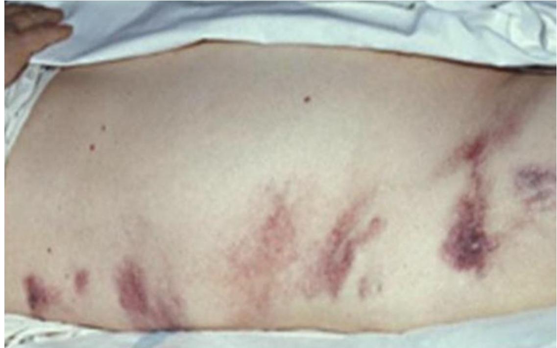
Past Exam Scenarios
- Scenario 1 (Laboratory Panel Recall): A 40-year-old female was admitted with sudden & severe epigastric pain. Investigations showed:
- Total bilirubin: ()
- ALT: ()
- AST: ()
- ALP: ()
- Amylase: ()
- Q1: What is the most likely diagnosis?
- Answer: Acute pancreatitis.
- Q2: What are TWO main causes of this condition?
- Answer: Gallstones, Alcohol.
- Scenario 2: A 45-year-old female was admitted with severe upper abdominal pain. Two days after admission she developed this flank skin lesion (Grey Turner’s sign).
- Q1: What are the initial FIVE steps in the management of this patient?
- Answer:
- ABC (Airway, Breathing, Circulation assessment).
- IV fluid resuscitation (aggressive crystalloid hydration).
- Analgesia (IV opioids).
- NPO (nil per os / bowel rest).
- Monitor vitals and urine output (via Foley catheter).
- Answer:
- Q2: Mention TWO local complications:
- Answer:
- Pancreatic pseudocyst.
- Pancreatic necrosis (or abscess).
- Answer:
- Q1: What are the initial FIVE steps in the management of this patient?
- Clinical Ecchymosis Signs:
- Grey Turner’s Sign: Flank ecchymosis from retroperitoneal hemorrhage tracking along tissue planes.
- Cullen’s Sign: Periumbilical ecchymosis tracking along the falciform ligament.
10. Colorectal & Anorectal Surgery
Station 42: Ascites Physical Examination
Past Exam Recall & Examination Steps
- Exam Task: Demonstrate special physical examination tests for ascites.
- Three Key Tests:
- Shifting Dullness:
- Patient supine. Percuss from umbilicus laterally toward the flank until resonant note turns dull.
- Keep finger at dull point, ask patient to roll onto opposite side; wait 20–30 seconds for fluid to shift.
- Percuss again: Note shifts from dull to resonant (confirms mobile intraperitoneal fluid).
- Fluid Thrill (Fluid Wave):
- Indicated in massive/tense ascites.
- Patient places ulnar border of hand firmly along the midline of the abdomen.
- Tap one flank sharply while feeling for a transmitted impulse with the palm on the opposite flank.
- Digital Rectal Examination (PR):
- Palpation reveals fullness or bulging in the rectovesical pouch (males) or pouch of Douglas (females).
- Shifting Dullness:
Station 43: Digital Rectal Examination (PR) & Proctoscopy
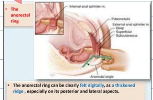
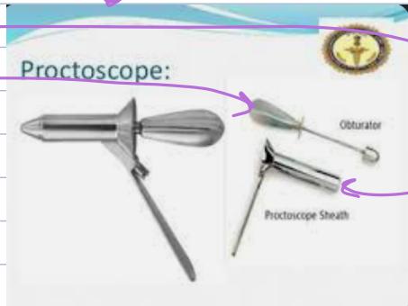
Past Exam Scenario
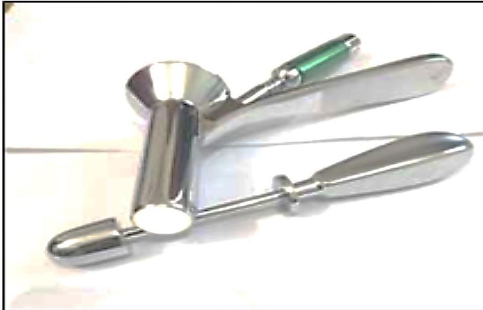
- Q1: What is this tool?
- Answer: Proctoscope (rigid tubular speculum with removable obturator and light source).
- Q2 (A): Mention TWO of its diagnostic uses:
- Answer:
- Examine the anorectal area to diagnose polyps / lesions.
- Take directed mucosal biopsies. (Also: visualize and grade internal hemorrhoids, inspect the dentate line).
- Answer:
- Q2 (B): Mention TWO of its therapeutic uses:
- Answer:
- Banding of internal hemorrhoids (rubber band ligation).
- Ligation or snare polypectomy of polyps (and injection sclerotherapy).
- Answer:
Clinical Digital Rectal Examination (DRE / PR) Guide
- Indications: Change in bowel habits, rectal bleeding, evaluation of prostate, urinary/fecal incontinence.
- Contraindications:
- Absolute: Absence or imperforate anus, severe anal pain (e.g., acute anal fissure), anal stricture, unwilling/non-consenting patient.
- Relative: Acute abdomen, severe coagulopathy, major rectal trauma, recent acute myocardial infarction.
- Complications: Pain and discomfort, perianal skin tearing, abrasion of hemorrhoids, transient bacteremia.
- Stepwise Clinical PR Procedure ():
- Preparation & Position: WIPES (introduce self, permission/consent, privacy, chaperone). Exposure from umbilicus to mid-thigh. Position in Sims’ position (left lateral decubitus with hips and knees flexed).
- Inspection: Separate buttocks gently; inspect perianal skin for external hemorrhoids, anal fissures, fistula external openings, perianal abscess, ulcers, and warts.
- Palpation: Place lubricating gel (e.g., 2% lidocaine) on gloved index finger; massage anal verge with finger pulp to relax sphincter; gently insert finger pointing towards the umbilicus; assess sphincter tone (ask patient to squeeze); sweep finger 360° feeling rectal mucosa (smooth vs granular, polyps, tumors, ulcers); palpate prostate anteriorly in males (feel median sulcus and lateral lobes: smooth enlargement in BPH vs hard nodular asymmetry/obliterated sulcus in carcinoma); palpate anorectal ring posteriorly; withdraw finger and inspect glove tip for blood, mucus, or pus.
’s Clinical Pearls on PR & Proctoscopy
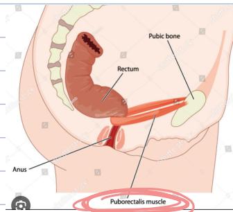
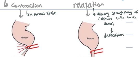
- Puborectalis Muscle: Originates from pubic bone and slings around anorectal junction; tonic contraction maintains the normal acute anorectal angle for continence.
- Direction of Insertion: Insert pointing towards the umbilicus conforming to anal canal axis determined by the puborectalis muscle.
- Why Normal Piles (Internal Hemorrhoids) Cannot be Palpated Digitally: They are soft, compressible vascular cushions of submucosal veins that empty under digital pressure; they can only be seen via proctoscopy (unless thrombosed or 4th-degree prolapsed).
Station 44: Sessile Colonic Polyp
Past Exam Scenario
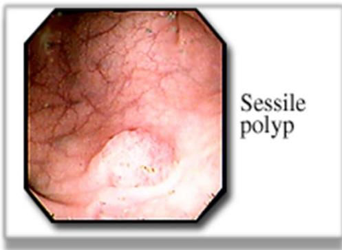
- Scenario: Colonoscopic view of a 55-year-old male who complained of rectal bleeding.
- Q1: What is the seen abnormality?
- Answer: Sessile colonic polyp (broad-based mucosal polyp without a stalk).
- Q2: What is the clinical significance of this abnormality?
- Answer: It has a high potential for malignancy / can lead to malignant transformation (adenoma-to-carcinoma sequence).
- Management: Complete endoscopic mucosal resection (EMR) with histopathological evaluation.
Station 45: Pseudomembranous Colitis
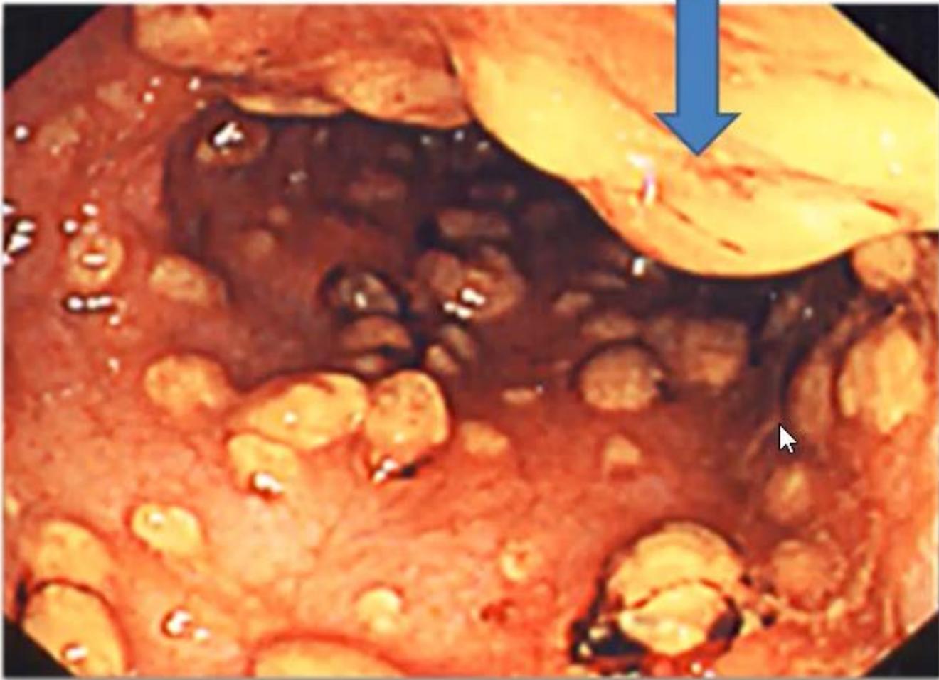
Past Exam Scenario
- Scenario: A perforated appendectomy patient receiving broad-spectrum antibiotics developed fever and foul-smelling greenish watery diarrhea on the 5th postoperative day. Sigmoidoscopy finding is shown.
- Q1: What is your diagnosis?
- Answer: Pseudomembranous colitis (Clostridioides difficile colitis).
- Q2: What is its specific treatment?
- Answer:
- Stop the offending antibiotic (or change to a narrower spectrum agent).
- Specific antimicrobial therapy: Oral Vancomycin (or oral fidaxomicin / metronidazole).
- Answer:
Station 46: Acute Perianal Abscess
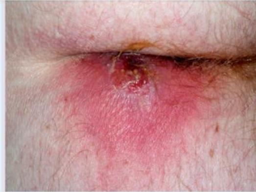
Past Exam Scenario
- Scenario: View of perianal area for a male patient who presented with perianal pain and swelling for 2 days.
- Q1: What is your diagnosis?
- Answer: Perianal abscess.
- Q2: Mention TWO steps in the management:
- Answer:
- Incision and drainage (I&D).
- Antibiotics.
- Answer:
- Complication: Formation of a fistula-in-ano (perianal fistula).
11. Urology
Station 47: Renal Cell Carcinoma (RCC)
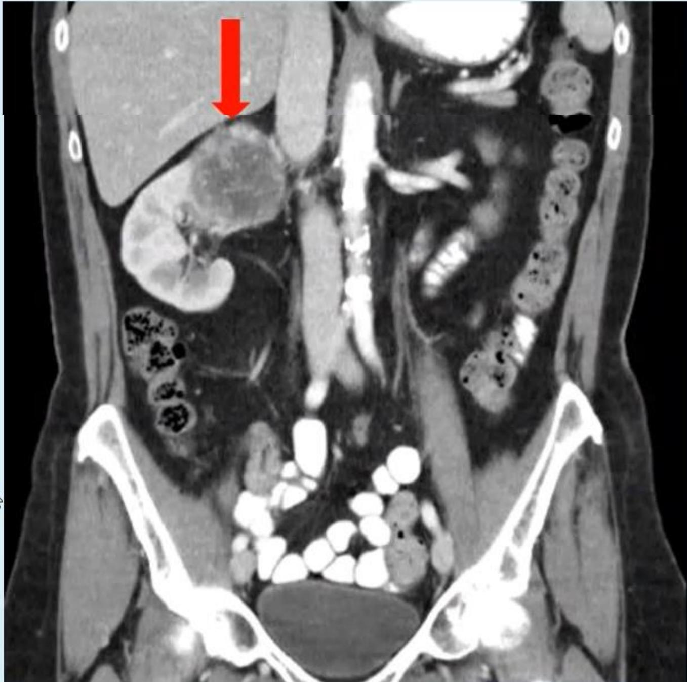
Past Exam Scenario
- Scenario: Coronal CT scan from a 62-year-old male presenting with right loin pain and hematuria.
- Q1: What are TWO possible renal neoplasms?
- Answer:
- Renal cell carcinoma (RCC).
- Angiomyolipoma (or oncocytoma).
- Answer:
- Q2: What is the main treatment for your 1st diagnosis?
- Answer: Radical nephrectomy (or partial nephrectomy if ).
Station 48: Urinary Bladder Carcinoma
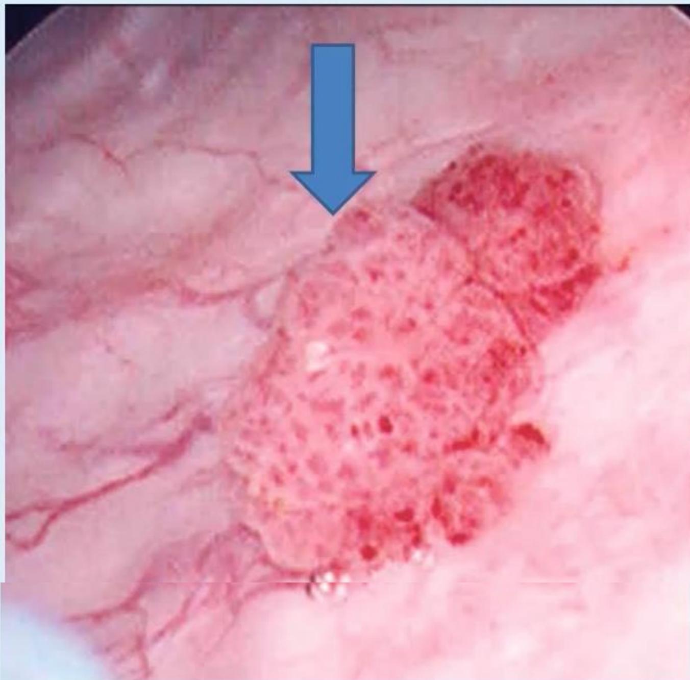
Past Exam Scenario
- Scenario: A 65-year-old male presented with painless hematuria. Cystoscopy finding is shown.
- Q1: What is the most likely diagnosis?
- Answer: Bladder cancer (Transitional cell carcinoma / Urothelial carcinoma).
- Q2: Mention TWO risk factors:
- Answer:
- Cigarette smoking.
- Chemical / industrial aromatic amine or aniline dye exposure.
- Answer:
- Q3: What is the main treatment of this condition?
- Answer: Resection: Transurethral Resection of Bladder Tumor (TURBT / TUR-BC), or radical/partial cystectomy for invasive disease.
Station 49: Hydrocele & Scrotal Examination
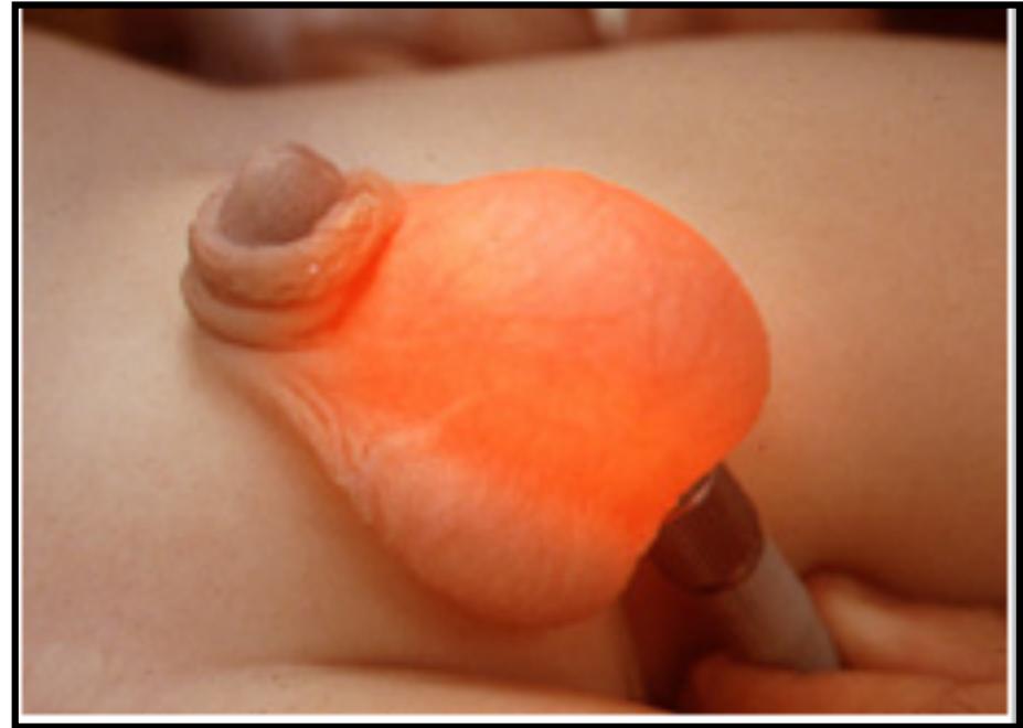
Past Exam Scenario
- Q1: What is the diagnosis?
- Answer: Hydrocele (abnormal collection of fluid within the tunica vaginalis).
- Q2: What is the name of the test? And what is the finding?
- Answer:
- Test Name: Transillumination test.
- Finding: Positive illumination (conduction and red shining of light throughout the fluid-filled scrotum).
- Answer:
- Q3: Give THREE further physical examinations / investigations:
- Answer:
- Ultrasound of scrotum with duplex.
- Cough impulse test (negative in hydrocele, positive in hernia).
- Fluctuation test (positive in hydrocele).
- Reducibility test (irreducible in hydrocele).
- Answer:
- Definitive Treatment: Lord’s plication (for small/thin-walled hydroceles) or Jaboulay’s eversion of the tunica vaginalis (for large thick-walled hydroceles).
12. Vascular Surgery
Station 50: Lower Limb Peripheral Vascular Examination
Complete Examination Checklist (‘s Framework)
- Preparation & WIPES:
- Expose both lower extremities completely from groins down to toes; position patient supine.
- Inspection:
- Trophic Skin Changes: Thin, shiny, atrophic skin; loss of hair on digits and lower leg; thickened, brittle toenails; muscle wasting (calf).
- Color Changes: Pallor, dependent rubor, cyanosis, mottled appearance.
- Lesions / Ulcers: Interdigital areas, tips of toes, heels, pressure points, surgical bypass scars.
- Palpation:
- Temperature: Use the dorsum of fingers to feel temperature gradient from toes proximally; compare bilaterally.
- Capillary Refill Time (CRT): Compress great toe pulp for 5 seconds; normal refill is seconds.
- Peripheral Arterial Pulses (Sequential Bilateral Palpation):
- Femoral Pulse: At the midinguinal point (midway between ASIS and pubic symphysis).
- Popliteal Pulse: Knee flexed 30–45°, thumbs on tibial tuberosity, fingertips pressing deep into midline of popliteal fossa against posterior tibia.
- Posterior Tibial Pulse: Midway between medial malleolus and Achilles tendon ( below and behind medial malleolus).
- Dorsalis Pedis Pulse: On the dorsum of the foot just lateral to the Extensor Hallucis Longus (EHL) tendon.
- Special Provocative Tests:
- Buerger’s Test: Elevate both legs to 45° for 1–2 minutes. Look for pallor (angle indicates critical ischemia). Have patient hang legs over bed edge: Ischemic foot becomes intense dusky red (“Sunset Foot” / Rubor of Dependency).
- Ankle-Brachial Index (ABI): Ratio of highest ankle systolic pressure to highest brachial systolic pressure ( normal; PAD; severe ischemia).
Station 51: History Taking for Peripheral Arterial Disease (PAD)
Structured History Checklist (‘s Past Exam Recall)
- Clinical Scenario: A 50-year-old patient presents with severe crampy pain in the left calf that started 3 months ago, worsening after walking 200 meters, rated 9/10 in severity.
- Key History Questions:
- Claudication Distance: Walking distance that consistently triggers crampy calf/thigh pain, relieved completely by standing still for minutes.
- Rest Pain: Severe, burning forefoot/toe pain worse at night when supine, relieved by hanging the foot over the bed (indicates critical limb ischemia).
- Vascular Risk Factors: Heavy smoking (packs/day, duration), Diabetes Mellitus, Hypertension, Dyslipidemia.
- Comorbidities: Previous myocardial infarction, stroke, transient ischemic attacks, family history of premature vascular disease.
- Medications: Antiplatelet therapy (aspirin, clopidogrel), statins, antihypertensives.
Past Exam Scenario: Intermittent Claudication (Batch 16 Recall)
- Scenario: A 60-year-old male comes to your clinic complaining of pain in the back of the leg after walking for about 500 meters, which forces him to sit down to relieve the pain.
- A — Three further questions to ask regarding the presenting complaint:
- Claudication distance & progression: Does the pain consistently appear after walking 500 meters, and has this distance been shortening over time?
- Relief by rest: Does the pain resolve completely within 2 to 5 minutes of standing still?
- Presence of rest pain: Do you experience severe, burning pain in the foot/toes at night while lying in bed, and is it relieved by hanging your foot over the edge of the bed? (Also: Inquire about cardiovascular risk factors: smoking packs/day, diabetes mellitus, hypertension, hyperlipidemia).
- B — Three clinical signs to look for on inspection of the affected lower limb:
- Skin color changes: Pallor on elevation, cyanosis, and dependent erythema/rubor (“sunset foot”).
- Trophic skin changes: Loss of hair over toes and shins, shiny atrophic skin, brittle/thickened toenails.
- Ischemic lesions: Ulcerations (punched-out ulcers on toes/heels) or gangrene (dry black necrotic tissue).
- Muscle wasting: Atrophy of calf muscles compared to the contralateral leg.
- C — Three clinical signs to look for on palpation of the affected lower limb:
- Skin temperature: Relative coolness/coldness compared to the contralateral limb (palpate using the dorsum of your hand).
- Peripheral arterial pulses: Diminished or absent pulses (dorsalis pedis, posterior tibial, popliteal, femoral).
- Capillary refill time: Prolonged refill () over the toes.
- Buerger’s angle of circulatory insufficiency: Elevation pallor developing at followed by delayed reactive hyperemic rubor on dependency.
Station 52: Acute Limb Ischemia (ALI)
Core Principles & Emergency Protocol
- The Classic “6 P’s” of Acute Limb Ischemia:
- Pain (sudden onset, severe, unrelenting).
- Pallor (waxy white initially, then mottled).
- Pulselessness (absent distal pulses).
- Paresthesia (numbness, tingling; earliest sign of nerve ischemia).
- Paralysis (loss of motor function; indicates impending muscle necrosis).
- Poikilothermia / Perishingly Cold (limb adopts ambient temperature).
- Immediate Emergency Management:
- Systemic Anticoagulation: Immediate IV Heparin bolus (5000 units) followed by continuous infusion to halt thrombus propagation.
- Urgent vascular surgical evaluation within the 6-hour golden window.
- Revascularization: Urgent surgical embolectomy with Fogarty balloon catheter, catheter-directed thrombolysis, or surgical bypass.
- Reperfusion Complications:
- Compartment Syndrome: Produce severe muscle swelling within rigid fascial compartments mandates prophylactic four-compartment below-knee fasciotomy.
- Reperfusion Syndrome: Release of toxic metabolites (hyperkalemia, severe lactic acidosis, myoglobinuria causing acute renal failure).
Station 53: Differential Diagnosis of Lower Extremity Ulcers
Comparison Table
| Feature | Arterial (Ischemic) Ulcer | Venous (Stasis) Ulcer | Neuropathic (Diabetic) Ulcer |
|---|---|---|---|
| Location | Distal: tips of toes, heel, lateral malleolus | Gaiter zone (medial lower leg above medial malleolus) | Pressure sites: Plantar surface of great toe, metatarsal heads |
| Edges | Punched-out, sharp, well-defined | Sloping, irregular, shallow | Punched-out surrounded by thick hyperkeratotic callus |
| Base / Floor | Pale, dry, gray/black slough, no granulation | Pink/red, moist, exudative, florid granulation | Deep, granular or sloughy; often penetrates to tendon or bone |
| Pain | Severely painful, worse with elevation/night | Dull ache, relieved by leg elevation | Painless (loss of protective pain sensation) |
| Pulses | Absent or diminished | Present (may be masked by edema) | Usually palpable and bounding |
Station 54: Diabetic Foot Ulcer & Infection
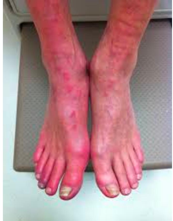
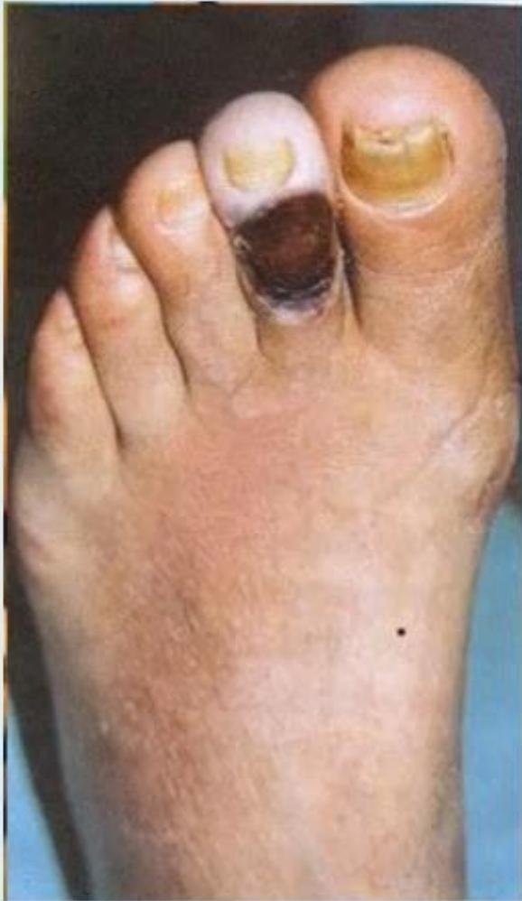
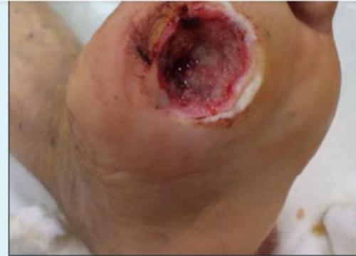
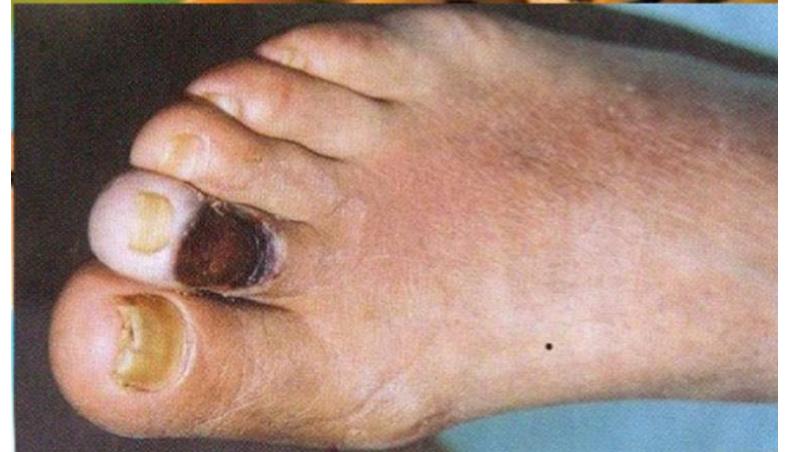
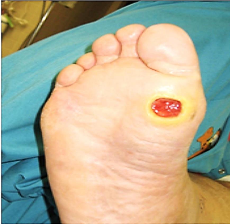
Past Exam Scenarios & Unified Q&A
- Clinical Presentation: Diabetic and vasculopathic patients presenting with lower limb ulcers, gangrenous toe lesions, claudication, or localized erythema and edema.
- Q1: What is the diagnosis?
- Answer: Diabetic foot ulcer (trophic/neuropathic ulcer over pressure points) or Arterial / ischemic ulcer (necrotic toe ulcer secondary to peripheral arterial disease).
- Q2: Mention TWO factors in the pathogenesis of this condition:
- Answer (Past Exam Key Points):
- Neuropathy (sensory loss of protective pain sensation leading to unrecognized repetitive trauma, motor intrinsic muscle wasting leading to foot deformities, and autonomic dry cracked skin).
- Vasculopathy / Ischemia (macrovascular and microvascular occlusive arterial disease impairing perfusion and wound healing).
- Answer (Past Exam Key Points):
- Q3: Mention major complications associated with this lesion:
- Answer (Past Exam Key Points):
- Chronic Osteomyelitis (deep bone infection).
- Charcot foot / joint (neuroarthropathy with joint collapse and rocker-bottom deformity).
- Wet gangrene and severe necrotizing soft-tissue infection requiring emergency debridement or amputation.
- Answer (Past Exam Key Points):
- Q4: Mention key clinical examinations to perform on palpation of the affected lower limb:
- Answer (Consolidated from Past Exam Batches):
- Peripheral arterial pulses: Dorsalis pedis, posterior tibial, popliteal, and femoral pulses.
- Capillary refill time: Comparing both feet.
- Skin temperature: Comparing both limbs using the dorsum of the hand.
- Palpation of ulcer base: Gently feeling the base (or probe-to-bone test; a crumbling biscuit sensation indicates underlying osteomyelitis).
- Sensation testing: Semmes-Weinstein 10g monofilament and vibration sensation.
- Presence of edema: Evaluating pitting vs non-pitting swelling.
- Answer (Consolidated from Past Exam Batches):
- Q5: Mention specialized / non-laboratory investigations to perform:
- Answer:
- Duplex ultrasound of the arterial and venous systems.
- Ankle-Brachial Index (ABI) using handheld Doppler.
- Plain X-ray of the foot (to rule out osteomyelitis, cortical erosion, or soft-tissue gas).
- Answer:
‘s Clinical Examination Pearls
- Golden Rule: Leave the ulcer for the very end of the examination; thoroughly inspect and palpate the rest of the limb first.
- Ulcer Floor vs Base: “The floor you see, the base you touch!” (Red granulation = healing; yellow = slough; black = necrosis).
- Bone Palpation (“Probe-to-Bone” test): If pressing or probing the ulcer base feels like “crushing a dry biscuit”, this confirms chronic osteomyelitis.
- The 4 “-pathies” of Diabetes: Neuropathy, Retinopathy, Nephropathy, Microangiopathy.
- Microbiology: Single commonest organism: Staphylococcus aureus (chronic ulcers are polymicrobial).
- Dry vs Wet Gangrene: Dry = sterile ischemic mummification (managed outpatient); Wet = active bacterial infection + tissue necrosis (surgical emergency requiring urgent IV antibiotics and debridement/amputation).
- Three Pillars of Treatment:
- Infection control (broad-spectrum IV antibiotics + radical debridement).
- Mechanical offloading (non-weight bearing / total contact cast).
- Revascularization (angioplasty or surgical bypass).
Station 55: Lower Limb Amputation & Flap Designs
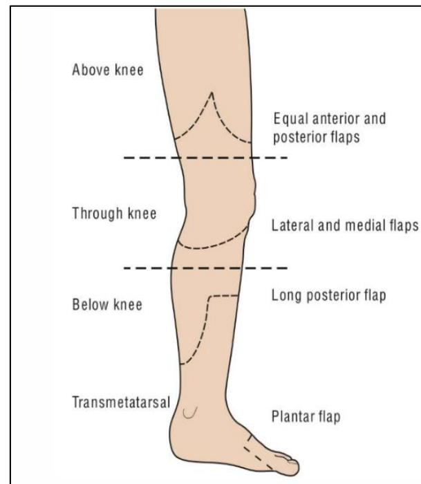
Flap Design Matching Table (Exam Recall)
| Amputation Level | Surgical Flap Design |
|---|---|
| Above-Knee Amputation (AKA / Transfemoral) | Equal anterior and posterior flaps |
| Through-Knee Amputation (Disarticulation) | Lateral and medial flaps |
| Below-Knee Amputation (BKA / Transtibial) | Long posterior flap (Burgess technique) |
| Transmetatarsal Amputation (TMA) | Long plantar flap |
Core Exam Questions
- Indications for Amputation (The “3 D’s” Rule):
- Dead: Irreversible extensive tissue necrosis or gangrene from vascular disease.
- Deadly: Rapidly spreading life-threatening sepsis (gas gangrene, severe necrotizing fasciitis).
- Dead loss (Damn nuisance): Severe intractable rest pain, paralysis, or malignancy (osteosarcoma, melanoma).
Station 56: Lower Limb Varicose Veins
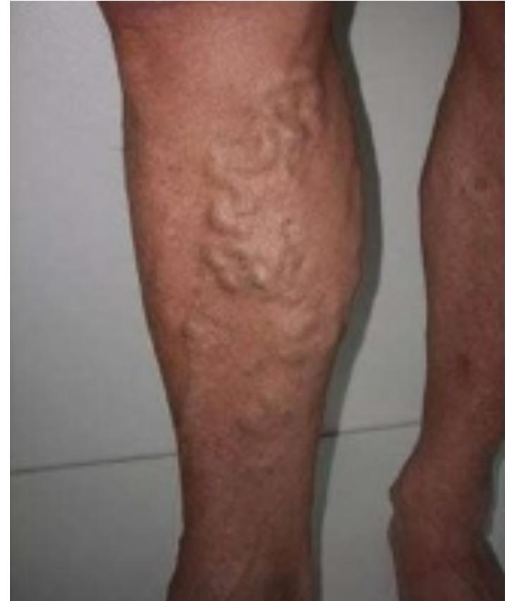
Past Exam Scenario
- Scenario: A patient presents with a dull ache in his leg on prolonged standing. Prominent, dilated, tortuous superficial veins are seen.
- Q1: Mention FOUR questions to ask in history:
- Answer:
- Prolonged standing (occupational or sports).
- History of immobility or prior Deep Vein Thrombosis (DVT).
- Family history of varicose veins.
- History of local leg trauma or previous venous surgery. (In females: oral contraceptive use, number of pregnancies).
- Answer:
- Q2: Mention TWO complications:
- Answer:
- Venous stasis ulceration (typically medial malleolus / gaiter area).
- DVT or superficial thrombophlebitis.
- Variceal bleeding / hemorrhage.
- Answer:
- Q3: Mention ONE specialized investigation you will initially order:
- Answer: Duplex ultrasound of lower extremity venous system.
- Treatment Modalities: Endovenous thermal ablation (EVLA / RFA), foam sclerotherapy, or surgical high ligation and stripping.
Station 57: Deep Vein Thrombosis (DVT)
Past Exam Scenario
- Scenario: A post-abdominal surgery patient complained of left lower limb pain and swelling. Photograph shows asymmetric left leg edema.
- Q1: What is your diagnosis?
- Answer: Deep Vein Thrombosis (DVT).
- Q2: How do you confirm the diagnosis?
- Answer: Doppler ultrasound (compression duplex ultrasonography) or venogram/angiogram.
- Q3: What are the three predisposing factors (Virchow’s Triad)?
- Answer:
- Venous stasis (post-op immobility, prolonged bed rest).
- Endothelial injury (surgery, trauma).
- Hypercoagulability (malignancy, surgery, thrombophilia).
- Answer:
- Treatment: Therapeutic anticoagulation (DOACs like Rivaroxaban/Apixaban, or LMWH bridged to Warfarin) for a minimum of 3 months.
13. Skin & Soft Tissue
Station 58: Carbuncle
Q&A & Clinical Principles
- Q1: Define a carbuncle and name the most common causative microorganism.
- Answer:
- Definition: An infective, necrotizing subcutaneous inflammatory mass formed by the coalescence of multiple adjacent infected hair follicles (furuncles).
- Causative Organism: Staphylococcus aureus (including MRSA).
- Answer:
- Q2: Explain how a carbuncle looks on inspection.
- Answer (Past Exam Key Points):
- A tender, painful, intensely erythematous, indurated subcutaneous inflammatory swelling occurring on thick, hair-bearing skin (classically the nape of the neck, upper back, or thighs).
- Features a necrotic sloughy center studded with multiple discharging cutaneous follicular sinuses releasing pus, producing a classic “sieve-like” or “honeycomb” appearance.
- Answer (Past Exam Key Points):
- Q3: What underlying medical condition must be actively investigated in every patient presenting with a carbuncle?
- Answer: Uncontrolled Diabetes Mellitus (check fasting blood glucose, HbA1c, and urine for glucose/ketones; carbuncles are classic markers of diabetic immunocompromise).
- Q4: Outline the treatment of a carbuncle (both conservative and surgical).
- Answer (Past Exam Key Points):
- Conservative Treatment:
- Strict glycemic control (sliding scale insulin therapy).
- High-dose broad-spectrum anti-staphylococcal antibiotics (IV Flucloxacillin or Cefazolin; IV Vancomycin if MRSA suspected).
- Analgesia and local antiseptic wound care.
- Surgical Treatment:
- Cruciate (cross-shaped) Incision and Drainage: Incise the mass across both axes down to deep fascia, unroof all loculi, debride devitalized necrotic slough and subcutaneous fat, saucerize the cavity, and pack with antiseptic ribbon gauze (e.g., povidone-iodine) to heal by secondary intention.
- Complete surgical excision of the necrotic infected core in extensive cases.
- Conservative Treatment:
- Answer (Past Exam Key Points):
Station 59: Acute Paronychia
Q&A & Clinical Principles
- Q1: Define paronychia and state the typical causative organism.
- Answer:
- Definition: A localized, acute superficial infection and abscess involving the soft tissue fold immediately adjacent to the lateral border or base of the fingernail (eponychium / paronychium).
- Causative Organism: Staphylococcus aureus; occasionally Streptococcus pyogenes or oral anaerobes (nail biting).
- Answer:
- Q2: How does management differ between early cellulitis and established fluctuant paronychia?
- Answer:
- Early non-fluctuant stage (cellulitis): Conservative management with warm water/saline soaks for 15 minutes 3–4 times daily, oral anti-staphylococcal antibiotics (Cephalexin or Amoxicillin-clavulanate), and finger elevation.
- Established fluctuant abscess: Surgical drainage: Elevate the lateral nail fold away from the nail plate using a small scalpel blade (#11 blade) or blunt elevator without cutting the skin. If pus extends beneath the nail plate (subungual abscess), partial or complete removal of the nail plate is required.
- Answer:
Station 60: Fournier’s Gangrene
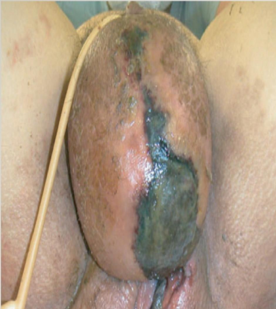
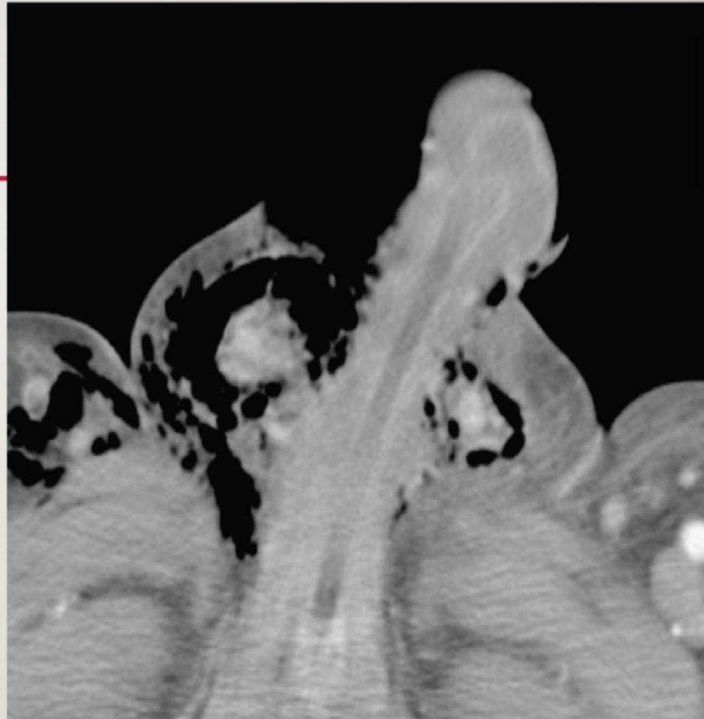
Q&A & Emergency Management
- Q1: Define Fournier’s Gangrene, its microbiological etiology, and typical predisposing factors.
- Answer:
- Definition: A fulminant, rapidly progressive, life-threatening necrotizing fasciitis of the perineal, perianal, and external genital regions.
- Microbiology: Polymicrobial synergy (Type I necrotizing fasciitis) combining aerobes and anaerobes: Gram-negative bacilli (E. coli, Klebsiella, Proteus), Gram-positive cocci (Staphylococcus aureus, Enterococci, Streptococci), and anaerobes (Bacteroides fragilis, Clostridium species).
- Predisposing Factors: Diabetes Mellitus (in ), chronic alcoholism, immunosuppression, end-stage renal disease, local trauma, perianal abscess, urethral strictures, or extravasation of infected urine.
- Answer:
- Q2: Describe the key findings visible on the pelvic CT scan.
- Answer: Extensive subcutaneous and fascial thickening, soft-tissue edema, and widespread pockets of gas (crepitus) dissecting along the fascial planes of the perineum, scrotum, and anterior abdominal wall (Buck’s, Colles’, and Scarpa’s fascia).
- Q3: Detail the emergency surgical and medical management protocol.
- Answer:
- Immediate aggressive fluid resuscitation and correction of electrolyte/acid-base derangements.
- Broad-spectrum empiric IV triple antimicrobial therapy: Vancomycin (for MRSA) + Piperacillin-Tazobactam / Carbapenem (for Gram-negatives and pseudomonas) + Clindamycin (inhibits bacterial protein/toxin synthesis and covers anaerobes).
- Emergency Radical Surgical Debridement: Immediate excision of all non-viable, necrotic skin, subcutaneous tissue, and fascia until healthy, bleeding, viable tissue margins are reached (testes usually spared due to separate testicular arterial blood supply from aorta).
- Mandatory re-exploration / second-look debridement in the operating room within 24 hours. Urinary diversion (suprapubic catheter) and fecal diversion (colostomy) if perianal/urethral involvement is extensive.
- Answer:
Station 61: Pilonidal Sinus Disease
Past Exam Scenario
- Scenario: A young male presented with recurrent purulent discharge and discomfort at the lower part of his back.
- Q1: What is your diagnosis?
- Answer: Pilonidal sinus (Sacrococcygeal pilonidal disease).
- Q2: Mention THREE important aspects of its treatment:
- Answer (Past Exam Recalls):
- Good local hygiene & hair removal with laser (permanent epilation to eliminate hair accumulation).
- Incision and drainage with curettage in acute cases (for acute pilonidal abscess).
- Excision with healing by secondary intention or flap reconstruction in chronic cases (e.g., Karydakis flap, Bascom cleft-lift, or Limberg flap to flatten the intergluteal cleft).
- Answer (Past Exam Recalls):
- Pathogenesis: An acquired condition resulting from friction, sitting, and deep cleft. Shed hairs drill into skin follicles due to the friction of walking buttocks, provoking foreign-body granulomatous reaction and secondary epithelialized sinus tracts.
Station 62: Malignant Melanoma
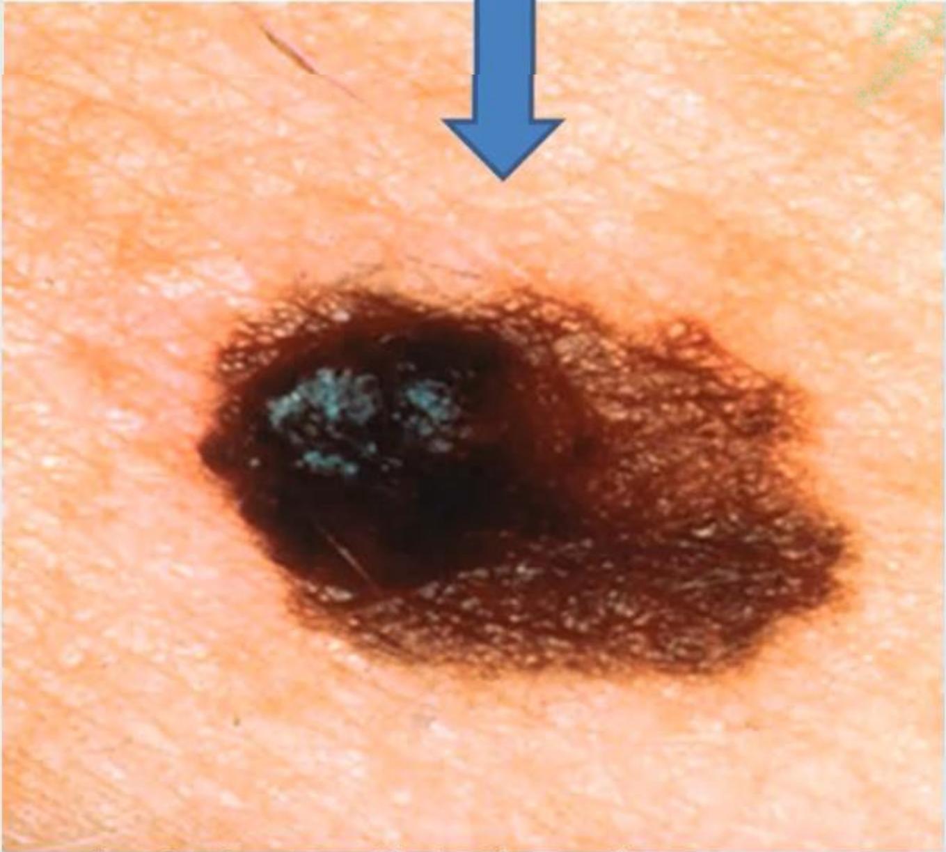
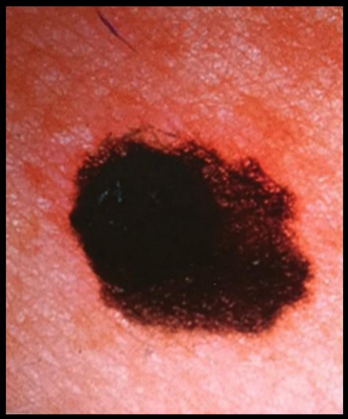
Past Exam Scenario
- Scenario: A patient presents with a pigmented skin lesion over the lower limb which has increased in size recently.
- Q1: What is the most likely diagnosis?
- Answer: Melanoma (Superficial spreading melanoma).
- Q2: How would you confirm the diagnosis?
- Answer: Complete excisional biopsy (deep excision with a narrow normal skin margin extending into subcutaneous fat; avoid partial/incisional biopsies).
- Q3: Mention TWO factors that determine the stage of the disease:
- Answer:
- Breslow tumor thickness (depth of invasion in mm) and presence of ulceration.
- Regional lymph node involvement (TNM staging / clinical stage).
- Answer:
- Clinical Criteria (ABCDE):
- A — Asymmetry: Unequal halves.
- B — Border Irregularity: Notched, scalloped, or blurred edges.
- C — Color Variegation: Multiple shades (brown, black, blue, red, white).
- D — Diameter: Greater than .
- E — Evolving: Rapid changes in size, shape, elevation, or bleeding.
- Definitive Wide Local Excision Margins:
- Melanoma in situ: .
- Thickness : .
- Thickness : .
- Thickness : .
- Sentinel Lymph Node Biopsy (SLNB): Indicated for Breslow depth or with adverse histological features.
14. Minimally Invasive Surgery (MIS) / Laparoscopy
Station 63: Laparoscopic Principles & Pneumoperitoneum Physiology
Core Principles & Q&A
- Q1: Which gas is used to create pneumoperitoneum and why?
- Answer: Carbon Dioxide (). Rationale:
- Highly soluble in blood and tissues (rapidly absorbed and eliminated by the lungs).
- Non-flammable and non-combustible (safe with electrocautery and lasers).
- Extremely low risk of fatal gas embolism if inadvertently injected intravascularly compared to air or oxygen.
- Answer: Carbon Dioxide (). Rationale:
- Q2: What is the standard intra-abdominal insufflation pressure maintained during laparoscopy?
- Answer: 12 to 15 mmHg in adults (ideal 15 mmHg; lower in pediatric and cardiopulmonary compromised patients, 8–10 mmHg).
- Q3: Differentiate open (Hasson) vs closed technique for peritoneal access.
- Answer:
- Open Technique (Hasson): Direct cut-down incision through the infraumbilical skin, fascia, and peritoneum under direct vision, followed by insertion of a blunt cannula anchored with stay sutures. Preferred in patients with prior abdominal surgery.
- Closed Technique: Blind puncture of the anterior abdominal wall using a spring-loaded Veress needle to establish pneumoperitoneum before first trocar insertion.
- Answer:
- Q4: What are the complications associated with laparoscopic access and gas insufflation?
- Answer:
- Access Complications: Injury of the skin and bleeding of abdominal wall layers (inferior epigastric vessels); puncture/injury of intra-abdominal viscera (bowel, stomach, liver); injury to major retroperitoneal vessels (aorta, IVC, iliac vessels).
- Gas Insufflation Complications: Hypotension (compression of IVC reducing venous return/preload); vagal bradycardia (from acute peritoneal stretch); hypercapnia and respiratory acidosis (systemic absorption); gas embolism; subcutaneous emphysema.
- Late Complications: Trocar site hernia (if fascia not closed), port-site infection.
- Answer:
Station 64: Veress Needle Insertion & Pneumoperitoneum Establishment
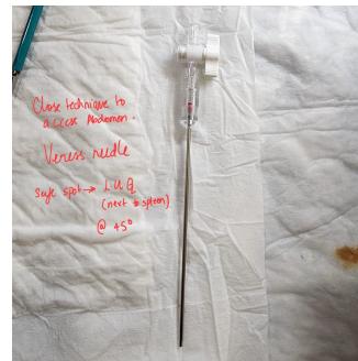
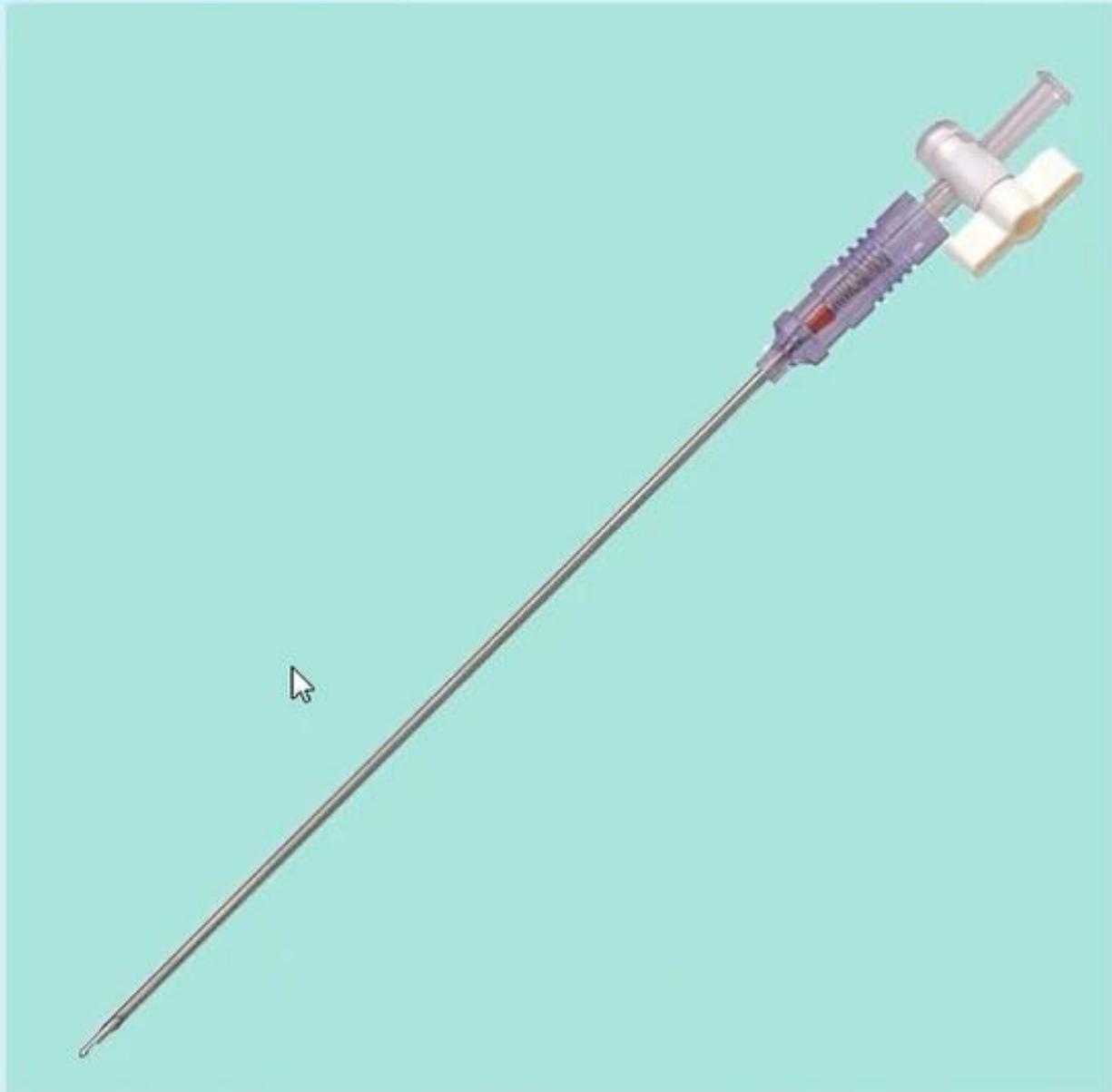
Q&A & Clinical Principles
- Q1: Name this instrument and its primary use.
- Answer: Veress Needle. Used for gas insufflation to create closed pneumoperitoneum in laparoscopic surgery.
- Q2: Explain its safety mechanism.
- Answer: It consists of a sharp outer cannula surrounding an internal, spring-loaded blunt-tipped stylet. When advancing through resistant abdominal wall fascia, the blunt stylet retracts inside, exposing the sharp needle tip. Once the needle enters the low-resistance peritoneal cavity, the blunt stylet immediately springs forward to shield the sharp tip, protecting underlying viscera and blood vessels from puncture.
- Q3: What safety checks confirm that the Veress needle is safely within the peritoneal cavity before connecting insufflation?
- Answer:
- Tactile / Auditory Feedback (“Two Clicks”): Examiner feels/hears two distinct mechanical clicks or pops during controlled advancement — the first when penetrating the rectus fascia/aponeurosis, and the second when piercing the parietal peritoneum.
- Saline Hanging Drop Test: Place a few drops of saline into the open hub of the needle; elevate the lower abdominal wall manually. The water drops disappear quickly (sucked down into the peritoneal cavity by negative intra-abdominal pressure).
- Aspiration Syringe Test: Connect a syringe containing 5 mL saline; aspirate. If blood, bowel contents, or urine is aspirated, the needle is in a major vessel, bowel, or bladder. If negative, irrigate and re-aspirate freely.
- Low Initial Insufflation Pressure: Connect tubing; initial resting intra-abdominal pressure at low flow () must be .
- Answer:
- Q4: What alternative safety site is used for Veress insertion in patients with previous midline scars?
- Answer: Palmer’s Point: Located in the Left Upper Quadrant (LUQ), below the left costal margin in the midclavicular line (safest alternate entry because it is free of adhesions in patients with previous lower abdominal surgery; ensure stomach is decompressed with NGT).
Station 65: Laparoscopic Trocars, Cannulas & Port Closure
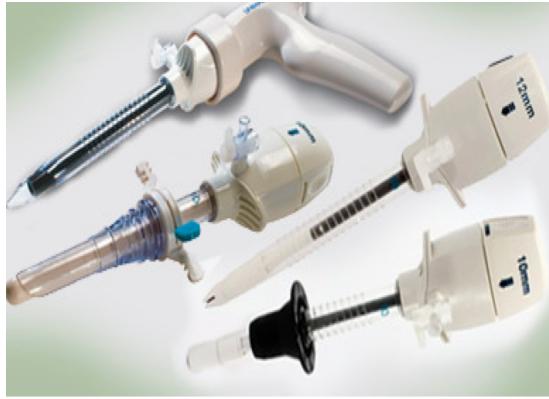
Q&A & Clinical Principles
- Q1: What is this instrument, its common sizes, and primary uses?
- Answer:
- Name: Trocar and Cannula (optical trocar also known as Visiport).
- Common Sizes: 5 mm and 10 mm / 12 mm.
- Primary Uses:
- Operative access conduit for introducing laparoscopic instruments (graspers, scissors, hook, stapler) and the laparoscope/camera during procedures such as laparoscopic cholecystectomy, appendectomy, or hernia repair.
- Side port with stopcock valve for continuous gas insufflation.
- Draining intraperitoneal fluid / suction irrigation.
- Answer:
- Q2: Describe the safe manual technique for secondary trocar insertion.
- Answer:
- Palm-Support & Index Finger Guard Technique: Hold the head of the trocar firmly against the palm of the dominant hand to deliver controlled axial thrust with gentle rotatory movement at 90° to the skin. Extend the index finger straight along the cannula shaft towards the tip as a mechanical stopper/guard; this prevents sudden, uncontrolled plunging into the abdominal cavity and protects against catastrophic injury to the abdominal aorta, IVC, or bowel.
- Secondary trocars must ALWAYS be inserted under direct laparoscopic visual control.
- Answer:
- Q3: What internal sealing mechanism prevents gas escape during instrument exchange?
- Answer: The trocar cannula contains two internal airtight valves (a primary flapper/duckbill valve and an elastic reducer seal) that maintain pneumoperitoneum when instruments are inserted or removed.
- Q4: Which trocar port sites require fascial closure at the end of the procedure and why?
- Answer:
- All trocar port sites require formal fascial closure (suturing the rectus sheath / aponeurosis) using heavy absorbable suture (e.g., #0 or #1 PDS/Vicryl).
- Rationale: Prevention of Trocar Site Hernia (TSH), which can lead to early Richter’s hernia, small bowel incarceration, and strangulation.
- Answer:
Station 66: Laparoscopic Hand Instruments & Advanced Energy Devices
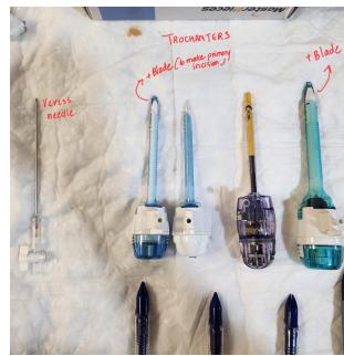
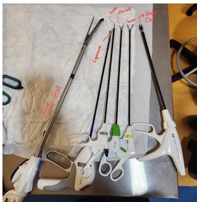
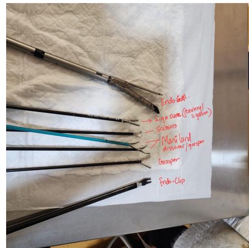
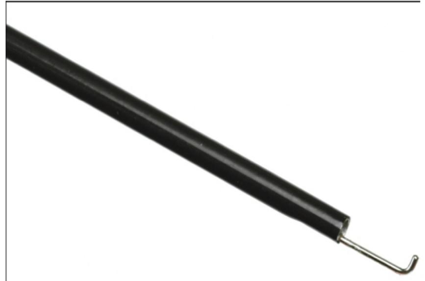
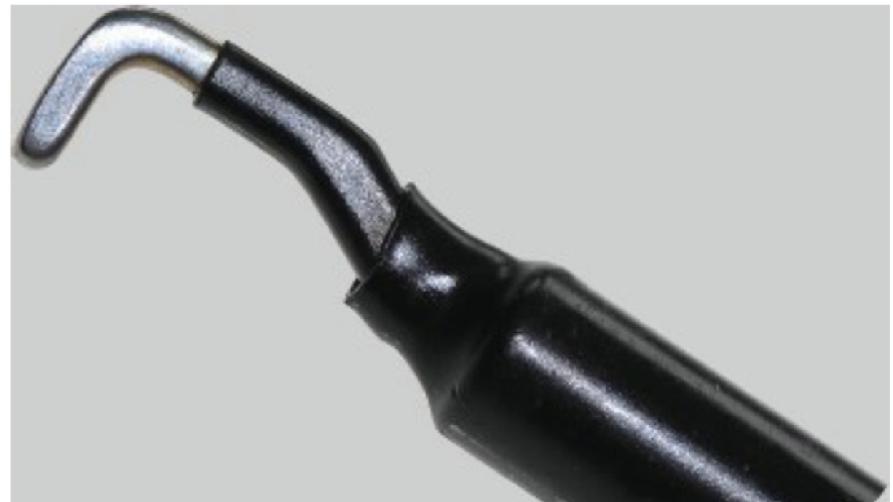
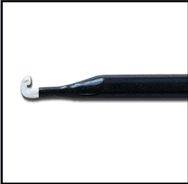
Comprehensive Instrument Guide & Q&A
- Q1: Identify the instrument and state its surgical functions.
- Answer:
- Instrument: Laparoscopic Hook (L-Hook) Electrode.
- Type of Surgery: Laparoscopic procedures.
- Functions:
- Precise dissection and separation of adhesions (adhesiolysis).
- Monopolar diathermy electrocautery for tissue cutting and hemostasis.
- Retraction and traction on tissues / peritoneal scoring.
- Answer:
- Q2: Describe the clinical functions of the following laparoscopic hand instruments:
- Maryland Dissector: Curved, tapering, fine-pointed double-action jaws with delicate serrations; used for fine blunt dissection, separating anatomical planes, and encircling tubular structures (e.g., cystic duct and cystic artery in Calot’s triangle).
- Atraumatic Laparoscopic Grasper (e.g., Bowel / Johan Grasper): Broad, flat, fenestrated jaws without sharp teeth; designed for gentle handling, retraction, and traction on delicate hollow viscera (small bowel, stomach, colon) without crush injury.
- Toothed / Traumatic Grasper (e.g., Claw / Gallbladder Grasper): Heavy, interlocking teeth; used for firm traction on resilient or discarded tissues, such as grasping the gallbladder fundus during cholecystectomy. (Crucial exam warning: Do NOT use on healthy viable bowel due to severe crush injury and perforation risk!)
- Laparoscopic Scissors: Sharp dual-action blades used for sharp mechanical cutting of tissues and dividing tubular structures.
- Endoscopic Clip Applier (Hem-o-lok / Titanium): Delivers individual titanium or polymer locking clips (sizes 5 mm, 10 mm). Primary uses: 1- Definitive ligation of the cystic duct in laparoscopic cholecystectomy; 2- Clipping the cystic artery or mesenteric bleeding vessels to secure hemostasis.
- Q3: What are the three core actions and primary application of LigaSure?
- Answer:
- LigaSure (Advanced Bipolar Vessel Sealer):
- Holds the tissue firmly with calibrated jaw pressure.
- Secures hemostasis by denaturing collagen and elastin to permanently seal vessels up to in diameter.
- Cuts the tissue via an integrated mechanical cutting blade.
- Clinical Application: Used during laparoscopic sleeve gastrectomy to divide the short gastric vessels and mobilize the greater curvature of the stomach away from the greater omentum.
- LigaSure (Advanced Bipolar Vessel Sealer):
- Answer:
- Q4: What is the Endo GIA Stapler and its two primary applications?
- Answer: An articulating laparoscopic linear cutter-stapler that places staggered rows of titanium staples while simultaneously dividing tissue between them.
- Two Primary Indications (Past Exam Recalls):
- Bowel Resection (e.g., dividing bowel loops during laparoscopic colectomy or enterectomy).
- Bowel Anastomosis (creation of gastrointestinal, jejunojejunal, or ileocolic anastomoses; also gastric sleeve transection).
15. Pediatric Surgery
Station 67: Undescended Testis (Cryptorchidism) & Empty Scrotum
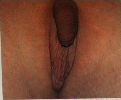
Past Exam Scenario
- Scenario: A 10-month-old child (or 1-year-old child) is brought by parents to your clinic with an abnormal scrotum.
- Q1: What clinical findings do you observe on inspection?
- Answer (Past Exam Recalls):
- Empty scrotum: Underdeveloped, hypoplastic, smooth hemiscrotum with loss of normal rugae and absence of palpable testis in the scrotal sac.
- Absence of inguinal swelling: Inguinal canal region appears flat without an obvious visible bulge or mass.
- Normal overlying skin: No erythema, signs of acute inflammation, sinuses, or scars.
- Answer (Past Exam Recalls):
- Q2: What TWO investigating tools can be used to establish the diagnosis?
- Answer:
- Ultrasound (US) of the inguinal canal and scrotum.
- Diagnostic Laparoscopy (gold standard for non-palpable intra-abdominal testis).
- Answer:
- Q3: What is the recommended age for treatment and the specific surgical procedure?
- Answer:
- Recommended Age: Between 6 and 12 months of age (classically performed around 10 months of age; spontaneous descent is rare after 6 months, and surgery before 18 months prevents irreversible germ cell degeneration).
- Specific Treatment: Orchidopexy (one-staged or two-staged Fowler-Stephens procedure according to the height and vascular tethering of the testis).
- Answer:
- Q4: Name major complications associated with untreated cryptorchidism:
- Answer (Past Exam Recalls):
- Infertility / Subfertility: Due to higher intra-abdominal temperature impairing spermatogenesis.
- Testicular Malignancy: 4- to 10-fold increased risk of germ cell tumors (Seminoma most common).
- Testicular Torsion: Increased mobility and abnormal mesentery predisposing to acute torsion.
- Associated Inguinal Hernia: Present in due to patent processus vaginalis.
- Answer (Past Exam Recalls):