SURGERY 2
Brain Abscess
Dr. Safdar Malik
Definition
Form within 2 weeks (short time)
become symptomatic
-
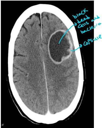
Brain abscess is a focal suppurative infection within the brain parenchyma, typically surrounded by a vascularized capsule.
-
Cerebritis: is often employed to describe a nonnencapsulated brain abscess.
Brain Abscess
Bone in the size and start compressing Should be removed
Pathophysiology
Risk Factors
Signs & symptoms
Evaluation
Treatment
Epidemiology
- Relatively uncommon
- Incidence~.3-1.3:100000 person per year
Etiology
Brain abscess may develop by
- Direct spread from a contiguous cranial site of infection
- Head trauma, neurosurgical procedures
- Hematogenous spread → Just like mets spread
(any problem in face, it go directly into brain)
25% cases : There is no primary source of infection
Predisposing conditions
-
Otitis media
-
Mastoiditis
-
Paranasal sinusitis
-
Pyogenic infection of chest or any other part of body
-
Penetrating head injury
-
Neurosurgical procedure
-
Dental infection → eg. from dental procedures
-
There is an artery that is communicating with intracranial arteries. So infection can spread
Cont.
In Immunocompetent persons:
- Streptococcus spp. (aerobic, Anaerobic and viridans) 40%
- Enterobacteriaceae (Proteus, E.coli, Klebsella) 25%
- Anaerobes (Bacteroides, Fusobacterium) 30%
- Staphylococci 10%
- Taenia solium(NCC)
- Mycobacterial infection (tuberculoma)
Cont.
In immuno-compromised host
- Nocardia
- T gondii
- Aspergillus
- Candida
- C. neoformans
→ in patients already suffering from brain tumors, HIV …
Stages (14 days)
- Early Cerebritis: 1-3 days → head ache, flu like symptoms
A perivascular infiltration of inflammatory cells around a central core of coagulation necrosis
- Late Cerebritis: 4-9 days
Pus formation in necrotic center which is surrounded by inflammatory cells and fibroblast
- Early Capsule Formation: 10-13 days
A capsule that is better develop on cortical then on ventricle side of lesion
- Late Capsule Formation: beyond 14 days
A well defined necrotic center surrounded by a dense collageous capsule
Clinical Presentation
- Typically presents as an expanding intracranial mass rather than as a infectious process
- Symptoms are gradual in onset
- Patients present in weeks to month
- Usually presents 11-12 days following onset of symptoms.
Symptoms
-
Classical triad: seen in <50% patients
-
Headache 75%
-
Fever 50% most common symptoms
-
Focal neurologic deficit 15-35%
Cont.
-
Focal neurologic deficit
-
Aphasia
-
Hemiparesis
-
Visual field defect
-
Ataxia
-
Nystagmus
-
Seizures → have some localization → Temporal, Frontal, Parietal lobes are all different
-
Raised ICP-Papilledema
-
Meningismus Uncommon unless abscess rupture in ventricle
vomiting, LOC, seizures, empyema Can form if abscess form between sub dural or extradural
- history taking is very imp. (ask about flu like symptoms)
Investigations
-
TLC, DLC
-
ESR, CRP
-
Blood cultures
-
Neuroimaging studies:
-
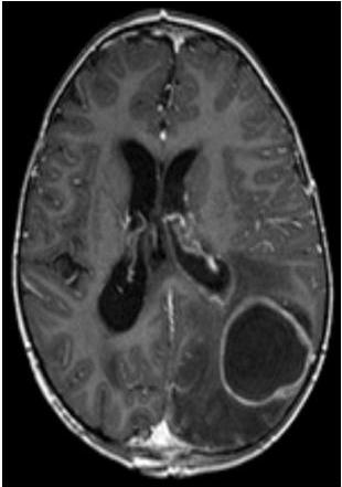
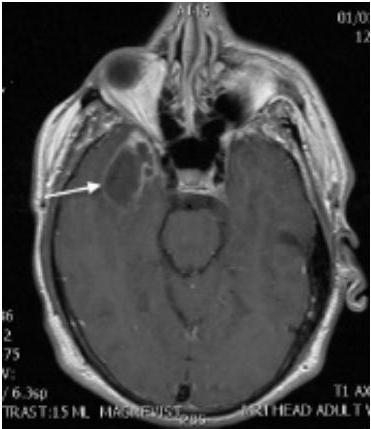
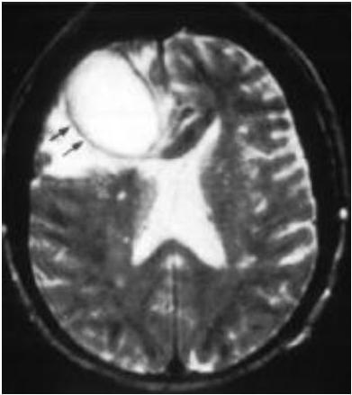
MRI: better esp can detect early stages of cerebritis
-
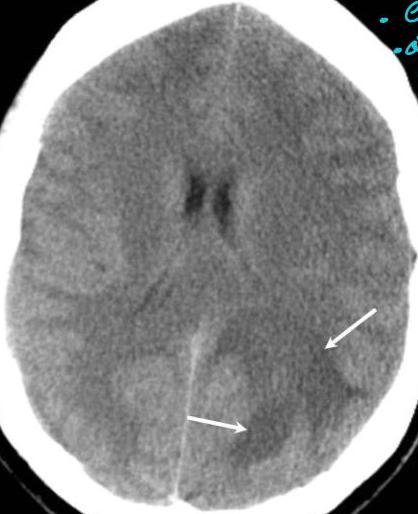
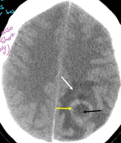
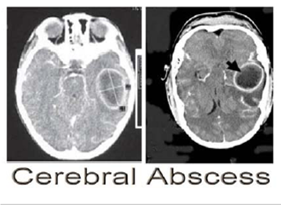
CT Scan: a focal area of hypodensity surrounded by ring enhancement with surrounding edema (hypodensity)
CT Scan
- • elementary
- • address is
- • similar to
- • mets
- • (both also
- • take short
- • history)
1/25/24
14
MRI
Microbiological Evaluation
CT-guided stereotactic needle aspiration
- Gram’s Stain
- Culture : Aerobic, Anaerobic, Mycobacterial and fungal cultures
Blood Culture
LP: do not perform
D/D
- Bacterial Meningitis
- Meningoencephalitis
- Acute disseminated encephalomyelitis
- Empyema
- Saggital Sinus Thrombosis
- Primary or Secondary brain tumor
- CVA
Treatment
- Combination of high dose parental antibiotics and neurosurgical drainage
- Third/fourth grneration cephalosporin+Metronidazole
- Patients with neurodurgery/Head trauma
- Vancomycin+Ceftazidine
- Meropenem+Vancomycin
- Modify antibiotics as per culture results
- Duration: Min 6-8 weeks
Cont.
-
Prophylactic anticonvulsant
- Should continue atleast 3 months after resolution of abscess
-
Role of steroids → becameful if patient is diabetic
-
dose with antibiotic not alone
-
Not given routinely
-
Usually reserved forof significant periabscess edema with mass effect and raise ICP
-
Dexamethasone 10 mg 6 hrly
-
Cont.
- Aspiration and Drainage of the abscess under stereotactic guidance
- Craniotomy and Complete excision of a bacterial abscess: reserved for multiloculated abscess or in those where aspiration is unsuccessful.
Prognosis
- Mortality rate <15%
- Neurological sequelae ≥20% of survivors
ENT
ENT
Brain Abscess
- Definition: Localized suppuration in the brain substance.
- It is the most lethal complication of suppurative otitis media.
- Incidence: 50% is otogenic brain abscess.
Pathology
- Site: Temporal lobe or, less frequently, in the cerebellum (more dangerous).
Diagnosis
- CT scans
- MRI
Treatment
- Medical:
- Systemic antibiotics
- Measures to decrease intracranial pressure
- Surgical:
- Neurosurgical drainage of the abscess
- Mastoidectomy