Burns
M K Zaky
Professor of surgery
ILOs
- Mechanisms
- Local & systemic effects
- Assessment - Degree & Surface area of burn, SPECIAL SITES
- Management- accident site & hospital
- Fluid requirements , ANALGESIA
- Wound management
- Prognosis & COMPLICATIONS
Introduction
- Trivial → OPD management
- Major → may need a long hospital stay +
Death & disability (disfigurement/ impaired function)
Introduction
Risk groups.
- Toddler
- Children
- Elderly
- Infirm & alcoholics
- Smokers
- WORKERS
Survival.
- Depends on the degree of the burn and the services provided
Epidemiology
- 66% - at home
- Death: predominantly in the extremes of age
Bimodal 1. Immediately after injury (in severe burn)
- Later from sepsis
Mechanisms of burn injury
- Sunburn
- Flame burn
- Scalds (children <5 years)
- Hot solids
- Friction
- Electricity
- Chemicals
- Irradiation
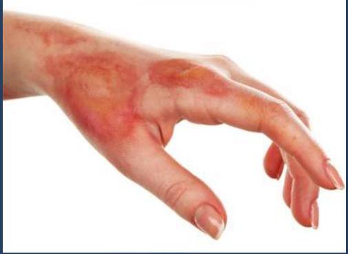
Burn compromises skin functions:
- Barrier to infection
- Temperature regulation
- Regulator of fluid loss
Local effects of burn injury
Destruction of tissues:
-
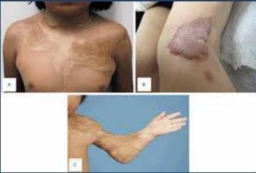
Superficial (1st degree) burn- epidermal remnants → healing with no effects.
-
Deep burn- epidermal loss → healing by scar formation (disfigurement, incomplete healing)
-
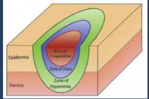
Inflammation of surrounding tissues → inflammatory cascade
-
Thrombosis of vessels
-
Fluid loss from the surface
Damaged capillaries permeable to protein- exudate
Exudation highest in first 12 hors
- Loss of barrier to infection- sepsis
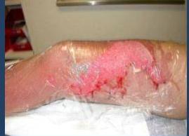
Systemic effects of burn injury
- Depends upon surface area & depth
Moderate - Large burn:
- Generalized capillary leak-max- 24 hrs. → Hypovolemia → AKI
- Increased metabolic rate- catabolism
- Hypothermia
- Systemic inflammatory response syndrome (SIRS)
- Immune suppression
- Lryngeal & Pulmonary oedema: inhalation burn
- Electrolyte disturbance
Burn Depth
- First-Degree (Superficial): Affects only the epidermis (outer layer). Symptoms include redness, pain, and no blisters (e.g., mild sunburn). Heals in 3–5 days.
- Second-Degree (Partial Thickness): Damages the epidermis and part of the dermis layer. Appears red, blistered, swollen, and is very painful. Superficial second-degree takes ~2 weeks to heal; deep second-degree may take longer and require surgery.
- Third-Degree (Full Thickness): Destroys all skin layers (epidermis and dermis). The area may appear white, leathery, or charred and is often numb due to nerve destruction.
- Fourth-Degree (Deep Full Thickness): Extends through the skin into fat, muscle, and sometimes bone
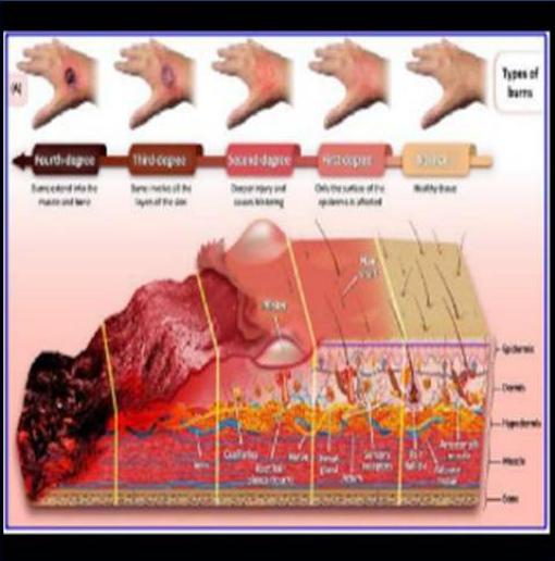
Depth of burn
Depth - depends upon degree of temperature & duration of contact
☐ 1st degree: (superficial layer of the epidermis)
- Limited to epidermis
- Skin red, painful, no blisters
- Heal spontaneously- 3-4 days
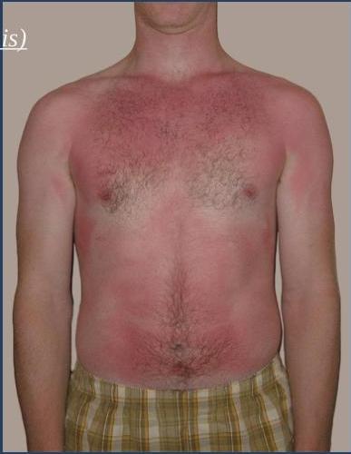
Depth of burn
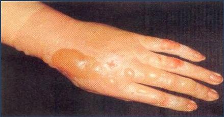
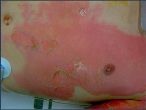
2nd degree: A) Superficial partial thickness burn
- Epidermis & superficial dermis Hot surface, hot liquids, chemical
- Blister, erythema, very painful, & swelling
- Fluid loss (depends on the area involved). Healing- from appendages (sweat glands, hair follicles, sebaceous glands) (\sim) 2-3 weeks Full function & good cosmetic result (pigmentation may occur)
- Infection can change a (2^{\text{nd}}) degree burn to a (3^{\text{rd}}) degree
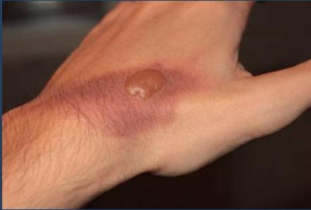
Depth of burn
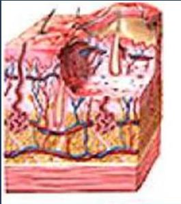
2nd degree: B) Deep-dermal burn
- Involve epidermis & much of dermis
- Nerve endings are damaged
- Blisters, pale white, absent pain sensation, swelling
- Marked fluid loss
- Healing takes longer, from few remaining epithelium
- Often heal with an ugly hypertrophic scar
- Infection- may convert it to full thickness injury & delay healing.
Depth of burn
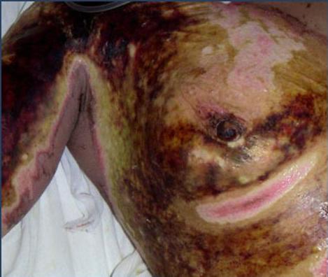
3rd degree: Full thickness burn
- Epidermis, whole dermis (with subcutaneous tissue) & all appendages destroyed.
- Coagulative necrosis & eschar formation.
- Eschar separates after 2-3 weeks.
- Healing: unless grafted, healing by wound contraction & fibrosis.
- Ugly contracture formation.
Depth of burn
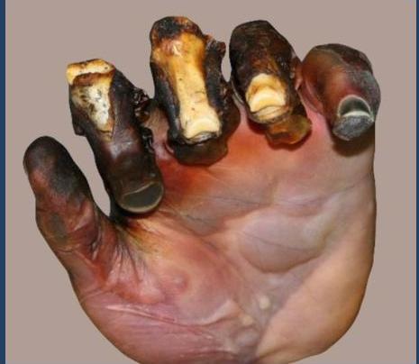
4 degree; (charring)
Deep fascia, muscle & bone
How to determine burn depth?
- No perfect method
- Accurate assessment- may take days
How to determine burn depth?
1.Mechanism of burn
- Scald- partial thickness.
- Flame- may be partial / more commonly full thickness
- Electricity- almost always full thickness
How to determine burn depth?
2. Appearance
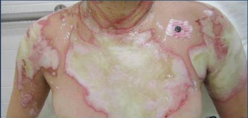
- Partial thickness: Erythema, blanching on pressure, blisters
- Deep dermal: Dead white appearance- burn
- Full thickness: Dry leathery with thrombosed veins.
How to determine burn depth?
3. Sensation
- Intact sensation- Superficial. Nerve endings intact. Very painful
- Loss of sensation- Deep burns
Pain decreases as burn depth increases (nerve destruction)
Rule of ‘9’ - % body surface areas (BSA) of a burn
Small area: patient’s hand- palm+ extended fingers = 1%
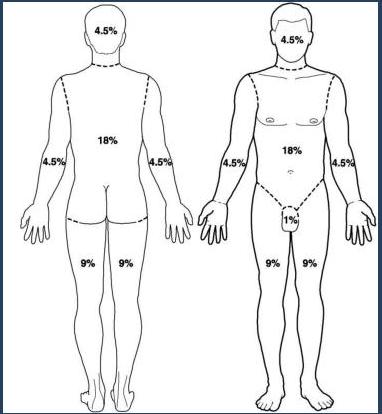
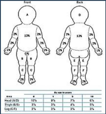
Minor: < 10 BSA, partial
Moderate: 10-20% BSA, partial
Severe: > 20% BSA, face, hand, airway,
Management- First aid
-
Stop burning process-
-
Cooling the burn area using running tap water for 20 minutes (avoid hypothermia)
-
Chemical burn - copious irrigation with running water.
-
Electrical - switch off or push patient with wooden object.
-
Ensure airway:
-
Move patient away from smoke.
-
Secure the airway/ventilation- if respiratory arrest.
-
Avoid contamination:
-
Cover with clean sheet
-
Quick transfer to hospital:
-
IV infusion & analgesic - on way to hospital.
Signs of inhalation injury
- Facial burn
- Singing of eyebrows
- Carbon deposits in oropharynx/ sputum
- Burns to head & trunk
- Confinement to burning environment
- All these patient will need respiratory support (intubation)
Hospital management
Severe burns → hospital admission, preferably to a specialized unit
Indications:
- Second degree burn > 20% BSA (adult), > 5% (children < 2y), > 10% (3-10 y), > 15% (10-15y), > 10% (> 65y)
- All full thickness burns (3rd degree)
- Burn involving face, hands, feet, genitals, joints
- Inhalation burn
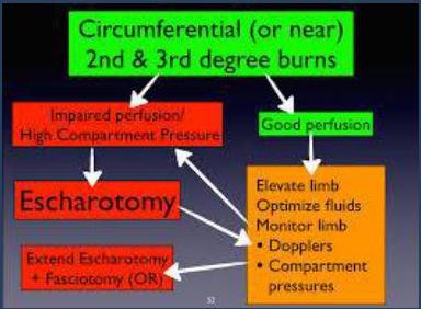
- Circumferential burn
- Chemical and electric burn
- Comorbidities
Use trauma approach (ATLS): ABCDE
Initial management
- A: Airway secured, humidified O₂ (100%), pulse oximetry, early intubation for inhalation burn
- B: assisted ventilation may be needed for inhalation or aspiration
- C: IV fluids (2ⁿᵈ-4ᵗʰ degree burn)- calculated from injury time, CBC, U/E, ABG, lactate, carboxyhaemoglobin, X-match, urinary catheter (monitor output)
- D: assess GCS
- E: remove clothes, jewellery, prevent hypothermia
- Analgesia- IV (morphine, pethidine), anti Tetanus prophylaxis (if needed)
- Assess the type, depth, BSA of burn and associated injuries
Fluid resuscitation
- Needed for burn > 10% in children, > 20% in adults. LR solution is given according to Parkland’s formula (first 24 h)
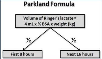
- Colloid solutions - may be added after 24 hours.
- Oral fluids- restrict for 48 hours (acute gastric dilatation and/or paralytic ileus (>25%)), then encouraged, consider PPI (Curling ulcer)
- Watch for:
Hyponatremia (effect of ADH which is secreted as a result of trauma) Hyperkalaemia (tissue destruction)
Resuscitation- monitor for organ failures
-
Respiratory- CXR, ABG
-
Renal failure- adequate fluid
-
Nutritional support (calorie & protein demand is high)
-
Monitor for sepsis: Routine antibiotics not recommended in the first 48 h
o Regular blood culture & swabs. o Organisms- Staph (most common), pseudomonas, hemolytic strep.
-
Gastric erosions & duodenal ulcer (Curling’s): Early feeding , H2 blockers. PPI
Local management of burn wound
- Shocked patients: Cover with sterile towel until stabilized.
- GA / IV analgesia- opiate (after stabilizing the patient)
- Wound- cleaned with mild detergent in saline
- Grossly devitalized tissue removed
- Better to keep blisters intact (evacuated with a sterile needle and left intact)
- Face, neck, perineum - left exposed, MEBO ointment may be used
- Other areas- covered with non-adherent dressing. Silver sulfadiazine cream, MEBO ont
- Dressing can be changed every 2-3 days
Local management of burns
-
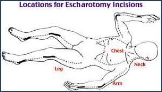
Escharotomy- full-thickness incision through insensate burn eschar for circumferential deep burn (neck, chest, limbs)
-
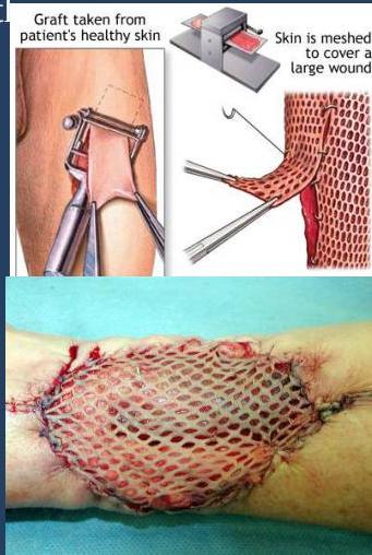
Full thickness burn- early (72 hrs.) excision & grafting
-
Extensive burn- delayed skin grafting once slough has separated
Local management of burns
- Skin cover by split skin graft within 2- 3 days for deep burn after excising eschar – small burn
- Large burn- staged skin grafting in < 3 months
- Wound clean & granulating before grafting
- Skin grafts & donor sites- moisturizing crème to keep it supple
- Splints – to prevent contracture
- Physiotherapy- mobilize joints
- Late reconstructive procedures
- Long term support & counselling
Prognosis
- Age: Extremes of age fare less well.
- Extent of burn: Hypovolemic shock, >15% in adults or >10 % in children
- Depth of burn: Superficial burn- heal rapidly , good cosmetic result.
- Deep burn- heal with scarring, risk of infection. Sepsis - life threatening.
- Site: Face, neck, hands, feet & perineum- threaten appearance & function.
- Respiratory injury: Inhalation of smoke from burning plastics- can be fatal
- Infection- higher morbidity & mortality
Complications
Local :
- Scarring
- Contracture
- Hypertrophic scar / keloid
General:
- Respiratory: smoke injury
- Renal failure: hypovolemia, haemoglobinuria, myoglobinuria
- Upper GI bleeding: Curling ulcer, gastric erosions
- DVT
- Sepsis
- Psychological problems