Complications of Abdominal Surgery
Dr. Ahmed Yousif Abdalla

Intended learning outcomes
- By the end of this interactive lecture every candidate should be able to:
- Define the complication as a concept.
- Differentiate between complication, side effect and medical error.
- Study common post op. abdominal complication.
- Analyze and interpret lap. Investigation to formulate a list of deferential diagnosis.
- Approach a patient with post op. complications.
Introduction
Definitions (terminology)
- Complication: unfavorable consequence of a disease, a health condition or a therapy — general term
- Examples: Diabetics and he developed Renal failure; trauma → compartment syndrome
- Adverse effect: undesired harmful effect resulting from a medication or other intervention such as surgery — specific
- Examples: NSAID → gastric ulcer; In rectal → drawnell leukar
- An adverse effect may be termed a “side effect”, when judged to be secondary to a main or therapeutic effect
- If it results from an unsuitable therapy, improper procedure, or incorrect dosage this is called a medical error and not a complication
- malpractice
Examples of medical errors (malpractice)
① trauma case after packing use & stop bleeding, he came with Fever and abdominal Pain, chest x-ray showed good Admixturei abdomen
② Pt undergo Pt nephrectomy for Pt kidney failure but the left kidney was taken.
③ Pt overweight the surgeon did for Pain show gastrectomy then he develop leakage (surgery was not indicated)
④ general surgeon did for his pt Rhinoplasty then pt had wound infection (privilege of surgeon - should be done by plastic surgeon)
⑤ if you don’t instruct your pit after surgery about wound care and taking of antibiotic
⑦ Pit I did for him. CT then he develop Renal failure then you found that he had his protein (no need to fit below)
⑧ pit was taken for removal of lipoma in his thigh, under GA you saw skin tag in his back and you decide to take it (no consent)
- But in case of endoscopy and you saw polyp you can take it because it’s a part of the procedure.
- Or in emergency, pit for laparotomy and you injured the bowel. you have to treat him even without consent
Key principles
- Medical knowledge about a disease, procedure or treatment usually entails a list of the most common complications.
- Complications can be foreseen, prevented or recognized more easily and speedily.
- Depending on the degree of vulnerability, susceptibility, age, health status, immune system condition, etc.
- Complications affect adversely the prognosis.
- Prompt identification of complications can significantly improve outcomes.
- Interdisciplinary approach is essential e.g. IR to help me I and d.
- Non-invasive, & minimally invasive medical procedures usually favor fewer complications in comparison to invasive ones.
Timing of complications
- Early, intermediate, late
- Early + intermediate + increased mortality
Grading of Postoperative complications
α not for your exam
| Grading | Grade | Definition/treatment performed |
|---|---|---|
| Minor complications (most of complications) | Grade I | Adverse event which alters the standard postoperative course without requiring a specific treatment |
| Can be treated easily | Grade II | Pharmacologic treatment or minor intervention required |
| Major complications (when patient needs to be taken for OR or ICU) | Grade IIIa | Surgical, radiologic, endoscopic treatment, or multitherapy required without general anesthesia |
| Needs intervention | Grade IIIb | Surgical, radiologic, endoscopic treatment, or multitherapy required with general anesthesia |
| Grade IVa | Intensive care unit treatment for single organ dysfunction required | |
| Grade IVb | Intensive care unit treatment for multiple organ dysfunction required (Stroke, MI…) | |
| Mortality | Grade V | Adverse event which leads to death (Brain death in ICU) |
Case 1: Generalized peritonitis (perforated appendix)
Presentation
- A 35-year old male presented with a 3 days history of abdominal pain. Examination: features of generalized peritonitis.
What are the features of generalized peritonitis?
- Systemic (general) signs:
- Unwell patient, swelling
- Disturbed vital signs (↓BP, tachycardia, fever, tachypnea)
- Specific (abdominal) signs:
- Guarding, rigidity
- Generalized tenderness and rebound tenderness
- Active or decreased bowel sounds (according to case)
- Fever
What next to do?
- Proper detail Hx, examination, relavent investigations
- Labs: CBC, RFT, LFT, LAC, Control
Differential diagnoses
- Perforated viscus, appendicitis
Management
- Laparotomy — surgical exploration (laparotomy or laparoscopic)
- He was prepared for surgery.
At laparotomy
- Perforated appendix was found
- Intraoperative features of generalized peritonitis:
- Pus collection + red inflamed peritoneum, pyogenic membrane covering the bowel, maybe blood
- Appendectomy & peritoneal lavage was done.
- Abdomen was closed and a drain placed
Postoperative care
- Postoperatively: NPO, IV fluid & broad spectrum antibiotics
- Then switch after C/S
- Why? Fear of leak + they will have paralytic ileus as we manipulated the abdomen
- Until when? they pass flatus or according to the procedure
- As a pt with cholecystitis, we hear bowel sounds after 6-10h then start feeding
Day 1-4
- Steady improvement.
- Afebrile, drain removed.
- Progressed from fluid to normal diet.
- When we should take out the drain:
- Amount: amount should be < 3.0 ml/day
- Content: content or color should be fluid (Haemoserous)
Day 5
- Temp. 38°C, loose motion- twice.
- Why? C-Diff colitis due to B-road spectrum antibiotic — high grade fever
- Possible causes of fever? Collection in the pelvic (abscess), drain removed so recollection; wound infection
- Loose motion?
- Next? Ct contrast
- Will x-ray help? Even if showed air under diaphragm? No, normal post op
- History: chest symptoms, urinary symptoms, abdominal pain, wound site pain, leg pain, nature & number of loose motion.
- Examination: General, IV sites, chest, abdomen (wound, abdomen), P/R, Lower limb (DVT)
Wound infection
Severity ladder:
- Early/ mild
- Moderate/ stitch = sheeth / dehiscence
- Sheeth + muscle + evicceration (burst abdomen)
| Severity | Involved (μg/d) | Typical Features | Management |
|---|---|---|---|
| Early, Mild | Skin, subcutaneous tissue | Restless, tenderness, minor discharge | Local site + antibiotics |
| Moderate, Mild/ Abscess | Subcutaneous, partial hernia | Pre-wound suture, partial dehiscence | Oestrogen, severe removal |
| Trained/Dehiscence | Traces + muscle | Hemangiomous discharge, wound sparing | Surgical repair |
| Burst abdomen (Extracerebral) | Skin, hernia, muscle, peritoneum | Wound spares, bowel perforation | Emergency surgery |
Postoperative fever
- Day 0 is the day of surgery = stress, anaesthesia = malignant hyperthermia with inhaled anaesthesia
| Category | Day | Description |
|---|---|---|
| Wind | POD 1-2 | the lungs, i.e. atelectasis, aspiration, pneumonia |
| Water | POD 3-5 | urinary tract infection, possibly catheter-associated (if a urinary catheter was inserted during surgery or remains in place currently i.e. Foley catheter) |
| Wound | POD 5-7 | infection of the surgical incision(s), either superficial or deep[4] |
| (W)abscess | POD 5-7 | infection of an organ or space[5] |
| Waking (or VEINS pronounced like “Weins”) (variant in Almaarefa deck: “Walking”) | POD 5+ (risk may persist for months post-operatively) | deep vein thrombosis or pulmonary embolism |
| Wonder drugs or “What did we do?” | Anytime | drug fever or reaction to blood products, either a febrile non-hemolytic transfusion reaction or transfusion-related acute lung injury |
| Wing/Waterway | Anytime | bloodstream infection, phlebitis, or cellulitis related to intravenous lines, either central or peripheral |
- Intermediate complications = Until they are out of the hospital/ few days
History & Examination (fever workup)
- History: chest symptoms, urinary symptoms, abdominal pain, wound site pain, leg pain, nature & number of loose motion.
- Examination: General, IV sites, chest, abdomen (wound, abdomen), P/R, Lower limb (DVT)
Standard work up
- Labs: CBC with diff, CRP — Septic work up: CBC
- CV/S risk factors
- EKG, ABG (Resp complications)
- Cultures:
- Sputum
- Blood
- Urine (and UA)
- Others: (Lines, wound)
- Radiology … To reach diagnosis + guide therapy in some cases:
- CXR
- CT scan/ spiral
- Treatment:
- Pan-culture everything (blood, urin, stool, drain)
Postoperative diarrhea
- Causes: MCA
- Pelvic collection: Cause irritation
- Passage of mucous with diarrhea is pathognomonic.
- Pseudomembranous colitis
- Food poisoning / gastro-enteritis
- Others
- Pelvic collection: Cause irritation
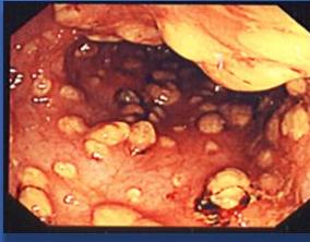
Case 1 continued: examination findings → pelvic collection
- GE: normal
- Chest clear
- Lower limb: no swelling or tenderness
- Abdomen: wound looks OK,
- P/R- bulging anteriorly, tenderness.
- Significance of this finding? Pelvic abscess
- What to do?
Investigations
- Labs. CBC, renal function, CRP
- Radiology:
- CXR, ?AXR
- U/S abdomen & pelvis
- CT scan
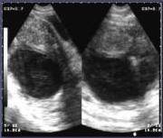
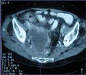
Pelvic collection (Collection in Pelvis)
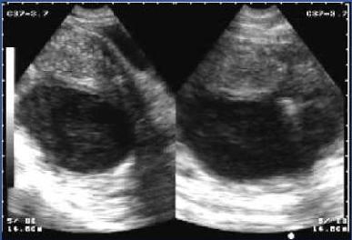
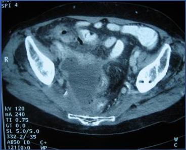
Management
- Conservative- very small collection may resolve
- Drainage:
- CT guided drainage: Trans-rectal, Percutaneous suprapubic
- Surgical
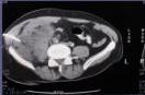
Case 2: Acute cholecystitis & post-cholecystectomy bile leak
Presentation
- A 40-year old female admitted with features of acute cholecystitis
- What are these features? URQ pain radiates to shoulder, increases after food
- CBC & LFT- normal. High total
- Diagnosis confirmed by ultrasound.
- What are US findings? Pericholestite fluid, wall thickness, presence of stone and its echolytic shadow
- Patient progress: Responded well to conservative management
- What is her further management? Stabilise and do cholecystectomy within 72h or wait until 6 weeks
Further management plan
- Cholecystectomy
- Time
- Approach
- Advantages vs disadvantages
Q: What are the preconditions?
- Stabilize the patient comorbidities
- Normal labs
- Anticoagulants
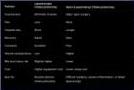
laprascopic: Injury to near by structure
Cholecystectomy on next list
- 2 days after admission.
- Difficult laparoscopic cholecystectomy.
- Left a drain at sub-hepatic area. Z
1st postoperative day
- Sips of water+ IV fluid
- Temp 38°C, pulse 100/ min.
- Abdomen mildly distended, sluggish bowel sounds. (hypoactive)
- Drain 90 ml darkish fluid. (Volum, Color)
- Most likely bile

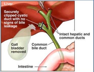
2nd postoperative day
- IV fluid + NPO
- Temp 37.8°C, pulse 90/ min.
- Vomited twice in last 24 hours.
- Abdomen mildly distended, non-tender, sluggish bowel sound.
- Drain 175 ml dark green. → bile
What next?
- Bile leak, why? Slip ligation, injury to the CBD or CHD, or anywhere.
- Accessory duct as ducts of Luchka — nothing usually to do as will close spontaneously, but as it is increasing we should manage
- Bismuth classification of injury
Post-cholecystectomy bile leak
- Biliary tract injury.
- Proximal bowel injury- ? Duodenum
- Bowel injury unlikely in this patient.
- Why?
Investigations
- ? CXR, AXR → Case: you saw Air under diaphragm and you took the pt to the OR (this is non-practice because normal to have air under diaphragm post op)
- U/S
- CT
- ERCP (diagnosis + therapy) + MRCP + PTC
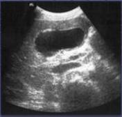
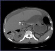
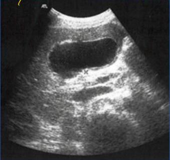
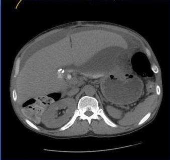
Ultrasound vs CT (VS)
- VS: only told me if there’s selection or not [only tells if there is collection or not]
- I Can Know the Source
Management of bile leak
- Establish and maintain adequate drainage.
- Drain was left in situ at the initial procedure- draining bile.
- Percutaneous guided drainage (USS guided)
- Antibiotics and a daily assessment of drain output.
- Drainage out put:
- <200 ml/day & reducing daily- likely to stop on its own.
- Persistent drainage > 200 ml/day – ERCP indicated
- ERCP: cystic duct leak- internal stent (5-7 cm)
- Abdominal drain removed once it stops drainage
- Stent removed in 6-8 weeks
- Other CBD injury- surgical intervention
- If ERCP is not possible – surgery in complicated (bypass hepatosejunostomy)
CBD stent
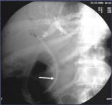
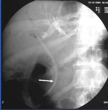
- Stent for 6 weeks to relife the Prusser [relieve the pressure]
- If it was major leakage sometime you need Recondition & biking free
Cystic duct leakage, post cholecystectomy
- ERCP
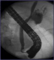
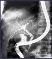
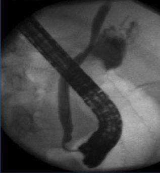
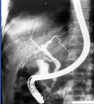
Causes of cystic duct leakage
- Clips not properly applied
- Pressure necrosis from too tight application
- Distal obstruction- stone in CBD
- Duct of luschka
- Backward pressure
- Accessory duct
Case 3: RTA victim — blunt abdominal trauma (splenic injury)
Primary & secondary survey
- RTA victim
- Conscious, talking
- Primary, & secondary survey
- A & B – stable
- C - hemodynamically unstable
- Rapid crystalloid infusion
- CBC, U/E, cross match
- FAST - splenic injury, free fluid in peritoneum
- Management ?
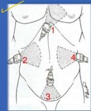
Supine patient
FAST views
- Standard views (standerded FAST):
- Subxiphoid/Subcostal: Pericardium
- RUQ: Morrison’s Pouch
- Pelvis: Pelvic Cul-de-sac (Douglas)
- Transverse
- Longitudinal
- LUQ: Splenorenal & perisplenic spaces
- Extended views (E-FAST): For pleural effusion
- Remember: Probe marker almost ALWAYS facing either patient’s right or patient’s head
Management
- Consent for emergency surgery.
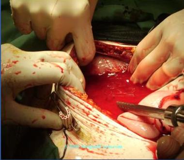
- Urgent laparotomy- midline incision.
- Splenic injury with massive hemoperitoneum.
- Perisplenic packing.
- Blood sucked out.
- Splenectomy.
- No other injury.
- Abdomen closed, drain placed
Day 1-3
- Hemodynamically stable
- Initially: NPO+ IV fluid,
- Later allowed oral feeding
- 2 units of PCV to raise Hb above 10g/dl
- Pneumococcal vaccination
- Vag implant in emergency splenectomy
- Antibiotics
Drain timeline
- Day 1 - bloody, 150 cc.
- Day 2 - serous 120 cc
- Day 3 - turbid fluid 150 cc,
- Day 4 - turbid fluid 230 cc
- Drainage fluid contained very high level of amylase → Pancreatic juice
- What is next?
Pancreatic fistula
- Causes:
- Don’t do ERCP in acute pancreatitis you will make it worse.
- ✓ Operative trauma,
- ✓ Complication of acute/ chronic pancreatitis
- Management:
- Skin care
- Management of electrolyte & nutrition:
- NPO+TPN
- Specific management:
- ERCP & pancreatic duct stenting
- Octreotide
- Repeated CT scan to rule any other collection
- PCD- for other collections
Case 4: Laparoscopic sleeve gastrectomy — early leak
- A 45- year male, smoker, complained of weight gain and obesity.
- Past Hx: DM (II), on OHA (in morning, now)
- Underwent laparoscopic sleeve gastrectomy
- Found to have HR : 120/ min — Early sign of leak = tachycardia
- Dx: Lackage [leakage] → to exclude it need Contrast → Reason, swallow, Gaital [Gastrografin] Good-grain
- Rixew Post of note
- How to treat? According to the site, size?
- Rx:
- CT with contrast = leak in peritoneal cavity
- Seal the leak + cover with omentum
- If mild = drain
- Some need bypass
- Bleeding to exclude it by CBC
- P.E. need CT to be excluded
- H2O (hematocrit) ↓ Hemoglobin
- How CT for hemolysis or to determine leakage
- Radiology
- The hemoglobin could be false high due to dehydration
- Or False positive if nurse take sample from arm which is his IV fluid
- No gain due to taste + loss for hematocrit
- Will leak have in day 07? Unlikely. From Day 2 +3
Case 5: Recurrent inguinal hernia after open mesh repair
- A 53- year male complained of right groin swelling for 3 months
- History of open mesh repair of right inguinal hernia 6 months ago — risk factors of recurrence
- Post operatively: wound infection
- 3 months later, presented with recurrent swelling at surgical site
- Past Hx: Unremarkable
- Clinical findings
- Dx:
first time give mesh
- Open, Now it should be laparoscopic:
- TAPP (TransAbdominal PrePeritoneal repair)+ TEP (Totally ExtraPeritoneal repair)
- Mesh infection: do US to confirm you will see collection and inflammatory process
- Open, Now it should be laparoscopic:
- Rx:
- When hernia should be laparoscopic? Bilateral + recurrence
- Take off the mish [mesh]
- If no response at Antibiotic
- Hestrocele [hydrocele] US to exclude
- Lymph node do US to confirm it
- Recurrence exclude it by US then CT
- Laparoscopic hernia repair (one of indication at laparoscopic) (second time)
Case 6: Post-thyroidectomy progressive neck swelling
- A 45- year female complained of neck swelling for 3 months
- Associated with difficulty in breathing and swallowing
- FNA: Benign
- Past Hx: Unremarkable
- Subtotal thyroidectomy
- Post operatively: Progressive neck swelling
- Rx