Esophageal Diseases
Esophageal Diseases
It includes :
- Esophageal motility disorder
- Esophageal diverticulum
- Benign esophageal tumors
- Malignant esophageal tumors
- GERD and Hiatus Hernia
- Esophageal perforation
- Caustic Injury
- Barrett’s Esophagus
1. Achalasia
Achalasia is an uncommon disease of esophageal motility disorder
It is characterized by degeneration of the myenteric neurons that innervate LES and esophageal body
lower esophageal sphincter
the pathogenesis :
all these factors autoimmune ? Viral ? Familial ?
Clinical features
most commonly presents in patients between the ages of 25 and 60 years. an equal male-to-female gender distribution.
both at the same time (not progressive)
Dysphagia to solids and liquids is the most common presenting symptom, experienced by greater than 90% of patients.
(unlike cancer esophagous)
- more in male
- older age group (>50) • but both have dysphagia in cancer, starts as:
- solid
- semisolid
- liquid
- saliva (pagressive dysphagia)
Clinical features
-
Regurgitation is the second most common symptom, occurring in approximately 60% of patients.
-
Nocturnal regurgitation of esophageal contents can lead to night time cough and aspiration.
Glead to aspiration Pneumonia
its nocturnal because LES is closed, and esophagous
is dilated, so it acts as reservoir (opposite to its function)
food will accumulate, and when laying down → aspiration of
- Weight loss occurs in end-stage disease.
- function of esophagous: Transient transmission of food from pharynx to stomach
Clinical features
- Chest pain is reported in 20% to 60% of patients
- Heartburn is reported in a large number of patients with Achalasia (30% of Achalasia patients). may be related to direct irritation of the Esophageal lining by retained food, pills, or acidic by products of bacterial metabolism of retained food.
• heart burn → from achalasia → from GERD: its from acidity from stomach because sphincter is open
• H / E
Diagnosis
-
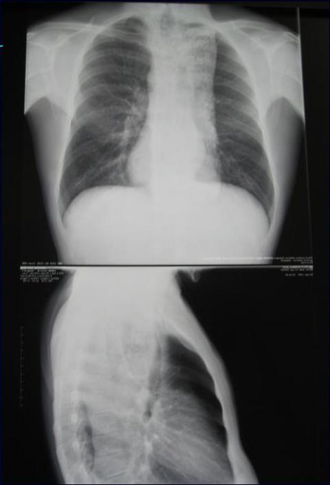
CXR may show air-fluid level
-
Barium study quite dilated, and an air-fluid level may be secondary to retained secretions. The classic finding is a gradual tapering at the end of the Esophagus, similar to a bird’s beak.
-
Upper endoscopy is the next diagnostic test in a patient with dysphagia or suspected Achalasia.
or cat’s tail or pencil shape
Diagnosis
Findings can include :
- dilated esophagus with retained food or secretions
- normal in as many as 44% of patients with achalasia
Difficulty traversing the GEJ should raise suspicion for pseudo-achalasia due to neoplastic infiltration of the distal esophagus.
- Prepare patient carefully before doing endoscopy (because esophagus is full of food) Keep NPO for few days irrigate Put NGT
Chest x-ray
okey dilated esophagous
barium swallow
bird’s beak / rat tail / pencil share
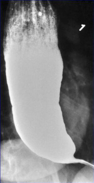
dilated esophagous
A
B
- if disease develop in patient and left untreated, its effect will be irreversible.
→ esophagous looks like haustration of colon
→ end stage achalasia
Diagnosis
golden standard in diagnosis of esophagous
• Esophageal manometry has the highest sensitivity for the diagnosis of achalasia :
3 findings:
-
- aperistalsis of the distal esophageal body
-
- incomplete or absent LES relaxation
-
- hypertensive LES
Manometric variants of achalasia exist
- The best known is vigorous achalasia
- defined by the presence of normal to high amplitude esophageal body contractions in the presence of a non-relaxing LES
Diagnosis
Manometric variants of achalasia exist
→ (anything raising amplitude of LES)
- vigorous achalasia may represent an early stage of achalasia
Chagas’ disease is a parasitic infection
caused by Trypanosoma cruzi which can cause secondary achalasia.
The most concerning secondary etiology
is cancer, which can present as achalasia through mechanical obstruction of the GEJ
Treatment
- The primary therapeutic goal in achalasia is to reduce the LES basal pressure
- Treatment options include medical therapy: botulinum toxin injection, pneumatic dilation, and surgical myotomy
- Symptom relief, particularly relief of dysphagia, is accepted as the primary desired outcome
Medical Therapy
Is inconvenient, only modestly effective, and frequently associated with side effects
It is reserved for patients who are awaiting or unable to tolerate more invasive treatment modalities
Pharmacologic therapies attempt to decrease the LES pressure by causing smooth muscle relaxation
Medical Therapy
① Nitrates were first recognized as an effective treatment of achalasia their systemic vasodilatory effects and headaches limit their tolerability by patients
② Calcium channel antagonists have a better side-effect profile when compared with nitrates
30% of patients report adverse side effects including peripheral edema, hypotension, and headache → from VD effect of drugs
Botulinum Toxin
3. Botulinum Toxin
→ not successful in high percentage
Response rates at 1 month following administration average 78% , By 6 months, the clinical response rate drops to 58% and by 12 months to 49% Given the limitations of the efficacy and durability of response, Botulinum toxin is generally reserved for use in patients who are not candidates for more invasive treatments.
4. Pneumatic Dilation
pneumatic dilation remains one of the most effective first-line therapies for achalasia
Long-term follow-up studies reported significant symptom relapse of 50% at 10 years
Complications of pneumatic dilation exist
:
- Gastroesophageal reflux 25-35% → if dilate more than needed
- Esophageal perforation 3%
treatment
Drug therapy:
- Smooth muscle relaxant (nitrate, calcium channel blocker, anticholinergic))
- 10% of pts. Benefit from this treatment (elderly)
Pneumatic dilation: → by endoscopy, put balloon in LES and inflate it, keep it for
a couple of minutes
- A balloon is insufflated at the level of the G.O junction to rupture the muscle fibre
- Success rate 70-80%
- 50% will require more than 1 dilation.
then repeat it
a few times.
and repeat
every 6 months
give some symptom relief but only temporarily, for 6 months to 1 year. so needs to be repeated
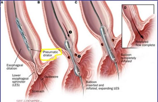
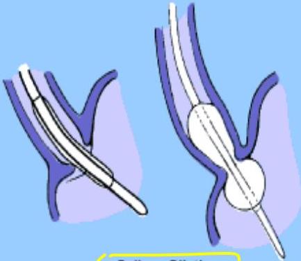
Balloon Dilation
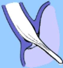
Bougie Dilation
Surgical Therapy
more preferred than medical therapy
has success rates in excess of 90% with hospital stays averaging only a few days acid exposure is a known complication of surgical intervention for achalasia
Even with a successful myotomy, it is expected that patients will have some degree of dysphagia as a consequence of esophageal peristaltic dysfunction
Surgical Therapy
Delayed recurrence of postoperative dysphagia is most commonly caused by development of a recurrent high pressure zone at the LES or a peptic stricture complicating acid reflux
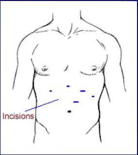
Laparoscopic Heller’s myotomy
demonstrated excellent results, with 98% of patients reporting symptomatic improvement at 5.3 years
98% Success rate done through scope, then open hiatus and isolate esophagous and cut the longitudinal and circular muscles, and expose the mucosa. then 60 a positive to prevent reflux in the future
Complications
The primary complications of achalasia are related to the functional obstruction rendered by the non-relaxing LES and include progressive malnutrition and aspiration.
Uncommon but important secondary complications of achalasia include the formation of epiphrenic diverticula and esophageal cancer → Squamous Cell Carcinoma
(*achalasia patient has risk to develop SCC more than normal population)
Complications
- (There is an established link between achalasia and esophageal cancer, most commonly squamous cell carcinoma)
- (The overall prevalence of esophageal cancer in achalasia is approximately 3% with an incidence of approximately 197 cases per 100,000 persons per year)
2.
Esophageal Diverticula
blind end sac
-
most diverticula are a result of a primary motor disturbance or an abnormality of the UES or LES
-
can occur in several places along the esophagus
The three most common sites of occurrence are pharyngoesophageal (Zenker’s), parabronchial (midesophageal), and epiphrenic
epiphrenic diverticula develop when there is motility disorder
Esophageal Diverticula
- can be classified into:
-
True diverticula involve all layers of the esophageal wall, including mucosa, sub-mucosa, and muscularis
-
A false diverticulum consists of mucosa and submucosa only
-
Pulsion diverticula are (false diverticula) that occur because of elevated intra luminal pressures generated from abnormal motility disorders
- can be classified into:
Esophageal Diverticula
Zenker’s diverticulum and an epiphrenic diverticulum fall under the category of false, pulsion diverticula.
Traction, or true, diverticula result from external inflammatory mediastinal lymph nodes adhering to the esophagus
usually in middle 1/3 (where LNs are present)
here, The management is not designed to treat the diverticulum itself because the diverticulum is not the main pathology its only a result. So removing diverticulum it will still reoccur. So treat the LNs, TB, and inflammatory process. and the diverticulum will heal by itself.
(LNs involved in inflammatory process, as in TB) (lead to adhesions)
Pull all layers of esophagus Traction True
Pharyngoesophageal (Zenker’s) Diverticulum
(False, pulsion)
Is the most common esophageal diverticulum found today
It usually presents in older patients in the 7th decade of life
found herniating into Killian’s triangle, between the oblique fibers of the thyro- pharyngeus muscle and the horizontal fibers of the crico-pharyngeus muscle
Symptoms and Diagnosis
Commonly, patients complain of a
-
sticking in the throat.
-
nagging cough, excessive salivation, and intermittent dysphagia often are signs of progressive disease
because its beside the airway (trochea)
when patient short eating, part of food pass through real lumen, part go in the diverticulum, once diverticulum is full of food, start to press on esophagus leading to dysphagia.
As the sac increases in size, regurgitation of foul-smelling, undigested material is common
because food stuck in diverticulum
Symptoms and Diagnosis
- → bad smell of breath
- Halitosis, voice changes, retro-sternal pain, and respiratory infections are especially common in the elderly population
- The most serious complication from an untreated Zenker’s diverticulum is aspiration pneumonia or lung abscess → due to aspiration of food from diverticulum
Symptoms and Diagnosis
Diagnosis is made by barium esophagram → Show diverticulum or different site
Neither esophageal manometry nor endoscopy is needed to make a diagnosis of Zenker’s diverticulum.
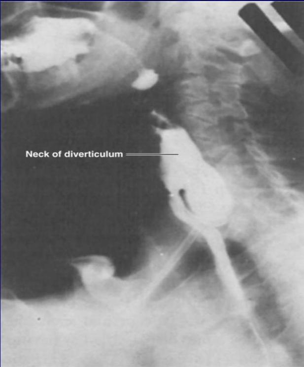
Neck of diverticulum
barium swallow
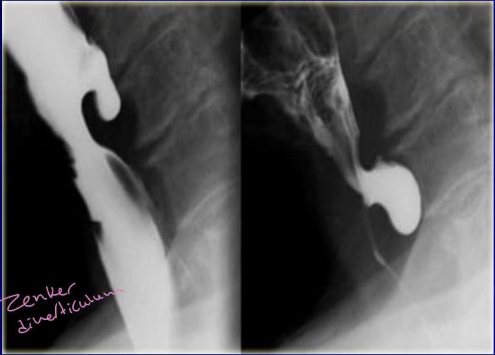
Zenker dineticulum
Treatment
excision → boys
Surgical or endoscopic repair of a
Zenker’s diverticulum is the gold standard of treatment
- if unfit for surgly
- put rigid phayrengoscope, esophagoscope and put fiber optic, and see real lumen, and blind end of diverticulum.
- retract fiber optic scope, adjust rigid scope until see the wall between real lumen and the diverticulum.
- put a Stapler at the end and divide the wall.
3. Barrett’s Esophagus
Barrett’s esophagus is a condition whereby an intestinal, columnar epithelium replaces the stratified squamous epithelium that normally lines the distal esophagus
- Chronic gastro-esophageal reflux is the factor that both injures the squamous epithelium and promotes repair through columnar metaplasia
→ although, these metaplastic cells are more resistant to injury from GERD, but more prone to malignancy → That’s why they develop adenocarcinoma
→ histo Pathological diagnosis
→ So need multiple biopsy by endoscopy
Barrett’s Esophagus
- Although these metaplastic cells may be more resistant to injury from reflux, they also are more prone to malignancy
- 10 % of patients with GERD develop Barrett’s esophagus
- the 40-fold increase in risk for developing esophageal carcinoma in patients with Barrett’s esophagus
Barrett’s Esophagus
- With continued exposure to the reflux disease, metaplastic cells undergo cellular transformation to low- and high-grade dysplasia → mild, moderate, severe dysplasia → one step away from becoming CIS then become adenocarcinoma these dysplastic cells may evolve to cancer
Barrett’s Esophagus
-
70% of patients are men aged 55 to 63 years
-
Men have a 15-fold increased incidence over women of adenocarcinoma of the esophagus, but women with Barrett’s esophagus are increasing in number as the differences in the Western lifestyle between men and women diminish
-
Smoking
-
Stress
Symptoms and Diagnosis
- Many patients harboring intestinal metaplasia in their distal esophagus are asymptomatic
- Most patients present with symptoms of GERD. Heartburn, regurgitation, acid or bitter taste in the mouth, excessive belching, and indigestion are some of the common symptoms associated with GERD
Symptoms and Diagnosis
- Recurrent respiratory infections, adult asthma, and infections in the head and neck also are common complaints.
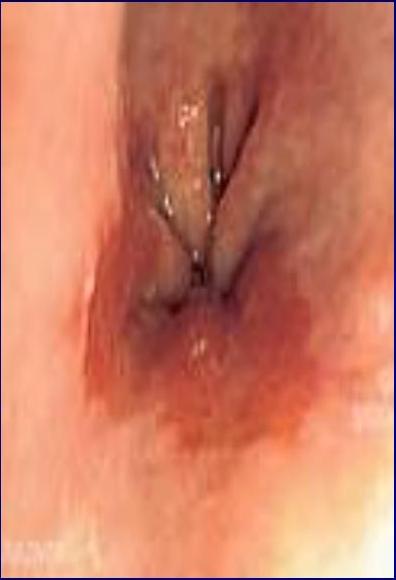
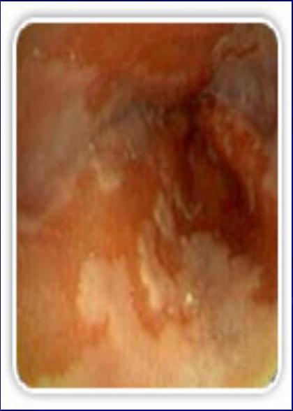
- The diagnosis of BE is made by endoscopy and pathology
- The presence of any endoscopically visible segment of columnar mucosa within the esophagus that on pathology identifies intestinal metaplasia defines BE
Symptoms and Diagnosis
lower esophagus
need, white, inflamed
Treatment
- Yearly surveillance endoscopy is recommended in all patients with a diagnosis of Barrett’s esophagus For patients with low-grade dysplasia, surveillance endoscopy is performed at 6-month intervals for the first year and then yearly thereafter if there has been no change
Treatment
Patients undergoing surveillance are placed on acid suppression medication and monitored for changes in their reflux symptoms. for life
Controversy surrounds the benefits of anti-reflux surgery in patients with Barrett’s esophagus Play role in treatment of early barrett esophagus
Treatment
Those in favour of surgery argue that medical therapy and endoscopic surveillance may treat the symptoms but fail to address the problem → acid reflux
The problem is the functional impairment of the LES that leads to chronic reflux and metaplastic transformation of the lower esophageal mucosa
Treatment
Surgery renders the LES competent and restores the barrier to reflux
Studies have demonstrated regression of metaplasia to normal mucosa up to 57% of the time in patients who have undergone antireflux surgery
Treatment
Photodynamic therapy (PDT) is the most common ablative method used to treat BE
Endoscopic mucosal resection (EMR) is gaining favor for the treatment of Barrett’s esophagus with low-grade dysplasia.
of severe dysplasia treated as CIS → subtotal resection
Treatment
(mucosal resection or esophagectomy)
→ aggressive therapy if moderate or severe dysplasia
Esophageal resection for Barrett’s esophagus is recommended only for patients in whom high-grade dysplasia is found
Pathologic data on surgical specimens demonstrate a 40% risk for adenocarcinoma within a focus of high-grade dysplasia
Benign esophageal tumor
Benign Esophageal Tumors and Cysts
Benign tumors are rare (< 1 %)
Classified in two groups
- Mucosal
- Extramucosal (intramural)
More useful classification:
- 60% of benign neoplasms are leiomyomas
- 20% are cysts
- 5% are polyps
- Others (< 2 percent)
4. Esophageal Cysts
Arise as diverticula of the embryonic foregut
¾ of this cyst present in childhood
Over 60% are located along the right side of the esophagus
Are often associated with vertebral anomalies (ex: spina bifida)
60% present in the first year of life with either respiratory or esophageal symptoms
Cyst found in the upper third of the esophagus present in infancy while lower third lesions present later in childhood
Pedunculated Intraluminal Tumors
(Polyps)
Benign polyps are rare
Usually occur in older men and may cause intermittent dysphagia
Are sometimes easily missed with barium swallow and esophagoscopy
6- Leiomyoma
Leiomyomas constitute 60% of all benign esophageal tumors
They are found in men slightly more often than women and tend to present in the 4th and 5th decades
They are found in the distal two thirds of the esophagus more than 80% of the time
Leiomyoma
They are usually solitary and remain intramural, causing symptoms as they enlarge.
*Tumor of the muscularis, under neath mucosa, so by endoscopy will find large, taking biopsy from mucosa will be normal.
Recently, they have been classified as a gastrointestinal stromal tumor (GIST)
GIST tumors are the most common mesenchymal tumors of the gastrointestinal tract and can be benign or malignant
need to take biopsy from tumor itself (arising from muscularis or submucosal)
Leiomyoma
Nearly all GIST tumors occur from mutations of the c-KIT oncogene, which codes for the expression of c-KIT (CD117).
All leiomyomas are benign with malignant transformation being rare
*esophagus is a distensible organ, any cause dysphagia with 2/3 of lumen is blocked. → That’s why presentation of patients with cancer esophagus is late
Symptoms and Diagnosis
Many leiomyomas are asymptomatic → if <2cm
during swallowing
Dysphagia and pain are the most
occur as lumen enlarge
common symptoms and can result from even the smallest tumors
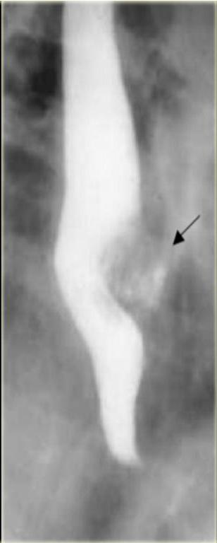
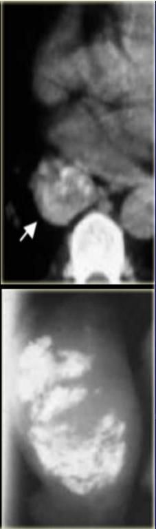
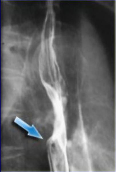
A chest radiograph is not usually helpful to diagnose a leiomyoma, but on barium esophagram, a leiomyoma has a characteristic appearance.
- filling defect
- pressure from outside
- normal mucosa
- smooth
barilum Swallow
[Non-Text]
[Non-Text]
[Non-Text]
[Non-Text]
[Non-Text]
[Non-Text]
[Non-Text]
[Non-Text]
[Non-Text]
[Non-Text]
[Non-Text]
[Non-Text]
[Non-Text]
[Non-Text]
[Non-Text]
[Non-Text]
[Non-Text]
[Non-Text]
[Non-Text]
[Non-Text]
[Non-Text]
[Non-Text]
[Non-Text]
[Non-Text]
[Non-Text]
[Non-Text]
[Non-Text]
[Non-Text]
[Non-Text]
[Non-Text]
[Non-Text]
[Non-Text]
[Non-Text]
[Non-Text]
[Non-Text]
[Non-Text]
[Non-Text]
[Non-Text]
[Non-Text]
[Non-Text]
[Non-Text]
[Non-Text]
[Non-Text]
[Non-Text]
[Non-Text]
[Non-Text]
[Non-Text]
[Non-Text]
[Non-Text]
[Non-Text]
Leiomyoma
During endoscopy, extrinsic compression is seen, and the overlying mucosa is noted to be intact
Diagnosis also can be made by an endoscopic ultrasound (EUS), which will demonstrate a hypoechoic mass in the submucosa or muscularis propria
biopsy → normal mucosa
FNA → if in doubt about tumor if its submucosa, leiomyoma, cancer, LN needle through endoscope, EUS guided
Treatment
Leiomyomas are slow-growing tumors with rare malignant potential that will continue to grow and become progressively symptomatic with time
Although observation is acceptable in patients with small (<2 cm)
asymptomatic tumors or other
significant co morbid conditions, in most patients, surgical resection is advocated
do CT, barium swallow, MRI, endoscopy, all to see exact location to know how to approach
Treatment
if in lower esophagus → approach from abdomen laproscoccally
Surgical enucleation of the tumor remains
the standard of care and is performed through a thoracotomy or with video or robotic assistance
The mortality rate is less than 2%, and success in relieving dysphagia approaches 100%
expose esophagus from outside, exercise tumor, leave mucosa intact, close remaining muscles don’t injure mucosa because its submucosal tumor.
Malignant esophageal tumor
CARCINOMA OF THE ESOPHAGUS
Esophageal cancer is the fastest growing cancer in the western countries Squamous cell carcinoma still accounts for most esophageal cancers diagnosed However, in the US, esophageal adenocarcinoma is noted in up to 70% of patients presenting with esophageal cancer
CARCINOMA OF THE ESOPHAGUS
Squamous cell carcinomas arise from the squamous mucosa that is native to the esophagus and is found in the upper and middle third of the esophagus 70% of the time
Smoking and alcohol both increase the risk for foregut cancers by 5-fold. Combined
• lower third → adenocarcinoma
CARCINOMA OF THE ESOPHAGUS
Food additives, including nitrosamines found in pickled and smoked foods, long-term ingestion of hot liquids caustic ingestion, achalasia, bulimia, tylosis (an inherited autosomal dominant trait), Plummer-Vinson syndrome, external-beam radiation, and esophageal diverticula all have known associations with squamous cell cancer.
CARCINOMA OF THE ESOPHAGUS
The 5-year survival rate varies but can be as good as 70% with polypoid lesions and as poor as 15% with advanced tumors.
esophageal adenocarcinoma now accounts for nearly 70% of all esophageal carcinomas diagnosed in Western countries
CARCINOMA OF THE ESOPHAGUS
There are a number of factors that are responsible for this shift in cell type:
Increasing incidence of GERD
Western diet
Increased use of acid-suppression medications
Not suppressed look so there is still harmful effect from the acid but patient not feel any symptoms.
Intake of caffeine, fats, and acidic and spicy foods all lead to decreased tone in the LES and an increase in reflux
& and Peppermint
CARCINOMA OF THE ESOPHAGUS
- As an adaptive measure, the squamous-lined distal esophagus changes to become lined with metaplastic columnar epithelium (Barrett’s esophagus)
- Progressive changes from metaplastic (Barrett’s esophagus) to dysplastic cells may lead to the development of esophageal adenocarcinoma
Symptoms
Early-stage cancers may be asymptomatic or mimic symptoms of GERD
Most patients with esophageal cancer present with dysphagia and weight loss
Because of the distensibility of the esophagus, a mass can obstruct two thirds of the lumen before symptoms of dysphagia are noted
Symptoms
Choking, coughing, and aspiration from a tracheo-esophageal fistula, as well as hoarseness and vocal cord paralysis from direct invasion into the recurrent laryngeal nerve, are ominous signs of advanced disease
Systemic metastases to liver, bone, and lung can present with jaundice, excessive pain, and respiratory symptoms.
Diagnosis
There are a plethora of modalities available to diagnose and stage esophageal cancer
Radiologic tests, endoscopic procedures, and minimally invasive surgical techniques all add value to a solid staging workup in a patient with esophageal cancer.
Esophagram
A barium esophagram is recommended for any patient presenting with dysphagia is able to differentiate intra-luminal from intramural lesions and to discriminate between intrinsic (from a mass protruding into the lumen) and extrinsic (from compression of a structures outside the esophagus) compression
Esophagram
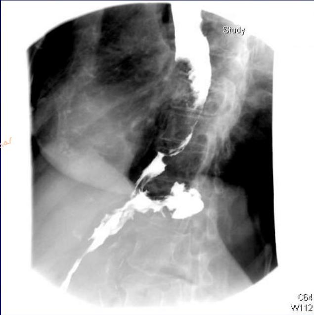
The classic finding of an apple-core lesion in patients with esophageal cancer is recognized easily
Although the esophagram will not be specific for cancer, it is a good first test to perform in patients presenting with dysphagia and a suspicion of esophageal cancer
barium swallow
apme core appearance →irregular
also seen in cancer colon
C64 W112
Esophageal carcinoma
Apple core lesion
Endoscopy
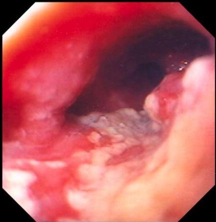
The diagnosis of esophageal cancer is made best from an endoscopic biopsy any patient undergoing surgery for esophageal cancer must have an endoscopy performed by the operating surgeon before entering the operating room for a definitive resection
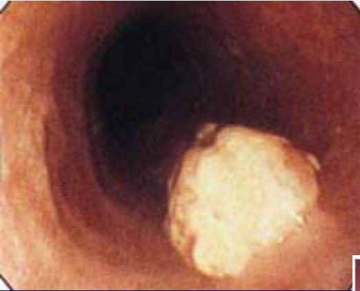

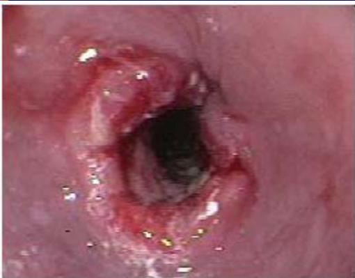
Squamous Cell Carcinoma of the Esophagus
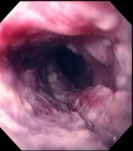
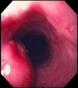
→ in middle 1/3
in lower 1/3
Adenocarcinoma of the Esophagus
Computed Tomography
9 for staging → To know treatment and prognosis
CT scan of the chest and abdomen is important to assess the length of the tumor, thickness of the esophagus and stomach, regional lymph node status and distant disease to the liver and lungs
CT-CAP
Positron Emission Tomography
To assess for distant metastasis PET scan evaluates the primary mass, regional lymph nodes, and distant disease
Its sensitivity and specificity slightly exceed those of CT; however, they remain low for definitive staging
Endoscopic Ultrasound
EUS is the most critical component of esophageal cancer staging.
The information obtained from EUS will help guide both medical and surgical therapy
biopsy samples can be obtained of the mass and lymph nodes in the paratracheal, subcarinal, paraesophageal, celiac region
- There is high mortality due to late presentation
Treatment
→ multi disciplining management.
Chemotherapy → neo adjuvant → 3-4 sessions Then restage by CT, MRI, PET
Radiation therapy
has effect in upper 1/3 SCC affect SCC more than adenocarcinoma
Chemo-Radiotherapy
Then put Patient in tumor board and refer to Thoracic Surgeon
Surgical resection → best one esp. in early stage
esophagectomy → and after 2-3 weeks, give adjuvant chemotherapy ± radiotherapy
GASTROESOPHAGEAL REFLUX DISEASE
LES has the primary role of preventing reflux of the gastric contents into the esophagus
GERD may occur when the pressure of the high-pressure zone in the distal esophagus is too low to prevent gastric contents from entering the esophagus
- There are barriers (anatomical) preventing acidity to come.
-
angle of Hiss
-
Crura
-
Presence of abd. part of esophagus in abdomen
→ if these barriers are gone ⇒ GERD
different from hiatus hernia
GASTROESOPHAGEAL REFLUX DISEASE
GERD is often associated with a hiatal hernia
the most common is the type I hernia, also called a sliding hiatal hernia → medical or surgical treatment
→ surgical treatment → mixed: Sliding and Para esophageal
Type II and III hiatal hernias are often referred to as para-esophageal hernias and they may be associated with GERD
asymptoms: dysphagia, early satiety, palpitation, chest pain, abd. pain
Type IV when there is other organ herniated into the chest (Spleen, Colon)
Treatment should be surgical because risk of stomach to be strangulated and get necrotized → ischemia
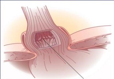
A
GE junction
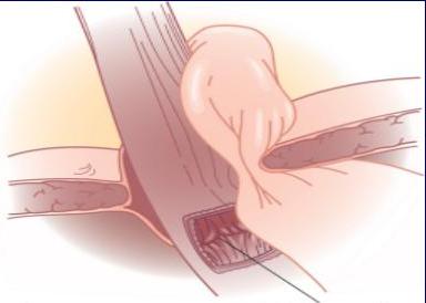
B
GE junction
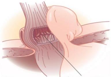
C
GE junction
GASTROESOPHAGEAL REFLUX
DISEASE
Definition :
Symptoms OR mucosal damage produced by the abnormal reflux of gastric contents into the esophagus
Often chronic and relapsing
May see complications of GERD in patients who lack typical symptoms
GASTROESOPHAGEAL REFLUX DISEASE
Epidemiology :
About 44% of the US adult population have heartburn at least once a month
14% of Americans have symptoms weekly
7% have symptoms daily
Clinical Presentations of GERD
1 Classic GERD 2 Extra esophageal/Atypical GERD 3 Complicated GERD
Clinical Presentations of GERD
Classic GERD :
Substernal burning and or regurgitation
Postprandial pain
Aggravated by change of position
Prompt relief by antacid → diagnostic factor
-
nexium
-
Pentobarbital
2 Extra-esophageal Manifestations of
GERD
These patients are wrongly treated for causes other than GERD
Pulmonary
Asthma
Aspiration pneumonia
Chronic bronchitis
Pulmonary fibrosis
from chronic aspiration
Other
Chest pain
Dental erosion
ENT
Hoarseness
Laryngitis
Pharyngitis
Chronic cough
Globus sensation
Dysphonia
Sinusitis
Subglottic stenosis
Laryngeal cancer
Clinical Presentations of GERD
3 Symptoms of Complicated GERD :
Dysphagia
- Difficulty swallowing: food sticks or hangs up
Odynophagia
- Retrosternal pain with swallowing
Bleeding
- Stricture formation
Diagnostic Tests for GERD
Barium swallow
Endoscopy
Ambulatory pH monitoring
→golden standard
↓ask patient to stop antineflux for 2-3 weeks,
Put probe scan above GEJ and leave for 24 hours
→ and will monitor the pH. and give patient
Esophageal manometry
booklet to write every time 1st symptoms
and what he was doing.
if in doubt, patient has GERD and at
the same time, has dysphagia symptoms.
do this (with ambulatory pH monitoring)
to rule out any motility disorders. → because they have different surgeries
Treatment
→ for GERD with or without sliding hernia
Lifestyle Modifications
Acid Suppression Therapy
Anti-Reflux Surgery
Endoscopic GERD Therapy
Treatment
Lifestyle Modifications
Elevate head of bed 4-6 inches
Avoid eating within 2-3 hours of bedtime
Lose weight if overweight
Stop smoking
Modify diet
- Eat more frequent but smaller meals
- Avoid fatty/fried food, peppermint, chocolate, alcohol, carbonated beverages, coffee and tea
OTC medications prn
Acid Suppression Therapy for GERD
H₂-Receptor Antagonists
(H₂RAs)
Proton Pump Inhibitors
(PPIs)
Cimetidine
(Tagamet®)
Ranitidine (Zantac®)
Famotidine (Pepcid®)
Nizatidine (Axid®)
Omeprazole
(Prilosec®)
Lansoprazole
(Prevacid®)
Rabeprazole
(Aciphex®)
Pantoprazole
(Protonix®)
Esomeprazole
(Nexium ®)
Anti-Reflux Surgery
Indication for Surgery :
- have failed medical management opt for surgery despite successful medical management (due to life style considerations including age, time or expense of medications, etc) have complications of GERD (e.g. Barrett’s esophagus; grade III or IV esophagitis) have medical complications attributable to a large hiatal hernia. (e.g. bleeding, dysphagia) have “atypical” symptoms (asthma, hoarseness, cough, chest pain, aspiration) and reflux documented on 24 hour pH monitoring
Endoscopic GERD Therapy
Endoscopic anti-reflux therapies → high recurrence rate
- Radiofrequency energy delivered to the LES Stretta procedure radiofrequency heating of GE junction
- Suture ligation of the cardia Endoscopic plication
- Sub mucosal implantation of inert material in the region of the lower esophageal sphincter Enteryx
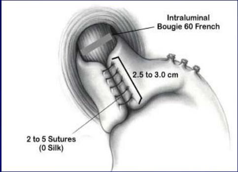
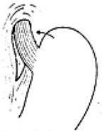
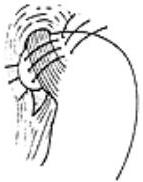
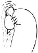
Nissen Fundoplication
→ Steps (?)
→ by laparoscopic
(a) Before
(b) Sutures
(c) After