SURGERY 2
HEAD INJURY
DR. SAFDAR H MALIK MD FCPS
MECHANISM OF HI
1-BLUNT INJURY
most common
*MOTOR VECHECLE ACCIDENTS
FALL
ASSAULTS
2-PENETRATING INJURY
GUN SHOTS INJURY
STABBING
EXPLOSIONS
9/1/2020
2
GCS
| EYE | SPONTANEOUS EYE OPENING OPENING TO VERBAL COMMOND ON PAIN FULL STIMULI NO EYE OPENING | 4 3 2 1 |
|---|---|---|
| VERBAL | ORIENTED & TALKING CONFUSED &DISORIENTED INCOMPREHSIBLE SPEECH/ WORDS SOUNDS NO VERBAL RESPONSE | 5 4 3 2 1 |
| MOTOR | OBEYS COMMAND LOCALIZE PAIN WITHDRAWL PAIN FLEXTION TO PAIN(DECORTICATE POSTURING) EXTENTION TO PAIN (DECEREBRATE POSTURING) NO MOTOR RESPONSE | 6 5 4 3 2 1 |
9/1/2020
3
TYPES OF HI ON BASIS OF GCS
- MILD HI – GCS FROM 13-15
- MODERATE HEAD INJURY-GCS 9-13
- SEVERE HEAD INJURY-GCS 8 OR LESS
need intubation
3 is lowest no zero
9/1/2020
4
Extent of injury
- SCALP INJURY
- SKULL INJURY
- BRAIN INJURY
9/1/2020
5
Scalp Injury
- Contusions
- Lacerations
- Avulsions
- Significant Hemorrhage
in children, hypovolemia and shock ( ttt: .stop bleeding . do CT . close wound
9/1/2020
6
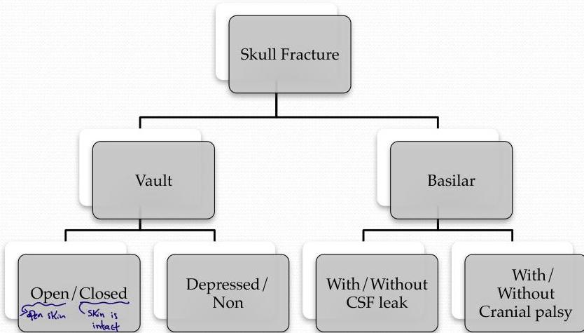
SKULL INJURY / FRACTURES
9/1/2020
7
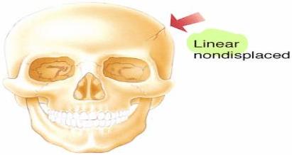
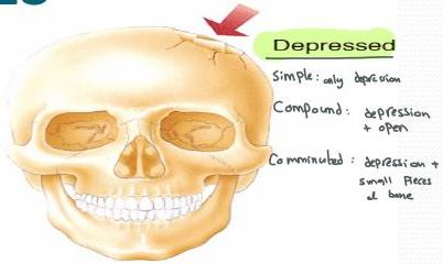
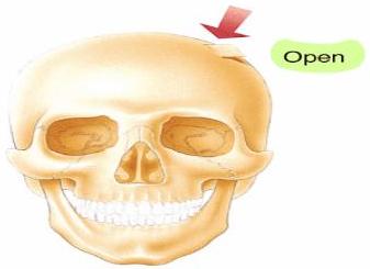
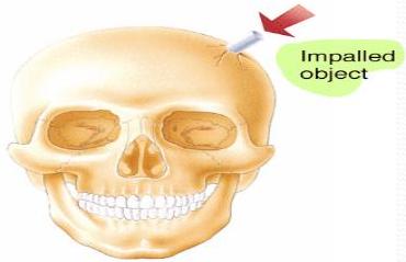
TYPES OF FRACTURES
9/1/2020
8
SKULL BASE FRACTURES SIGNS/SYPMTOMS
1-ANTERIOR SKULL BASE
RACCOUN EYES-BILATERAL PERIORBITAL ECCYMOSIS
CSF RINORRHEA
2-MIDDLE SKULL BASE
BATTELS SIGN -RETO AURICULAR ECCYMOSIS
CSF OTORIA
CRANIAL NERVES INJURIES
- Cribriform plate → Loss of smell
9/1/2020 4. Temporal bone → CN 7,8
9
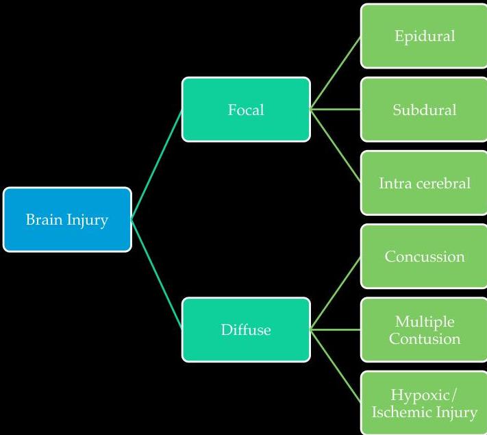
BRAIN INJURY CLASSIFICATION
ON MORFALOGY
PRIMARY
Primary injury caused by forces of trauma.
could progress to
SECONDARY
Secondary injury caused by factors resulting from the
primary injury
brain edema, herniation
hyponatremia ↓ 132
FOCAL INJURY
DIFFUSE INJURY
9/1/2020
10
SYSTEMIC INSULTS
- HYPOXIA (PaO2 less than 60mmhg)
- HYPOTENTION(systolic BP dropped < 90mmhg)
- HYPO/HYPER CAPNIA (Pco2 <32 0r >35)
- BLOOD LOSS ANEMIA
9/1/2020
11
Cont.
- Seizures
- Electrolytes imbalance
- Coagulopathy
- Infection
- Hyponatremia
- Iatrogenic (under resuscitation)
- INTRACRANIAL INSULTS
- Intracranial hypertension
- Intracranial lesion
- Cerebral edema
9/1/2020
12
9/1/2020
13
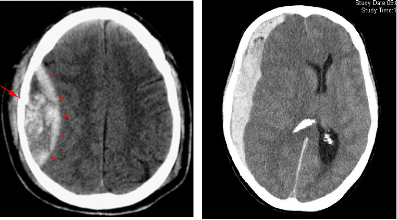
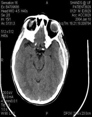
Epidural Hematomas
very dangerous
- Associated with skull fracture
- Classic: Middle meningeal artery tear
- Lenticular / biconvex
- Lucid interval
- Can be rapidly fatal
- Early evacuation essential.
- Outcome: 5% mortality.
- Head Injury w/ LOC + Lucid Interval followed by deterioration
- we have to do CT scan *
9/1/2020
14
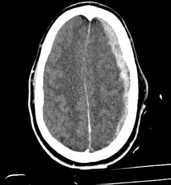
Subdural Hematomas
- Venous tear / brain laceration
- Occur in 15% of severe head trauma.
- Morbidity / mortality due to underlying brain injury
- Rapid surgical evacuation recommended, especially if > 5 mm shift of midline.
- Outcome: 35% mortality.
- Increased risk in elderly and alcoholics due to decreased brain volume
9/1/2020
15
DIFFUSE AXONAL INJURY
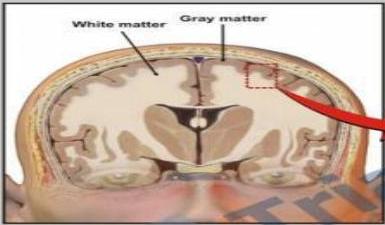
Sudden acceleration-deceleration forces cause injury to the brain.
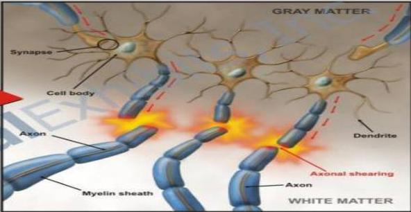
The injury is greatest in where the density difference is greatest. Most tearing occurs at the gray-white matter junction.
- Neuronal disconnection secondary to severe brain injury.
- Clinical: Low GCS without obvious lesion on CT Scan.
- Location: Corpus callosum – Internal capsule- Midbrain (Tectum)-Descending corticospinal Tract.
- Radiological: Punctate hemorrhage on MRI.
- Outcome: Poor
9/1/2020
16
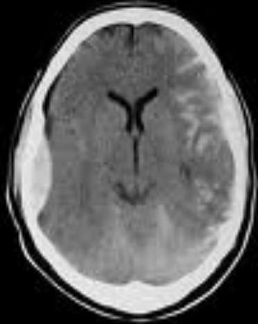
Subarachnoid Hemorrhage
- Bleeding beneath the arachnoid membrane on the surface of the brain.
- Most common cause of SAH is Traumatic.
- SAH is a form of Stroke.
- Symptoms:
- Severe Headache(Thunderclap)
- Confusion or decrease LOC/ COMA.
- Diagnosis:CT Brain without contrast-LP-cerebral Angio.
9/1/2020
17
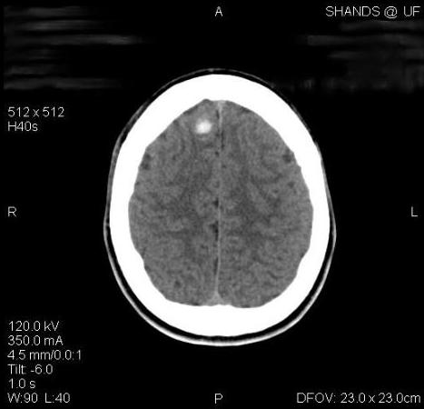
Intracerebral Hematoma “ICH”
- Focal areas of hemorrhage within the parenchyma
- Classified into Traumatic & Spontaneous.
- Outcome: 40% mortality.
- Symptoms depend on the site of bleeding.
- Diagnosis: CTA- MRA-
- Radiological: Spot sign.
9/1/2020
18
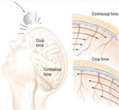
Direct Brain Injury Types
Coup
Injury at site of impact
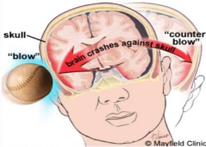
Countercoup
Injury on opposite side from impact
9/1/2020
19
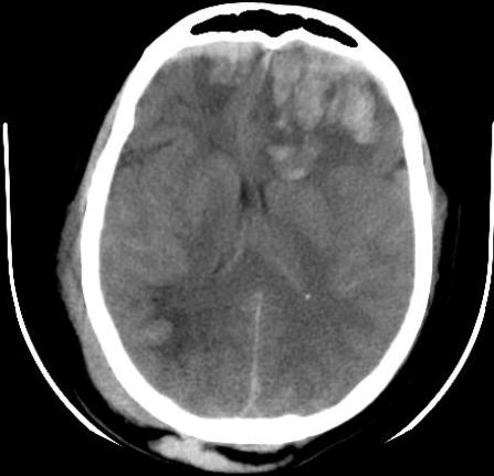
Large frontal
contusion with shift
9/1/2020
20
MANAGEMENT OF HI
- INTIAL QUICK ASSESSMENT
ABCDF
- FINAL ASSESSMENT
- TYPE OF HI ON BASIS OF GCS
- EXAMINATION OF HI
- CT TRAUMA SURWAY
9/1/2020
21
FOCUS EXAMINATION
-
GCS
-
PUPILS EXAMINATION
-
LATERALIZING SIGNS
-
Hemiplegia, hemiparesis, Facial asymmetry.
-
Seizures focal or generalized
-
Memory
-
Visual disturbances
9/1/2020
22
Neurologic Exam interpretation
Pupillary Size + Reactivity
Fixed Dilated Pupil = Ipsilateral Intracranial Hematoma resulting in uncal herniation
Bilateral Fixed + Dilated = Poor Brain Perfusion, bilateral uncal herniation or severe hypoxia
Indicative of very poor neurological outcome
Neurological Posturing
Decorticate Posturing = Upper extremity flexion with lower extremity extension Cortical Injury above the midbrain
De cerebrate Posturing = Arm extension and internal rotation with wrist flexion Indicative of brainstem injury Very Poor predictor of outcome
9/1/2020
23
Moderate Head Injury
- GCS Score = 9–13
- Initial evaluation same as for mild injury
- CT scan for all
- Admit and observe
- Frequent neurologic exams
- Repeat CT scan
Admit and observe
Frequent neurologic exams & Repeat CT
- Specialized Subset = “Talk and Die Syndrome”
- Initially, talkative and without significant signs of external injury
- Within 48 hours of injury, rapidly deteriorate
- Epidural Hematoma is cause in 78-80% of cases
- Patients with “talk and die syndrome” who present with a GCS > 9 but who deteriorate have been shown to have a worse outcome than patients who present with severe TBI at outset
9/1/2020
24
Severe Brain Injury
-
GCS Score = 3–8
-
Evaluate and resuscitate
-
Intubate for airway protection & admit in ICU
-
Focused neurologic exam
-
Frequent reevaluation
-
Identify associated injuries
9/1/2020
25
CONT.
- ICP MONITOR
- TREATMENT FOR BRAIN EDEMA
- RAISED HEAD END OF PARIANT BED.
- SEDATION
- DVT CARE
- ANTI CONVULSANT
- MANITOL IV / Furosemide osmotic duration check Nat emb 3h
- 3% NORMAL SALINE
- CONTROLED VENTILATION
- DECOMPRESSIVE CRANIOTOMY
9/1/2020
26
Operative management.
- ICP & OR EVD INSERTION
- CRANIOTOMY FOR EVACUATION OF EDH
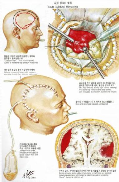
- WIDE DECOMPRESSIVE CRANIOTOMY & EVACUATION OF ACUTE SUBDURAL HAEMATOMA /IC HAEMATOMA
- WIDE DECOMPRESSIVE CRANIOTOMY FOR BRAIN EDEMA IN DIFFUSE BRAIN INSULT
9/1/2020
27
Operative
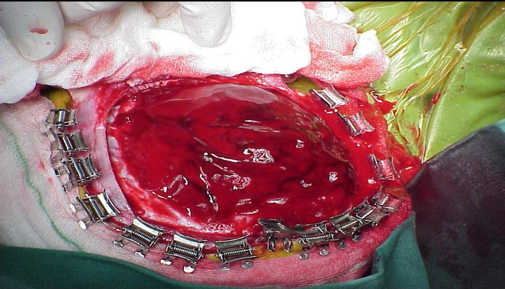
Craniotomy
- Salvage operation used to manage increasing ICP
- Removal of part of skull and underlying dura.
- Decreases ICP, improves cerebral perfusion, prevents ischemia
- Serves to limit secondary insults
- Literature divided on true benefit
9/1/2020
28
EDH
9/1/2020
29
Intracranial Pressure (ICP)
10 mm Hg = Normal
20 mm Hg = Abnormal
40 mm Hg = Severe
Many pathologic processes affect outcome
Sustained ↑ ICP leads to ↓ brain function and outcome
9/1/2020
30
POST HI SEQUALE
- POST TRAUMATIC SYNDROME
- POST CONCUSSION SYNDROME
- EPILEPSY
- PERSISTENT VEGETATIVE STATE
9/1/2020
31
SUMMARY –WHAT TO DO
- KEEP HEAD END AT 30 DEGREE P
- MAINTAIN MAP AT 90 mmHg
- MAINTAIN PCO2 AT 35 mmHg
- Use isotonic saline for euvolemia
- Frequent neurological exams
- Liberal use of CT scan brain.
- Early Neurosurgical consult
9/1/2020
32
SUMMARY –WHAT SHOULD NOT DO
- Allow patient to become hypotensive
- Over aggressive hyperventilation
- Use hypotonic IV fluids
- Long acting paralytics
- Paralyze before complete examination
- Depend upon clinical exam alone
9/1/2020
33
REFERENCES:
- National Head Injury Foundation.
- Atls manual of Head Trauma.
- Tarascon Neurosurgery Pocketbook.
THANK YOU
9/1/2020
34
SURGERY 1
Head injury severity
| Severity | GCS |
|---|---|
| Mild | ≥ 13 |
| Moderate | 9- ≤ 12 |
| Severe | ≤ 8 |
Traumatic brain injury (TBI)-
is the leading cause of death in trauma patients- 50% of all traumatic deaths.
Primary injury the anatomic and physiologic disruption that occurs as a direct result of trauma
Secondary injury -extension (sequelae) of the primary injury, result from local swelling, increased ICP, hypoperfusion, hypoxemia, or other factors.
Aim: detection and treatment of primary injury and prevention of secondary injury
management
- Maintain BP >90 mmHg, PaO2 >60 mmHg (75-100mmHg)
- Assess GCS and lateralizing signs- pupil and motor function
- a neurological symptom that suggests an issue with one side of the brain or body
- Pupillary asymmetry >1 mm suggests intracranial injury
- Larger pupil is on the side of the mass lesion
- Extremity weakness- detected by testing motor power
- CT scan head- accurate localization of the lesion
- Epidural or subdural hematoma: Treatment: evacuation
- Intracerebral hematoma & contusion
- Diffuse axonal injury: maintain brain perfusion & prevent rise in ICP.
(Image: Epidural Hematoma & Subdural Hematoma)