Surgery 2
Large Bowel Obstruction & colostomies
Moneer Almadani MD, FACS, F.MAS
Objectives
- Introduction
- Large bowel obstruction
- Causes
- Diagnosis
- Treatment
- Complications
- Diverticular disease
- Colon volvulus
- Colostomies
Introduction
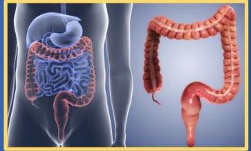
General Anatomy
- The colon is a 5-6–ft long, inverted, U-shaped.
- Cecum (and appendix) and ano-rectum, which are parts of the large intestine, are not included in the colon.
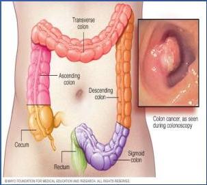
Ascending Colon
- Starts from a proximal blind end (pouch) called the cecum.
- The ascending colon takes a right-angled turn just below the liver (hepatic flexure) and becomes the transverse colon
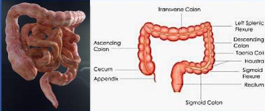
Transverse & Descending Colon
- Transverse colon: has a horizontal course from right to left, occupying the right hypochondrium, epigastrium, and left hypochondrium.
- At splenic flexure, it is attached to the diaphragm by the phrenocolic ligament) and becomes the descending colon,
- Splenic flexure is higher (cranial) to hepatic flexure.
- Descending colon: leads to the inverted V-shaped sigmoid colon, which then becomes the rectum at the S3 level
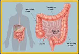
Peritoneal Relations
- Ascending, & descending parts of the colon are retroperitoneal
- The transverse colon and the sigmoid colon have a mesentery
- Cecum is intraperitoneal but uses the mesentery of the ileum.
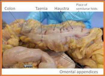
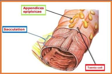
Teniae Coli & Omental Appendages
- Teniae coli are present in large bowel, but not present in the rectum.
- Appendages of fat, containing small blood vessels, called omental appendages (appendices epiploicae) are attached to colon.
Paracolic Gutters
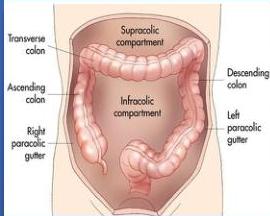
- Lateral to ascending and descending colon are the right and left gutters of the peritoneal cavity,
- Fluid/pus in the upper abdomen can trickle down into the pelvic cavity.
Blood Supply
- Superior mesenteric artery through its right colic and middle colic branches
- Inferior mesenteric artery through its left colic and multiple sigmoid branches.
- Vasa recta are terminal branches of these arteries entering the colonic wall
Layers of the Colon Wall
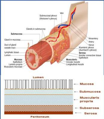
- The colon has 4 layers:
- Mucosa:
- Columnar epithelium with a large number of mucus secreting goblet cells
- No villi,
- Submucosa
- Contains the blood vessels and Meissner nerve plexus.
- Muscularis propria
- Contains the inner circular and outer longitudinal
- Muscles and myenteric (Auerbach) nerve plexus
- Serosa outermost layer
- visceral peritoneum.
Functions
- Functions: you have to provide these after colonic surgery
- Absorption of water, and electrolytes (Na, K+)
- Normal flora manufacture-
- Formation of vitamin K, & B complex (colonic bacteria)
- Storage and expulsion of feces
Large bowel obstruction
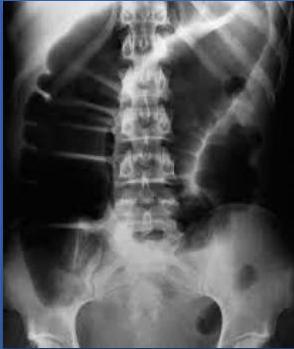
Definition & Classification
- Impedance to the normal propulsive action through intestine
- It is an emergency condition that requires early identification and intervention.
- Colonic obstruction is more common in elderly individuals
✓ Acute Vs chronic Subacute: slowly but new progress
- ✓ Complete vs Partial
- Mechanical vs pseudo-obstruction
Symptoms
- Crampy abdominal pain, poorly defined
- Abdominal distention
- Altered bowel habits (Constipation) = in complete obstruction
- Nausea and vomiting
- Symptoms of complications( perforation, peritonitis)
- Other symptoms that may be diagnostically significant include the following:
- Abrupt onset of symptoms (acute obstruction)
- Recurrent left lower quadrant abdominal pain over several years (suggestive of diverticulitis, a diverticular stricture)
- Chronic constipation, long-term cathartic use, and straining at stools (diverticulitis or carcinoma)
- Changes in stool caliber (suggestive of carcinoma)
- Rectal bleeding ⇒ tumor in easterly
- Incomplete evacuation ⇒ tumor
Signs
- Abdomen (inspection, auscultation, percussion, and palpation)
- Evaluate bowel sounds, tenderness, rigidity, guarding, and any mass or fullness
- Inguinal and femoral regions, look for a possible incarcerated hernia
- Rectum, contents of anal vault, and stool consistency; perform fecal occult blood testing as appropriate
Pathophysiology
- Increased contraction of bowel proximal to obstruction
- To overcome the obstruction, proximal dilatation, and distension
- Mucosal edema and impaired venous and arterial blood flow
- Increase the mucosal permeability of the bowel
- Bacteria translocation makes Large bowel obstruction more serious
- Electrolytes imbalance, gross distension, 3rd space fluid loss
- Ileus formation+ dehydration
- Toxemia
- Further vascular compromization
- Perforation
Etiology (Different Classifications)
- Neoplasm (benign or malignant) 60%
- Stricture (diverticular or ischemic) 10 %
- Volvulus (Sigmoid, Caecal) 5%
- Hernia
- Fecal Impaction
- Adynamic
Dynamic
- Intraluminal
- Fecal impaction
- Foreign body
- Bezoars
- Gall stones
- Intramural
- Stricture
- malignancy
- Extramural
- Adhesions & bands
- Hernias (internal – external)
- Volvulous
- Intussception
Adynamic
- Paralytic ilus
- Mesenteric vascular occlusion
- Pseudo obstruction
Diagnosis: Laboratory Tests
- Complete blood count (CBC) : WBC, HB, Hematocrit
- Coagulation
- Electrolytes
- LFT
- Serum lactate (if bowel ischemia is a consideration)
- Urinalysis
- Stool guaiac test
Diagnosis: Imaging & Endoscopy
- Radiology Imaging
- Plain radiography (flat and upright)
- Contrast radiography with enema
- Computed tomography (CT)
- This is the imaging modality of choice in any
- Able to confirm the diagnosis!!
- Localize the level of obstruction
- Able to identify the cause.
- Complications
- and assess the vascularity
- Colonoscopy / Bx Biobsy
- ↓ Cause more distention I may Cause perforation
- → so you have to do cleansing enema.
- no role of of Clonoscopy in acute case of obstruction
- → no role of endoscopy in acute case of small bowel obstruction
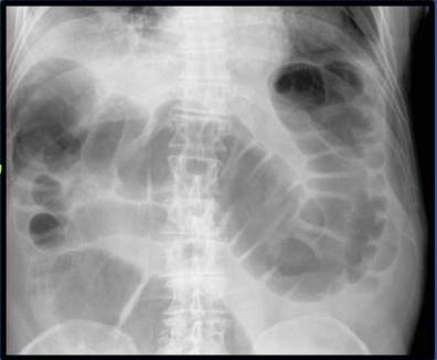
Imaging Findings (Early & Advanced Stages)
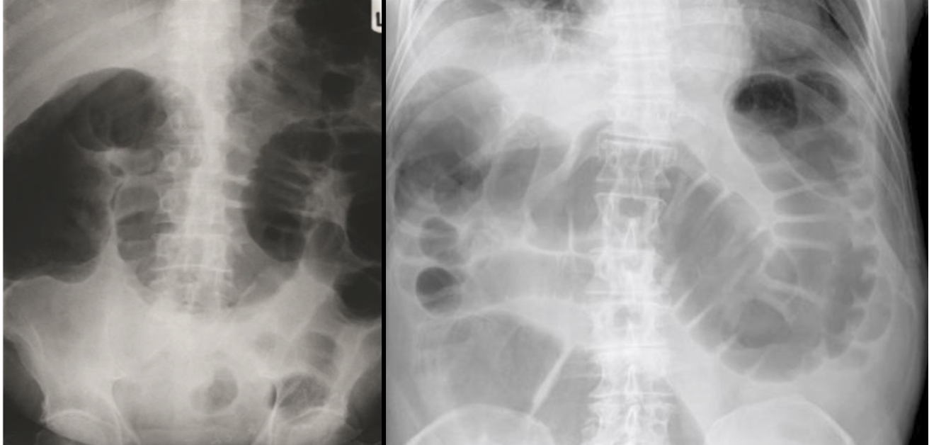
- In early stages: colonic distension:
- Gaseous secondary to gas-producing organisms in faeces
- Collapsed distal colon
- ve rebound tenderness
- In advanced cases one may see the stigmata of an ischaemic colon, namely:
- Intramural gas (pneumatosis coli)
- Portal venous gas
- Free intra-abdominal gas (pneumoperitoneum)
Management: Supportive Measures
- Nil By Mouth
- Intake- output charts
- IV lines, and rehydration (IV crystalloid with K⁺)
- Foley catheter
- NG Tube to aspirate content for ‘decompression’
- TED stockings, DVT prophylaxis
- Antibiotics
- Antiemetics
- Analgesia
Management: Surgery
- exploratory laparotomy.
- Surgery: indication:
- Perforation
- Closed loop obstructions
- Sepsis
- Bowel ischemia
- Volvulus
- Hernia, stone or foreign body
Ogilvie syndrome
- Loss of peristalsis and results in the accumulation of gas and fluid in the colon.
- Unclear pathophysiology
- Autonomic imbalance
- Decreased parasympathetic tone
- Excessive sympathetic output.
- Medical or surgical illnesses.
- Complications: colonic ischemia or perforation
- The right colon and cecum are most commonly involved.
- Risk of perforation (3% -15%)
Diverticular diseases
Diverticulosis
- Includes a spectrum of conditions
- Asymptomatic vs symptomatic
- Acute vs chronic
- Complicated vs non complicated
- Diverticulosis:
- Small pouches due to herniation of the mucosa into colonic wall
- Commonest pathology responsible for lower GI bleeding
- False diverticula
- because not all layers involve
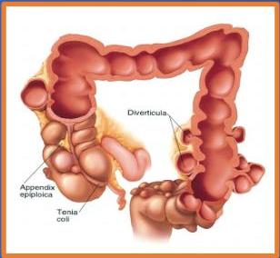
Diverticulitis
- Diverticulitis: Pain in LL abdomen
- Inflammation of diverticula, due to obstruction by fecalith
- Usually affects elderly
- 20% of patients with diverticulitis are younger than 50 years.
- In chronic form, patients may have recurrent low-grade diverticulitis.
Signs and Symptoms
- Depends on the location, the severity of the inflammatory process, and the presence of complications.
- Abdominal pain:
- Left lower quadrant: (70% of patients), as most diverticula occur in the sigmoid colon
- Right lower quadrant tenderness, mimicking acute appendicitis, can occur in right-sided diverticulitis
- Change in bowel habits
- Nausea and vomiting
- Constipation
- Diarrhea
- Flatulence
- Bloating
- Abdominal pain:
Complications
- Bleeding
- Perforation/ Abscess:
- Generalized tenderness with rebound and guarding
- Distended and tympanic abdomen
- Bowel sounds can be diminished or absent
- Tender palpable mass (Abscess formation)
- Fistula:
- Colovaginal : purulent vaginal discharge
- Colovesicular: urinary tract symptoms (eg, suprapubic, flank, or costovertebral angle tenderness) or pneumaturia, and sometimes fecaluria
- Stricture/ obstruction
Diagnosis: Laboratory Tests
- Usually made on the basis of history and physical examination,
- Laboratory tests
- CBC: high WBC, Hb,
- Electrolytes,
- Renal function,
- LFT
- Urinalysis / culture
- Blood cultures
- Pregnancy test
Diagnosis: Radiology Imaging
- Plain abdominal radiograph series
- Contrast enema,
- Water-soluble medium,
- Mild-to-moderate uncomplicated cases
- CT abdomen with contrast gold standard
- Best imaging method to confirm the diagnosis.
- Sensitivity and specificity 97%
- Bowel wall thickening
- Soft-tissue inflammatory masses
- Complications, exclude other pathology
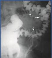
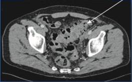
Hinchey’s Classification
- Clinical staging
- To chose the proper management option
| Hinchey grade I | Localised para-colonic abscess |
|---|---|
| Hinchey grade II | Distant abscess (e.g. pelvic, sub-phrenic) |
| Hinchey grade III | Purulent peritonitis |
| Hinchey grade IV | Faecal peritonitis |
Management
- Conservative: (mild cases)
- Initiate bowel rest and intravenous fluid hydration
- Broad-spectrum intravenous antibiotic Within 2-3 days of hospitalization, fever, pain, and leukocytosis should begin to resolve
- Start on a clear liquid diet and advanced as tolerated
- CT-guided percutaneous drainage
- Surgical intervention:
- Hinchey’s stage III or IV
- Complications
- Inability to rule out carcinoma
- Failure of medical therapy
Colonic volvulus
Definition & Types
- Derived from the Latin word volvere (“to twist”).
- Part of the colon twists on its mesentery, resulting in colonic obstruction.
- The main types:
- Sigmoid volvulus (counterclockwise)
- in canary
- Cecal volvulus (clockwise direction)
- in young
- Important in antiviral
- by colonoscopy
- Sigmoid volvulus (counterclockwise)
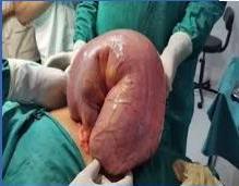
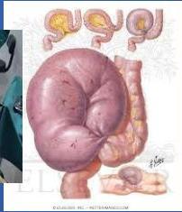
Sigmoid vs Cecal Volvulus
Definition
- Sigmoid volvulus is the twisting of the sigmoid colon of the large intestine.
- Cecal volvulus takes place due to the torsion caused in the cecum area
Extension of the Volvulus
- Sigmoid volvulus can only move upwards and goes to the right upper quadrant.
- Cecal volvulus can move in any direction and can be observed in the pelvic area as well.
- at any direction
Affected Population
- The elderly population is mostly affected by sigmoid volvulus.
- The young population is mostly affected by cecal volvulus.
Causes
- High residue diet
- Bulky stool
- Chronic constipation
- Laxatives abuse
- Organic/ Anatomical:
- Tortious elongated colon
- Adhesive band
- Overloaded colon
- Long mesocolon
- Narrow colonic attachment
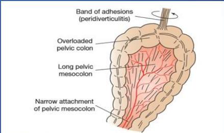
Management
- In patients with no evidence of peritonitis or ischemic bowel
- Resuscitation
- Detorsion:
- Sigmoid volvulus (success rate 90%)
- Sigmoidoscopy or Colonoscopy
- Rectal tube placement
- Cecal volvulus (success rate 15-20)
- Cecostomy
- Sigmoid volvulus (success rate 90%)
- Surgery:
- if record or details
- Open vs Lap
- Sigmoid resection/ Hartman’s (Sigmoid)
- Right hemicolectomy (Cecal)
- Cecopexy alone is associated with volvulus recurrence in 20-30% of patients
- Function
- Unstable patient: surgery
Colostomies
Definition
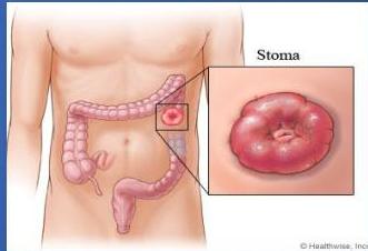
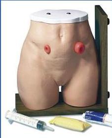
- Stomas are artificial opening to connect a body cavity to the outside
- Named according to anatomic location (Colostomy, Ileostomy..)
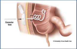
- A colostomy is a surgical procedure where a portion of the large intestine is brought through the abdominal wall to carry stool out of the body.
- Temporary vs. Permanent
- Emergency vs. Elective
Types of Colostomies
1. Loop Colostomy
- Proximal and the distal limbs are opened into the common opening
- Two opening in one stoma, with
- Artificial devise between the openings, to keep bowel from slipping back
- Transverse colon/ Sigmoid colon
- Typically temporary, and an emergency procedure
2. Double Barrel Colostomy
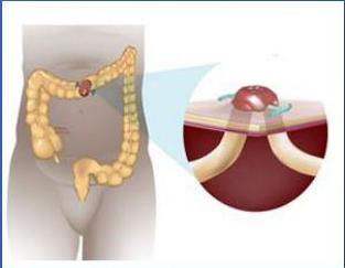
- Two opening
- Proximal opening is functional, but distal one as mucous fistula
- Temporary or permanent
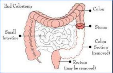
3. End Colostomy
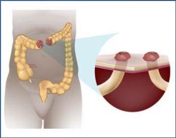
- Created from the proximal end of the colon
- Distal end remains as a blind pouch, or it can be exteriorized separately
- Hartmann procedure:
- Proximal end colostomy
- Distal end of colon closed.
Indications
- To relieve distal obstruction
- Colorectal carcinoma
- Large bowel obstruction
- Protection of distal anastomosis
- Traumatic perineal injury
- High anal fistulae
- Diverticular disease/ IBD
- Ischemia
- Congenital anomalies (Hirschsprung’s disease, Meconium ileus, Imperforate anus)
Complications (+ Nutritional Complication)
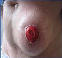
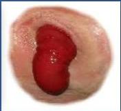
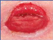
- Edema/ Prolapsed
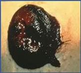
- Necrosis/ Ischemia
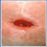
- Retraction
- Bleeding
- Obstruction
- Wound infection
- Leakage
- Stenosis
- Parastomal hernia
- Related to output (Metabolic/ hydration)
Surgery 1
Large bowel obstruction
- It is an emergency condition that requires early identification and intervention.
- Acute Vs chronic
- Complete vs Partial
- Mechanical vs pseudo-obstruction
Symptoms:
- Crampy abdominal pain
- Abdominal distention
- Nausea and vomiting
Other symptoms that may be diagnostically significant include the following:
- Abrupt onset of symptoms (acute obstruction)
- Recurrent left lower quadrant abdominal pain over several years (suggestive of diverticulitis, a diverticular stricture)
- Chronic constipation, long-term cathartic use, and straining at stools (diverticulitis or carcinoma)
- Changes in stool caliber (suggestive of carcinoma)
Signs:
- Abdomen (inspection, auscultation, percussion, and palpation)
- Evaluate bowel sounds, tenderness, rigidity, guarding, and any mass or fullness
- Inguinal and femoral regions, look for a possible incarcerated hernia
- Rectum, contents of anal vault, and stool consistency; perform fecal occult blood testing as appropriate
Etiology:
- Neoplasm* (benign or malignant) 60%
- Stricture (diverticular or ischemic)
- Volvulus (colonic, sigmoid, cecal) 5%
- Fecal Impaction
Diagnosis
Labs:
- Complete blood count (CBC) : WBC, HB, Hematocrit
- Coagulation
- Electrolytes
- LFT
- Serum lactate (if bowel ischemia is a consideration)
- Urinalysis
- Stool guaiac test
Diagnosis
Radiology Imaging
- Plain radiography (flat and upright)
- Contrast radiography with enema
Computed tomography (CT) –
- This is the imaging modality of choice
Colonoscopy/Bx
Management
Initial therapy
- Volume resuscitation
- Appropriate preoperative broad-spectrum antibiotics
- Timely surgical consultation
- Consideration of a nasogastric tube for severe colonic distention and vomiting
The following are emergencies that call for surgical intervention:
- Closed loop obstructions
- Sepsis due to complicated diverticular disease
- Bowel ischemia
- Volvulus
Supportive measures:
- Nil By Mouth
- Intake- output charts
- IV lines, and rehydration (IV crystalloid with K+)
- Foley’s catheter
- NG Tube to aspirate content for ‘decompression’
- TED stockings, DVT prophylaxis
- Antibiotics
- Antiemetics
- Analgesia
Surgery:
Closed loop obstructions, Sepsis, Bowel ischemia, Volvulus