جامعة المعرفة ALMAAREFA UNIVERSITY

Leenah Turjoman
نرتقي مع المعرفة
جامعة المعرفة^{}[] ALMAAREFA UNIVERSITY
LOWER GIT
BLEEDING
نرتقي مع المعرفة
جامعة المعرفة ALMAAREFA UNIVERSITY
Objectives
At the end of this presentation students will be able to:
Define lower GI haemorrhage (LGIB).
Enumerate the causes of LGIB.
Describe the pathophysiology of LGIB
Describe the symptoms and signs
Describe the diagnostic work up.
Describe the resuscitative measures.
Describe the management- non-surgical & surgical.
Introduction
- Definition: Bleeding distal to DJ flexure- ligament of Treitz
- Frequent cause of hospital admission, morbidity & mortality.
- 20-33% of all gastrointestinal bleeding.
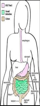
- Proximal to caecum- melena
- Right colon- maroon color
- Left colon- bright red bleeding (hematochezia)
- Brisk UGI bleeding- bright red color

Causes of Lower G I T Bleeding in Adults
Percentage of Patients
•Diverticular disease → most common
60%
•Inflammatory bowel disease
13%
•Crohn disease
•Ulcerative colitis
•Benign anorectal diseases
→ more common but not too much bleeding
11%
•Hemorrhoids
•Anal fissure
•Fistula-in-ano
•Neoplasia → less common
9%
•Malignant neoplasia of colon, rectum, SI, anus
Coagulopathy
4%
Arteriovenous malformations
3%
TOTAL
100%
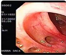
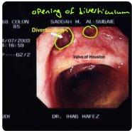
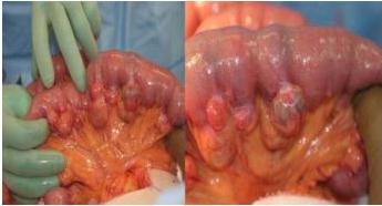
→ Treatment: (according to Hinchey Classification) I, II → Conservative III, IV → Surgery (hartmann Procedure) → resection of area of diverticulosis, and do end colostomy of remaining descending colon, and closure of rectum. later can do anastomosis of descending colon with rectum
Diverticular Disease
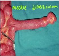
→ most common → false (true is in right colon) → mechle’s diverticulum bleeding occur in diverticulosis more than diverticulitis → present just like acute appendicitis but on left side diverticule is filled with contents and when complications occur, lead to bleeding • Greater involvement in left colon 90%. occur due to low fiber diet → Const. Pation → bulging of mucosa outside, to the entrance of bilis (mucosa only that’s why its false diverticulum) • Most bleeding from right 60-80%. → mechles diverticulum → main and major cause of GI bleeding (huge amount) • Diverticulosis - not cause of occult bleeding → its a cause of visible bleeding • Diverticulitis - rarely associated with significant bleeding
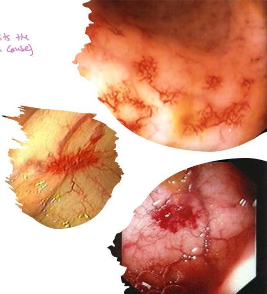
Angiodysplasia
(in research its the most common cause)
- Most common A-V malformations found in the GIT
- Most common site- cecum and ascending colon
- Acquired lesions
- Elderly >60 years
- Bleeding is venocapillary in origin.
- Generally, less vigorous than diverticular bleeding.
- 80% untreated angiodysplasia experience rebleeding.
❖ LOWER GI HEMORRHAGE IN CHILDREN & ADOLESCENTS
✓ Meckel diverticulum → → → → → → →
✓
Juvenile polyps and polyposis
✓
Peutz-Jeghers syndrome
✓ FAP + melanosis
Gardner’s syndrome GFAP + osteomas
✓
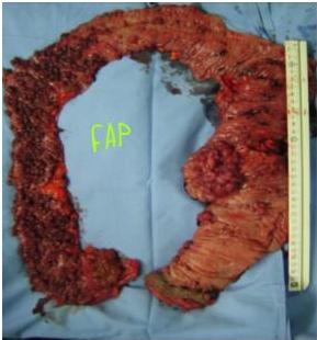
Familial adenomatous polyposis (FAP) → → → → →
✓ > 100 Polyps
✓ Premalignant → so should
✓ Should do surgery, but if refused then follow up
Crohn’s disease or Ulcerative colitis
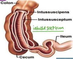
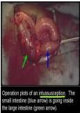
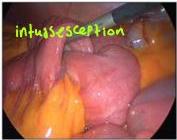
✓ Intussusception → → → →
✓ Fred Current jelly stool
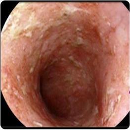
Colitis (IBD)
-
Massive hemorrhage-due to IBD is rare
-
Ulcerative colitis: Bloody diarrhea in most.
smore malignant
- Mild to moderate LGIB in up to 50%.
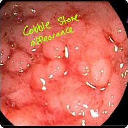
- Crohn disease: LGIB is not as common.
binhole all layers
-
Bleeding more common with colonic involvement.
-
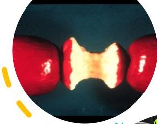
Ischemic colitis: Elderly, pain abdomen, bloody diarrhea. Involves splenic flexure and the rectosigmoid.
-
❖ Not associated with significant blood loss or hematochezia.
❖ Neoplasms
- Polyps and carcinoma.
- Occult bleeding, Low grade and frequent bleeding- common
- Massive bleeding- unusual
❖ Benign anorectal disease
Hemorrhoids, anal fissures, anorectal fistulas cause intermittent rectal bleeding.
- 11% of LGIB- from anorectal disease.
❖ Small intestinal conditions
Peutz-Jeghers syndrome, hemangiomas, & adenocarcinomas usually cause occult bleeding.
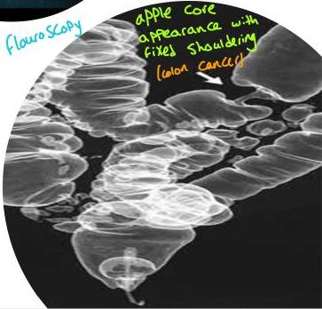
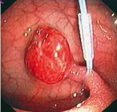
Pedunculated Polyp (narrow neck) → Remove by Snaring (by endoscope)
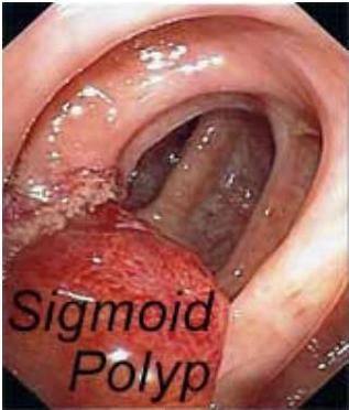
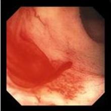
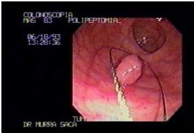
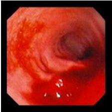
Sessile Polyp → wide base
Diagnosis
-
History & physical examination
-
Nasogastric tube →
because the cause could be upper but bleeding going down (1/3-7) eg bright red color could be from heavy ugIB or transient time (too much bowel movement → diarrhea) eg millers
-
Digital rectal examination, anoscopy / proctoscopy
-
Complete blood cell (CBC) count
-
Serum electrolytes levels
-
Coagulation profile: aPTT, PT, platelet count
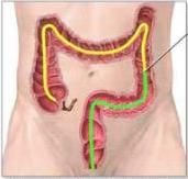
End of sigmoidoscopy
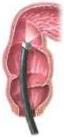
Colonoscopy examines the entire length of the colon; sigmoidoscopy examines only the lower third
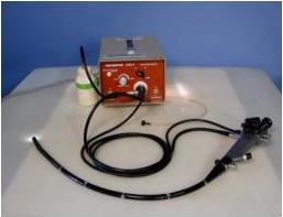
Diagnosis- UGIT Endoscopy
then COLONOSCOPY
4 cuz Cause could be upper GI
of but if know the patient, and sure that cause is lower then start with colonoscopy.
- Flexible colonoscopy: Initial diagnostic method of choice. Hemodynamically stable.
- Colonoscopy following a rapid bowel preparation.
- Bowel prepared colonoscopy- higher diagnostic/therapeutic yields than unprepared colonoscopy. To see the site of bleeding
- Successfully identify the origin of severe LGIB in 80-90%.
CT Angiography
- Detects bleeding at rates of 1-1.5 mL/min.
- Indications:
- Brisk ongoing LGIB,
- Hemodynamically unstable,
- with or without a preceding radionuclide scan & failed colonoscopy.
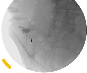
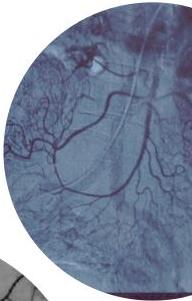
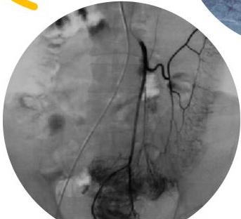
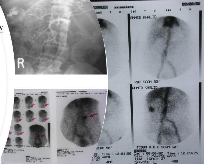
- Tc⁹⁹RBC scan: Detects hemorrhage at rates as low as 0.1-0.5 mL/min.
- CT Angiography: Routine workup failed- contrast extravasation, bowel wall enhancement, vascular dilatation
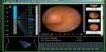
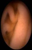
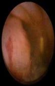
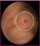
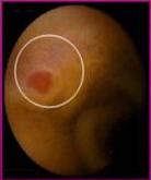
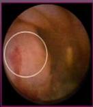
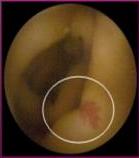
Wireless capsule endoscopy (WCE) Technical aspects
- 3 components
- Capsule endoscope
- External receiving antenna with attached portable hard drive
- Computed workstation for review and interpretation of images
To know site of bleeding


3 steps
Posted after 3-5 days
-
Swallowed by the patient
-
propelled by peristaltism
-
Images captured at 2 frames per images. Total (50000-75000 frames) reviewed on A work station
Contra indications to WCE
- Small bowel strictures
- Pregnancy
- Pace Maker ?
- MRI
- Diverticula (relative) $ capsule can go in the diverticula and not come out



Capsule Endoscopy
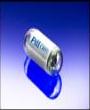
- Diagnostic, not therapeutic
- 8-hr test
- Risk of retained capsule
- False-negative rate
THE UNIVERSITY OF CALIFORNIA LET THERE BE
THE UNIVERSITY OF CALIFORNIA 1868
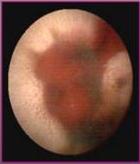
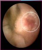
WCE
small bowel angiodysplasia
Obscure GI Bleeding
(can’t find source of bleeding)
-
Bleeding of unknown origin that persists or recurs after negative colonoscopy and negative upper endoscopy
-
May be occult or overt not visible only RBCs and can’t find source visible but can’t find source
-
10% - 20% of GI bleeding without identifiable etiology
-
5% GI bleeding recurrent without identifiable etiology
-
Majority have small bowel source
Multiple episodes of LGIB without a known source
See: esophagus, stomach, duodenum
See: rectum, sigmoid, Descending, Transverse, ascending
- Upper and lower endoscopy
So still not see the small bowel
-
CT angio
-
Elective mesenteric angiography
-
Meckel scanning (Tc⁹⁹)
-
Upper GI series with small bowel enteroclysis: is an examination of the small bowel
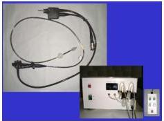
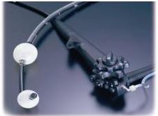
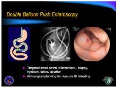
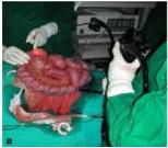
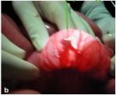
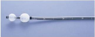
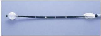
done in the OR under anesthesia. (Prepared for Surgery) in tertiary hospital, Push enteroscope → (it has 2 ballons) during laparotomy Push the enteroscope inside and inflate the 2 ballons. and see if filling of blood occur in that segment. (more clear than milking by hands). until reach site of bleeding
In Poor Country: take segment of jejunum and do milking, and wait, if there is bleeding in that segment, it will be filled. if not filled then go to other segment until reach segment where there is filling after milking → Then open that area, see the problem and deal accordingly
| Colonoscopy | Tc^{99}RBC scan | Angiography | CT scanning (A & P) | Exploratory laparotomy |
|---|---|---|---|---|
| Initial diagnostic method of choice (in Hemodynamically stable). (Bowel prepared colonoscopy) Done after empty the colon. -HIGHER diagnostic and therapeutic than unprepared colonoscopy | Detects bleeding at rates as low as 0.1-0.5 mL/min. | Detects bleeding at rates of 1-1.5 mL/min. Indications -Brisk ongoing LGIB, -Hemodynamically unstable, with or without a preceding radionuclide scan -Failed colonoscopy | Routine work up failed Findings : ( contract extravasation, bowel wall enhancement, and vascular dilatation) | (Rarely) Intraoperative push enteroscopy (hemodynamically unstable) |
Exploratory laparotomy (rarely): Intraoperative push enteroscopy in hemodynamically unstable patients.
*at the end, if still can’t find source of bleeding → do Subtotal colectomy
Principles of Management
-
Resuscitation and initial assessment
-
Localization of the bleeding site by: - upper endoscopy
- lower colonoscopy
- enteroscopy
-
Therapeutic intervention to stop bleeding
- Large-bore IV access
- Crystalloid infusion.
- CBC, electrolytes, coagulation profile, crossmatch.
- Blood loss / hemodynamic status ascertained.
- Severe bleeding-invasive hemodynamic monitoring.

2
Localization of the bleeding site
-
Upper GI endoscopy
-
Flexible colonoscopy - hemodynamic stable patient.
-
RBC isotope scan
-
Angiography
-
CT scan
③
Therapeutic interventions
defend on the cause
- Diverticular bleeding: Colonoscopic bipolar probe coagulation, epinephrine injection, or metallic clips. Recurrent bleeding- resection of the affected bowel segment.
- Angiodysplasia: Thermal therapy (electrocoagulation, argon)
- Ischemic colitis : NPO and IV hydration.
- Bleeding site cannot be determined: Vasoconstrictive agents used.
- Superselective embolization angiography.
Surgery
Indications :
- Active persistent bleeding with hemodynamic instability refractory to aggressive resuscitation
- Persistent, recurrent bleeding
- Transfusion of >4 units PRBC in a 24-hours with active or recurrent bleeding
- Transfusion of >6 units of PRBC during the same hospitalization
Too much blood transfusion and there is still bleeding lead to DIC
What To Do
- ✓ Segmental bowel resection following precise localization of the bleeding.
- ✓ Subtotal (total abdominal) colectomy with temporary end ileostomy is the procedure of choice in patients who are actively bleeding from an unknown source.
Then anastomosis
ترتيقي مع المعرفة
920011909
@in almaarefa
@almaarefa
f almaarefa.sa