Diseases of the Mediastinum & Mediastinal Masses
Dr. Waseem HAJJAR MD, FRCS,
Objectives
- Normal anatomy and contents of the Mediastinum
- Clinical manifestations produced by diseases and lesions in the region
- Diagnostic evaluation and techniques for imaging the mediastinal contents and methods for obtaining tissue for cytologic and histologic examination
- The Mediastinum is the region in the chest between the pleural cavities that contain the heart and other thoracic viscera except the lungs
- Boundaries
- Lateral - parietal pleura
- Anterior - sternum
- Posterior - vertebral column and paravertebral gutters
- Superior -thoracic inlet
- Inferior - diaphragm
Mediastinal Anatomy
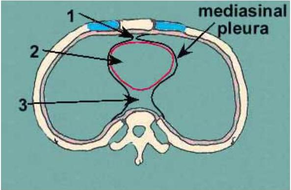
MEDIASTINUM IN CROSS SECTION
Normal Mediastinum
-
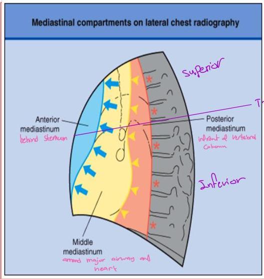
Anterior mediastinum
-
Everything lying forward of and superior to the heart shadow
-
Boundaries
-
Sternum, first rib, imaginary curved line following the anterior heart border and brachiocephalic vessels from the diaphragm to the thoracic inlet
-
Contents
-
Thymus gland, substernal extension of the thyroid and parathyroid gland and lymphatic tissues
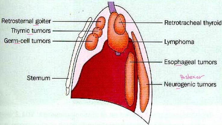
any tumor in superior or Anterior mediastinum is one of these 5 t’s
thyroid, thymic, T cell lymphoma, teratoma, T lymph adenitis
Normal Mediastinum
- Middle mediastinum
- Dorsal to the anterior mediastinum, extends from the lower edge of the sternum along the diaphragm and then cephalad along the posterior heart border and posterior wall of the trachea
- Contents normal or abnormal
- Heart, pericardium, aortic arch and its major branches, innominate veins and superior vena cava, pulmonary arteries and hila, trachea, group of lymph nodes, phrenic and upper vagus nerve
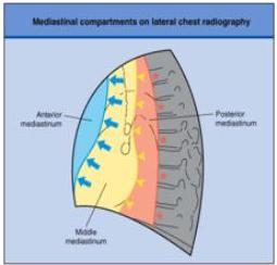
Normal Mediastinum
-
Posterior Mediastinum
-
Occupies the space between the back of the heart and trachea and the front of the posterior ribs, and paravertebral gutter
-
It extends from the diaphragm cephalad to the first rib
-
Contents
all neurogenic or sympathetic chain intercostal nerves
tumor originate from posterior Mediastinum
Esophagus, descending aorta, azygos and hemiazygos vein, paravertebral lymph nodes, thoracic duct, lower portion of the vagus nerve and the sympathetic chain
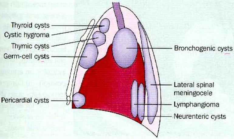
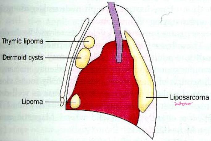
Mediastinal masses
Solid masses
Cystic masses
Fat density masses
Clinical Presentation
-
Asymptomatic mass
-
Specific disease entities according to anatomical, and embryologic origin
-
50% of all mediastinal mass are asymptomatic
-
80% of such mass are benign
-
Incidental discovery – most common (routine CXR)
-
Silent in early phase
-
Mainly cause pressure symptoms
-
More than half are malignant if with symptoms
-
Pluritic Chest Pain
-
Cough if compress on airway
-
Hemoptysis, stridor
-
Plural effusion
Clinical Presentation
- Effects on Compression or invasion of adjacent tissues
- Chest pain, from traction on mediastinal mass, tissue invasion, or bone erosion is common
- Cough, because of extrinsic compression of the trachea or bronchi, or erosion into the airway itself
- Hemoptysis, hoarseness or stridor
Clinical Presentation
- Pleural effusion, invasion or irritation of pleural space
- Dysphagia, invasion or direct invasioin of the esophagus
- Pericarditis or pericardial tamponade
- Right ventricular outflow obstruction and cor pulmonale
Clinical Presentation
-
Superior vena cava
-
Vulnerable to extrinsic compression and obstruction because it is thin walled and its intravascular pressure is low, and relatively confined by lymph nodes and other rigid structures
-
Superior vena cava syndrome obstruction chronic
it happens
because one
of these 2
Pathologies
-
Results from the increase venous pressure in the upper thorax, head and neck
-
characterized by dilation of the collateral veins in the upper portion of the head and thorax and edema and phlethora of the face, neck and upper torso, suffusion and edema of the conjunctiva and cerebral symptoms such as headache, disturbance of consciousness and visual distortion
-
Bronchogenic carcinoma and lymphoma are the most common etiologies
end stage
Clinical Presentation
- Hoarseness, invading or compressing the nerves
- Horners syndrome, involvement of the sympathetic ganglia
- Dyspnea, from phrenic nerve involvement causing diaphragmatic paralysis
- Tachycardia, secondary to vagus nerve involvement
- Clinical manifestations of spinal cord compression
Clinical Presentation
- Systemic symptoms and syndromes : Constitutional symptoms
- Fever, anorexia, weight loss and other non specific symptoms of malignancy and granulomatous inflammation
Techniques for visualizing the mediastinum and its content & obtaining tissue Bx
Initial
-
Chest PA & Lateral
-
Chest Ct with oral & i/v contrast
-
Fluoroscopy
-
Bronchoscopy
-
Esophagogram (Barrium swallow)
-
Isotope Scanning
-
FNA
-
True cut Needle Bx
-
Medistinoscopy
-
VATS
-
Thoracotomy
Lateral chest xR or CY to localise which mediastinum
Ant. mediastinal mass
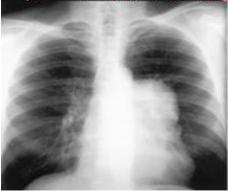
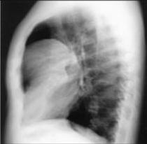
to confirm the location
do later! x-Ray
or CT
high diaphragm
CT chest mediastinum window
benign Teratoma
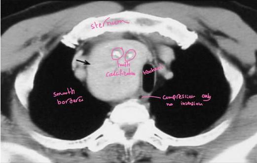
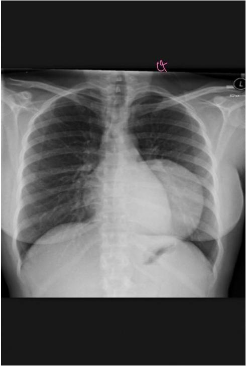
43 y/o female w/ Hx of asthma presents with progressive SOB, dysphagia, fatigability for 5 months. No wheeze or cough, EKG normal. CXR showed
easy
all Matheria gravis pit need to be done for them CT to exclude thymoma
MG:
-
Generalized fatigability
-
Ptosis
-
bulbar type symptoms:
-
dysphagea
-
dystonia
-
nasal reggargation
-
dysphonia
-
nasal speech
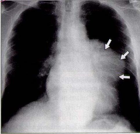
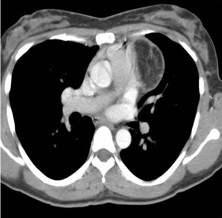
• FILM FINDINGS: -Mass just lateral to main pulmonary artery
- thick-walled smoothly-marginated
- No calcification
Thymoma
- Anterior mediastinum
- Most common (20%) of mediastinal tumor in adults but rarely seen in children
- Equal frequency in males and females
- 30 – 50 yrs
- 50% are asymptomatic
- Various Classification : Lymphocytic, Epithelial, Spindle Cell
- Most encapsulated; 35% invasive (but histologically benign!)
- Parathymic syndromes – 30-50% myasthenia gravis,
- less common– hypogammaglobulinemia (10%), pure red cell aplasia (5%)
Thymoma (Staging)
- Stage I : contained within an intact capsule very well capsulated
- Stage II: extension through the capsule to surrounding fat, pleura, pericardium
- Stage III : Intrathoracic metastasis droplet
- Stage IV: Extrathoracic Metastasis
Thymoma(Treatment)
- Stage I : Surgical resection → Recurrence 2-12%
- Stage II & III : Surgery + Radiotherapy
- Stage IV : Multimodality → Induction chemotherapy, surgery + post op Radiotherapy
- complete surgical resection – usually good prognosis
- 2-12% of resected encapsulated thymomas recur
- invasive thymoma has much worse prognosis– 50% 5-yr survival, compared to 75% in noninvasive.
- Survival not affected by the presence of Myasthenia Gravis very slow growing tumor
-23year-old female had a 8-week history of fever and night sweats accompanied by a 8kg weight loss
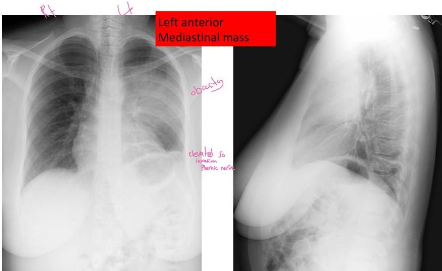
A CT was ordered to further characterize the mass:
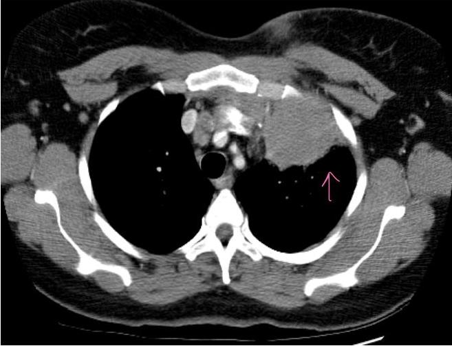
- Film findings:
- large, Inhomogeneous solid, antero left mediastinal Mass. No calcium. No fat.
PATHOLOGY
- A percutaneous CT guided truecut biopsy was performed
- Pathology reported the presence of Reed-Sternberg cells.
- What is the diagnosis?

Lymphoma
- 5-10% is mediastinal primary
- Second most common Anterior Mediastinal Mass in Adults
- Malignant > Hodgkin’s & non-Hodgkin’s
- Surgeon’s primary role is to provide sufficient tissue for diagnosis and to assist in pathologic staging.
- Dx: Mediastinoscopy, thoracotomy ,True cut Bx
- Rx: Chemotherapy or XRT
- Prognosis: Varies with tumor histology
anything infront of heart any behind a sternum this is Anterior medistinum
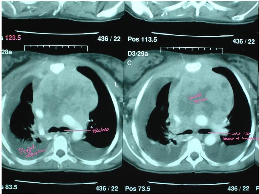
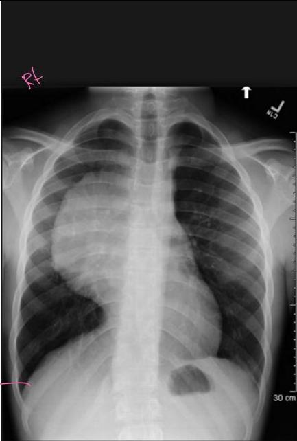
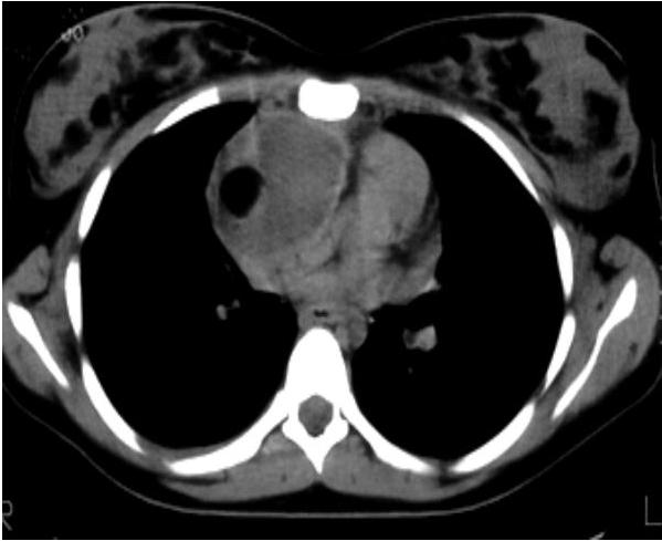
18 y/o female with R upper chest and shoulder pain x 1 month. Exacerbated by movement and inspiration. No findings on PE. Working Dx is musculoskeletal injury. A CXR done
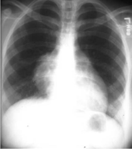
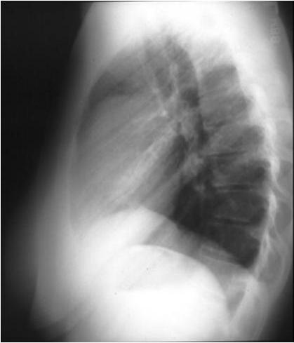
CHEST CT
-
CT shows mass with areas of:
-
fat
-
fluid
-
soft tissue
Likely diagnosis ?
Germ Cell Tumors
-
The mediastinum is the most common location for extragonadal germ cell tumors (GCTs) in adults
-
GCTs can be either benign (teratomas, dermoid cysts) or malignant (seminomas, non-seminomatous GCTs).
-
Mature teratoma – most common mediastinal germ cell tumour.
-
All ages – particularly young adults (F>M)
-
Presentation – mostly asymptomatic, incidentally diagnosed on X-ray, CT., may cause cough, dyspnea, pain
-
CXR: well-circumscribed, round or lobulated, calcifications in up to 26%
-
CT: well-marginated, lobulated, cystic component 88%, fat 50-75%, calcification 25-50%, fat-fluid levels diagnostic, but rare (<10%)
-
Surgical excision is curative
Malignant Nonteratomatous Germ Cell Tumors
- Usually in the third and fourth decades of life
- Symptoms: chest pain, cough, dyspnea, and hemoptysis
- The superior vena cava syndrome occurs commonly
- Diagnostic imaging: A large anterior mediastinal mass
- Serologic measurements (α-fetoprotein and β-hCG) useful for:
- differentiating seminomas from nonseminomas tumors,
- assessing response to therapy,
- diagnosing relapse or failure of therapy
- Seminomas rarely produce β-hCG and never produce α-fetoprotein
- More than 90 of nonseminomas secrete one or both of these hormones
- seminomas are radiosensitive and nonseminomas are relatively radiosensitive
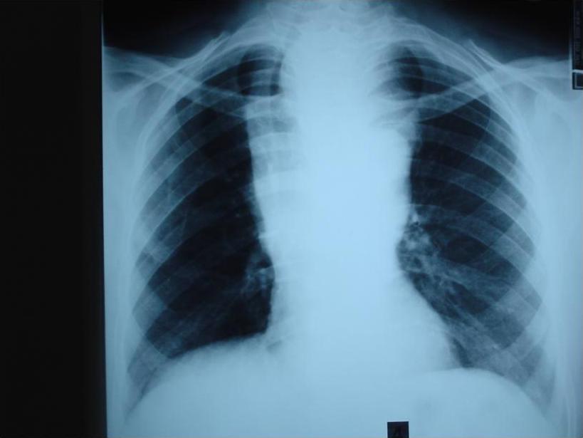
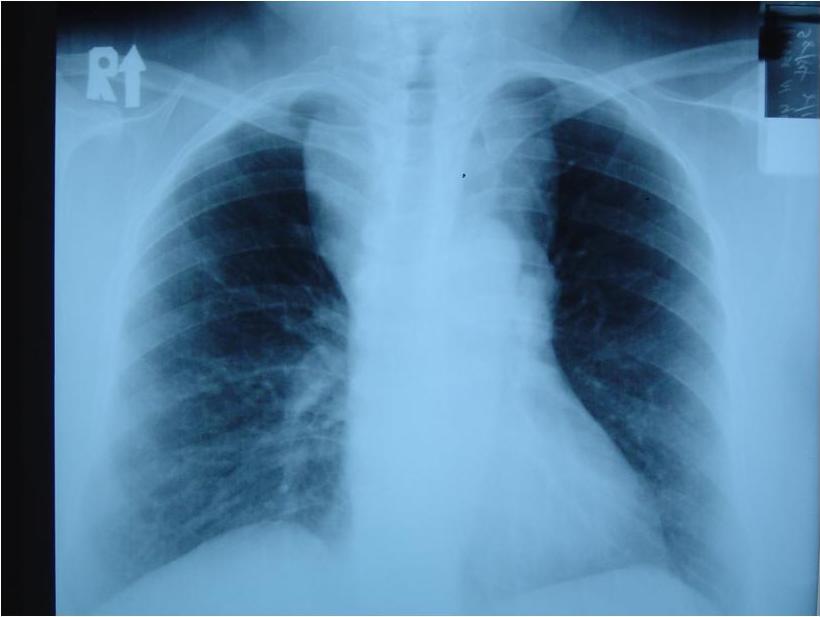
A 46-year-old woman came to you with complain of a persistent cough for the past 3 weeks and mild dysphagia. O/E she has no respiratory distress. There is an enlarged left lobe of the thyroid gland, without any cervical adenopathy.
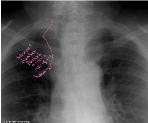
-Film Findings : -Trachea deviated to right. Left anterosuperior Mediastinal mass extending into Cervical region
Substernal Thyroid Tissues
- Goiters usually are considered substernal (also referred to as mediastinal, intrathoracic, or retrosternal) when more than 50% of the thyroid parenchyma is located below the sternal notch
- Mediastinal goiters are classified as primary or secondary
- Primary mediastinal goiters, also referred to as ectopic or aberrant goiters, uncommon, 1% of all surgically excised goiters
- Secondary mediastinal goiters are a much more common, 5–15% of all goiters demonstrate some extension into the mediastinum
• Radiographic:
- Chest x-ray → mediastinal mass, superior mediastinal widening, tracheal deviation or compression
- Chest CT scans → define the full extent and anatomic relationships of the substernal thyroid to surrounding structures and to facilitate preoperative planning
• serum thyroid-stimulating hormone measurement → If hyperthyroidism is present → antithyroid medications and beta blockade should be undertaken before elective resection
1
سورة النساء
Retrosternal goiter
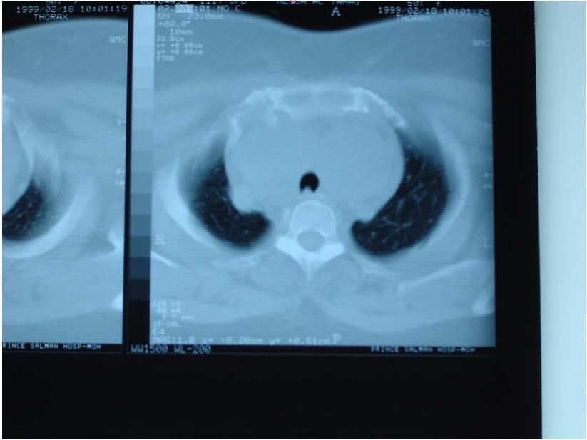
A 25-year-old man incidentally discovered, asymptomatic, isolated, rounded paravertebral mass on CXR and further CT scan showed following findings. The most likely diagnosis is
“Dumb-bell” Tumor
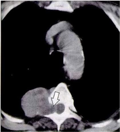
Neurilemmoma(Schwannoma)
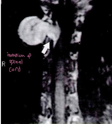
Neurogenic tumours
- most common tumours to arise in the posterior mediastinum.
- peripheral nerves – neurofibroma, schwannoma, malignant tumours of nerve sheath origin.
- Tumours arising from sympathetic ganglia.
- Peripheral nerve tumours typically originate in an intercostal nerve in the paravertebral region.
- Neurofibromas and Schwannomas present as well-defined round or oval posterior mediastinal masses.
Mesenchymal Tumors
- Lipoma, Fibroma, Mesothelioma
- Superior or Anterior mediastinal location
- Diagnosis with CT scan, MRI
( to see if there’s extention to the spinal canal
Benign Cysts
- Most Common in Middle mediastinum
- 20% of mediastinal masses
- Usually asymptomatic
- Bronchogenic cyst(32%), pericardial cyst(35%), enteric cyst(12%), thymic cyst, and thoracic duct cyst
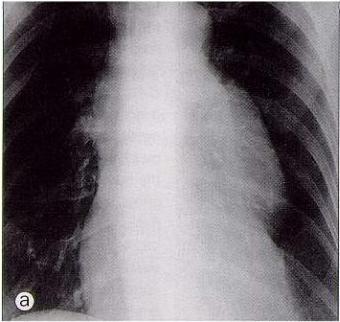
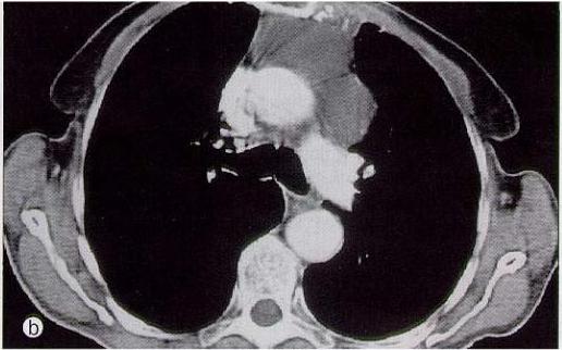
Pericardial Cyst
- Thin-walled, mesothelial cell lining
- most common in Right C-P angle
- Simple cysts are almost always asymptomatic
- Rare cardiac impingement
Pericardial Cyst
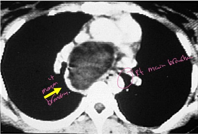
Bronchogenic Cysts
has low places
subcarina
tracheal
- 30 - 60% of all mediastinal cysts
- Lined by ciliated respiratory epithelium
- May contain cartilages or mucous
- Communicate with tracheobronchial trees
- May become infected
- ✓ Wheezing, dyspnea, recurrent pulmonary infections it has to be excise
Bronchogenic Cyst
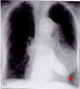
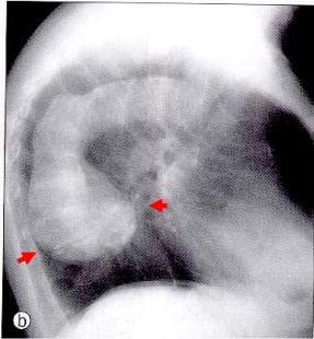
Aortic Aneurysm
posterior meristinum
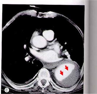
Paratracheal Lymphadenopathy
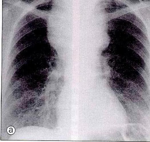
Could be because of TB should be diagnosed by Isolate scanning
Pleuro-pulmonary infection
Infectious
A. Lung Abscess
Causes
Clinical Features
- Copious production of foul smelling sputum
Investigation
- C X R
Air Flowed Level
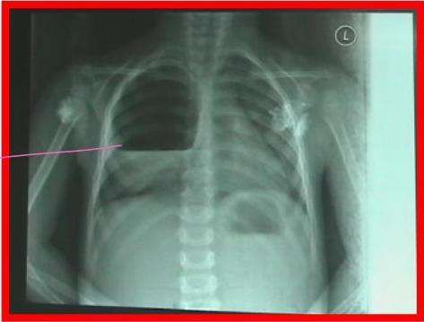
➤ Treatment
Abx
Drainage
- Internal
- External
Pulmonary resection
▪ Indications
- Failure of medical RX
- Giant abscess ( >6cm)
- Haemorrhage
- Inability to R/O carcinoma
- Rupture with resulting empyema
▪ Type of Resection
- Lobectomy
if unable to exclude
large masses are SCC
and presented by lung abscess
So you have to excise it
B. Bronchiectasis
types: Cystic → this is what we dealing with Cylindrical → usually it’s bilateral disease
Def.
Bronchial dilatation
Cause
Congenital Cystec Fibrosis
Infection messes - but dec. by vaccination
Obstruction by forien body
Clinical Features
Cough
Dyspnea
Haemoptysis (50%)
Clubbing
Investigation
Bronchogram
CT
Bronchoscopy
Treatment
Medical
- Resolve most cases
Surgical
Failure of medical Rx Patient with localized disease
non Perfused seen by VQ scan (cystic type usually non perfused)
Bilateral lung disease → need lung transplant
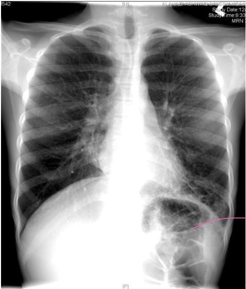
cystic type
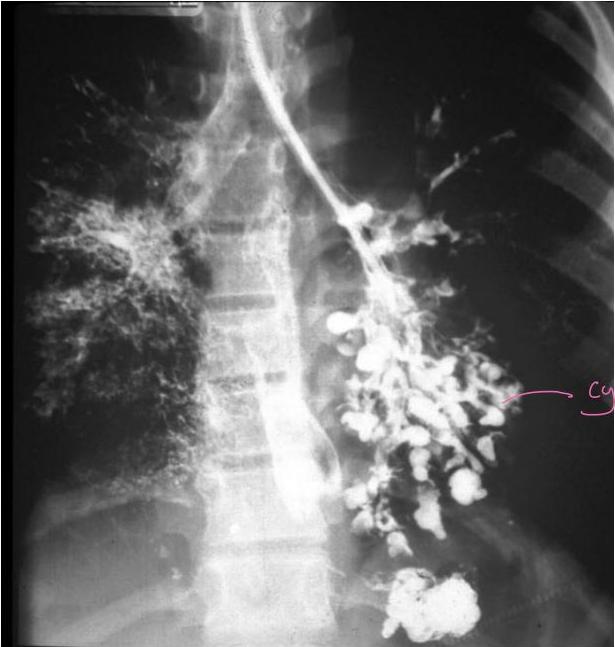
cystic
morning productive cough - Hemiplegia - failure to thrive
دنه طوله الليل يتجمع
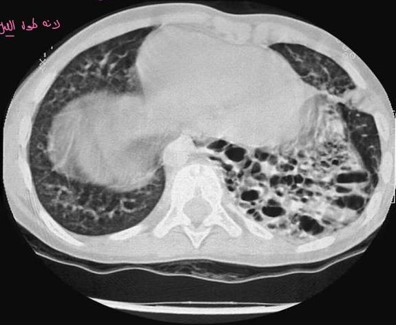
cyhic
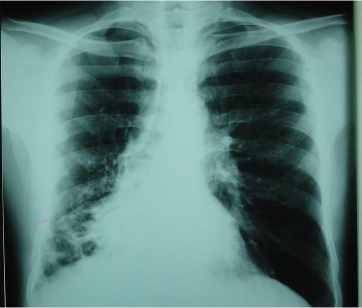
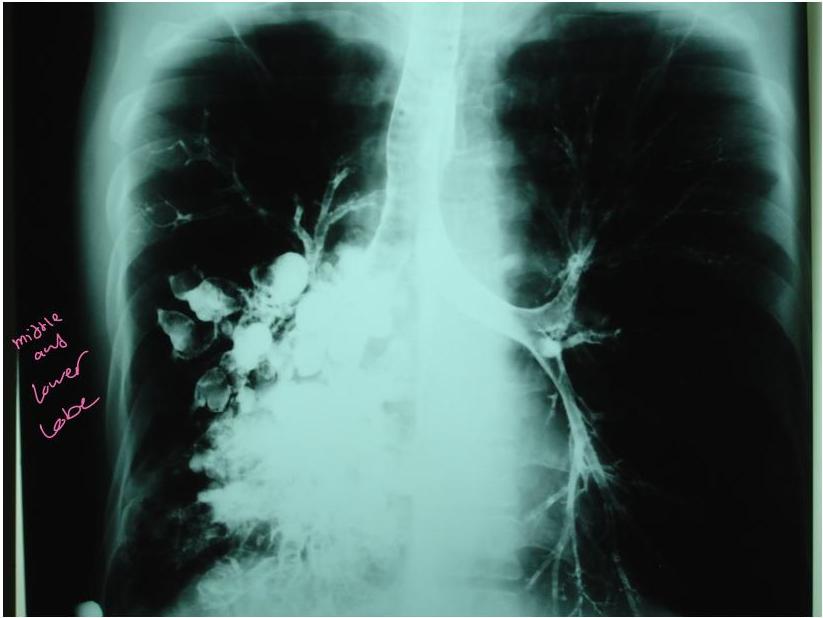
middle and lower lake
C. Tuberculosis
- 30,000 new cases occur annually in U.S.A
-
Cause
-
Pulmonary
-
Extra-pulmonary
-
Investigation
-
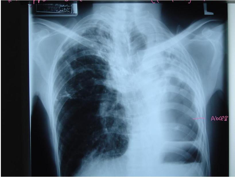
C X R
Plura and lymphadenitis
TB empyema
Rt upper lobe
Lt lung distroid
DFOV 29.0cm LUNG
R
kV 120
mA 230~
Smart mA 216
Large
10.0mm/1.5:1
Treatment
-
Medical anti TB
-
Surgical Lobectomy or Pneumonectomy
✓ Failure of medical Rx ✓ Destroyed lobe or lung ✓ Pulmonary haemorrhage massive hemoptysis ✓ Persistent open cavity with + ve sputum ✓ Persistent broncho pulmonary fistula
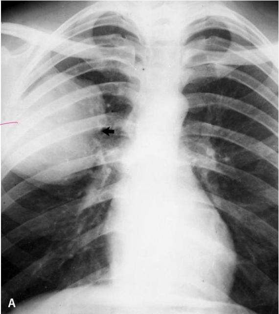
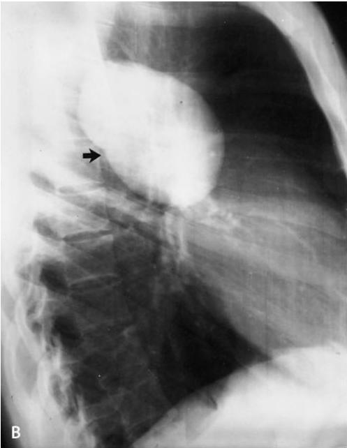
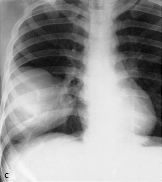
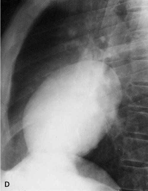
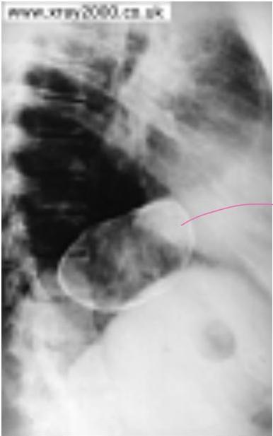
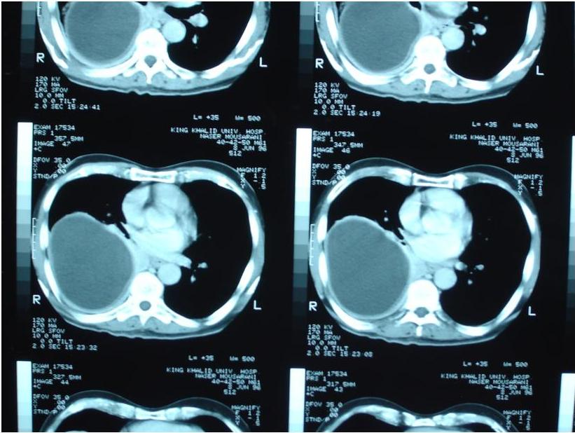
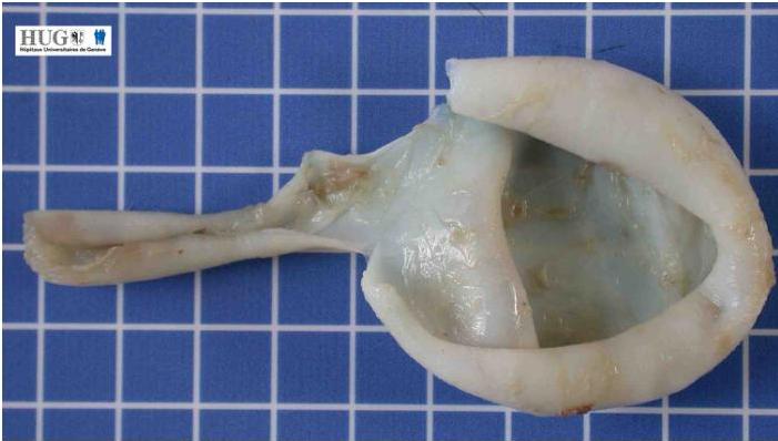
Hydatid cyst
Cause
eating of raw liver
Echinococcus granulosus
Diagnosis
Treatment
if pit has Liver or lung Hydatid cyst we have to screen for their other organ Liver ⇐> Lung
large cyst
if ruptured may cause
-
Anaphylactic shock or
-
empyema
B
C
D
calcofide
inject hypertonic Saline
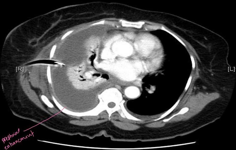
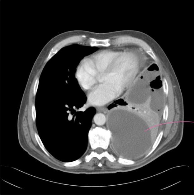
Thoracic empyema
- Thoracic empyema is the collection of pus or the presence of infected fluid within the pleural cavity.
- Pleural empyema affects a large number of patients and may lead to severe and disabling sequelae in cases of inappropriate diagnosis or treatment.
Striffeler H, Gugger M, Im Hof V, Cerny A, Furrer M, Ris HB.
Ann Thorac Surg 1998;65:319-323.
Striffeler H, Ris HB, Würsten HU, Im Hof V, Stirnemann P, Althaus U.
Eur J Cardiothorac Surg 1994;8:585-588.
difference in opacity
Se:44499
Im:1
[H]
MOHAMMED
Study Date:2/8/2007
Study Time:6:37:10 AM
MRN-t
[R]
[L]
[F]
C5411
W2859
Se:2
Im:27
[A]
SALMA
Study Date:2/3/2007
Study Time:10:35:08 AM
MRN:
ROUTIN CHEST 5X5
100ML OMNIP 300
[P]
C52
W354
collapsed lung
stage 1: Aspiration
stage 2: chest tube
last stage “Fibrotic”:
Decortication surgery
by scope not open surgery