جامعة المعرفة ALMAAREFA UNIVERSITY
Skin, & Subcutaneous Tumors
Dr. Ahmed khan MBBS, FCPS
Assoc. Professor, Dept. Surgery
Leenah Turjoman ♥
2/3/2024
Dr. M. Almadani
1
Objectives
- Skin structure & functions
- Type of skin wounds & its management
- Different methods to provide skin coverage
- Diagnosis & management of common lesions
2/3/2024
Dr. M. Almadani
2
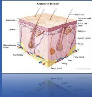
Skin consists of:
Skin structure, & function
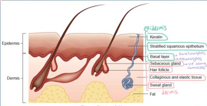
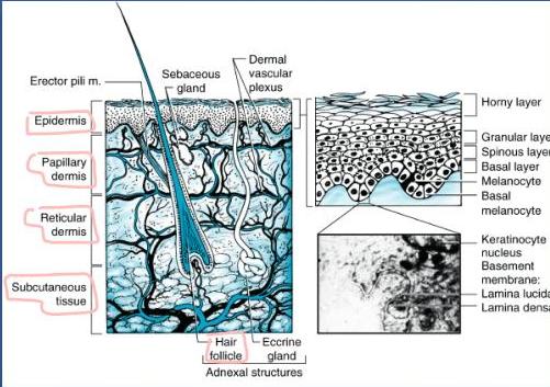
① Epidermis: → Thin layer consists of keratinocytes (keratinized layer)
- The epidermis is derived primarily from surface ectoderm
- Keratinized stratified squamous epithelium.
- Basal germinal layer generates:
a. Keratinocytes- migrates superficially to form keratinized SE and finally shed. → form keratin b. Pigment cells (Melanocytes)- melanin passed to keratinocytes. → form melanin
2/3/2024
Dr. M. Almadani
3
Skin structure, & function
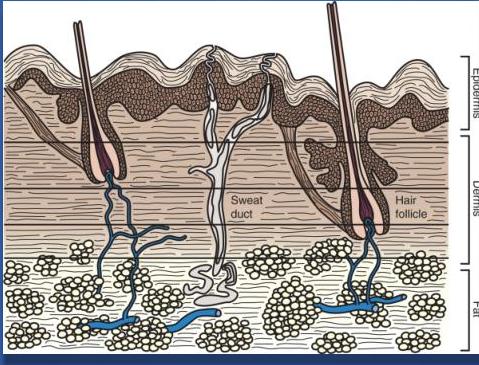
②. Dermis: → Thicker part
- Derived primarily from mesoderm
- Collagen, elastic fibers & fat, thus to sustain and support the epidermis
- Supports blood vessels, nerves, lymphatics and epidermal appendages.
- Has appendages (hair follicles, sweat & sebaceous glands)
- Sweat glands:
- Eccrine: Major glands, (excessive sweating → loss of salt and water). Mostly in palms, & soles
- Apocrine: Musty smelling fluid in the axilla, perineum, ears & areola
- Composed of 2 layers: - the more superficial papillary dermis and
- the deeper reticular dermis
2/3/2024
Dr. M. Almadani
4
the hair follicle, and the external surface
2/3/2024
Dr. M. Almadani
5
2/3/2024
Dr. M. Almadani
6
2/3/2024
Dr. M. Almadani
7
Skin function
- The skin covers the entire external surface of the human body and is the principal site of interaction with the surroundings.
- It serves as:
- Protective barrier (anatomical)
- Prevents internal tissues from exposure to trauma, ultraviolet (UV) radiation, temperature extremes, toxins, and bacteria.
- Sensory perception
- Immunologic surveillance
- Thermoregulation
- Control of insensible fluid loss → incase of burns → lose skin → lose fluid
- Protective barrier (anatomical)
2/3/2024
Dr. M. Almadani
8
Wounds
- Disruption of normal continuity of bodily structures due to trauma.
- Types:
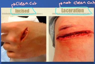
Incised (Tidy): Clean cut, inflicted by sharp object (knife, glass), no devitalized tissue. eg. Surgical wound
Lacerated (Untidy): Irregular margin. Caused by crushing, tearing, blunt knife contains devitalized tissue. → main source of wound infection
Abrasions: Friction damage. Superficial bruising & loss of varying skin thickness. → depending on the force applied
2/3/2024
Dr. M. Almadani
9
Wounds
For TTT: Cut the skin because it will not survive, there will be necrosis within 24-48 hours and there will be gangrene of affected part.
Degloving: Shearing forces, extensive section of skin is completely torn off the underlying tissue, severing its blood supply. Skin remain intact but deprived of blood supply (Skin may be attached by the edges but its blood supply is severed)
Fenetrating Knife and gun shots. High velocity wounds are associated with extensive deeper tissue injuries.
Leg, intra abdominal organs
2/3/2024
Dr. M. Almadani
10
AEC (DEH)
Types of wound healing
Primary (First intention)
best type of healing approximation of skin, using sutures or clips bring 2 edges together
- Wounds are sealed immediately, may done for wounds <24 h
- Clean type → neat and clean wounds (no devitalized tissue)
- Simple suturing & Skin graft /flap
Secondary (Second intention)
- No closure
- Highly contaminated wounds
- Re-epithelialization > Wound contracture
leave area to heal, do dressings slowly and gradually, treat infection and healing occurs by 2ry intention There will be a scar, because no approximation of skin occurs.
Tertiary (Third intention)
- Delayed primary closure (use Closure Suture)
- Contaminated wounds treated first, once the wound is clean then closed → done after debridement and cleaning of wound
2/3/2024
Dr. M. Almadani
11
Wounds managements
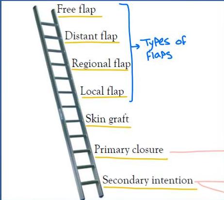
→ for larger wound or cosmetically exposed
▲ mostly
▲ Small wounds esp. for non cosmetically exposed
2/3/2024
Dr. M. Almadani
Wounds managements
-
Incised wound:
-
Primary closure
-
Lacerated wound:
-
Wound debridement.
-
Primary or Tertiary.
-
Grossly contaminated/infected:
-
left to heal by secondary intention.
-
Degloving injury: → (devitalized hanging skin) (can reach extent of bones being visible)
-
Excision of devascularized skin, skin grafting/flap.
later do
2/3/2024
Dr. M. Almadani
13
difference between graft and first
Wounds managements
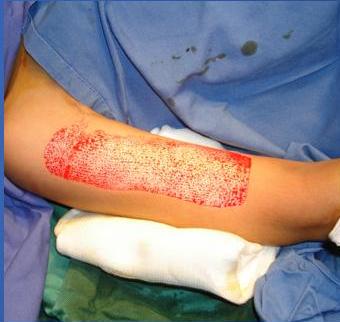
Thin slice of epidermis and part of dermis taken from donor area and transplanted to recipient area
Grafts: are the tissues that are completely detached from the body before it is transplanted to other host bed.
recipient area
-
DONOR AREA → eg. from thigh
-
RECIPIENT AREA → eg. to arm
-
not contain blood supply (blood supply is not transferred)
-
graft depend on blood supply of recipient area
-
grafts applied to wound that is healed and red, and granulation tissue appear on the floor
2/3/2024
Dr. M. Almadani
14
Wounds with skin loss
- Small skin defect:
- Aesthetically or functionally unimportant
- Allowed to heal by secondary intention.
- Large skin defects:
- Skin “graft”:
- All vessels nourishing the graft are cut
- Types: Full thickness, Partial thickness → both are detached from blood supply
- Skin “flap”:
- Some aspect of the blood supply to the segment of tissue has remained intact during transfer. → blood supply remain attached until revascularization occurs
- Skin “graft”:
2/3/2024
Dr. M. Almadani
15
grafts:
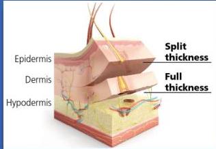
Split skin (Thiersch) graft (Partial thickness)
- Epidermis with a portion of the dermis
- Graft taken by electric dermatome → an instrument
- Donor site: Heal by epithelialization from skin appendages → so epithelium grows from inside and covers it by itself
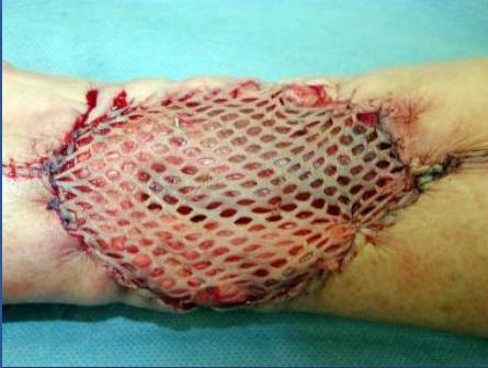
- Graft can be expanded by ‘meshing’ to cover larger area.
- Thinner graft take more easily on imperfect vascular bed.
- Used for covering granulating areas and burns.
- Do not take on bare bone, cartilage and tendons → only put it on granulation tissue not on bare areas
- Appearance not as good as full thickness graft. (because bare areas not have blood supply) because its a very thin layer
2/3/2024
Dr. M. Almadani
16
Split skin (Thiersch) graft
There should be no infection and area should be healing and presence of granulation tissue
↓
ready recipient area to receive graft
2/3/2024
Dr. M. Almadani
17
Dermatomes
epithelium will grow in those holes
→ used to take Skin so it can be applied on recipient area
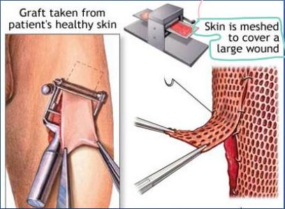
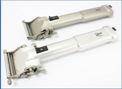
②
Full thickness graft
- Full portion of the dermis with the epidermis.
- Grafts survival:
- Absorption of nutrients from recipient area capillary beds & angiogenesis.
- 5 days are required for definitive vascular ingrowth to occur.
- Dressings left on the recipient site for this period of time.
more time because it is full thickness
So good healing occurs and it develops its blood supply
2/3/2024
Dr. M. Almadani
19
Full thickness graft
- Do not shrink, looks better and stronger than partial thickness
- Graft retain functional hair follicles and sweat glands.
- Commonly used in “reconstructive surgery.”
- Donor area covered by suture or graft.
- Chosen sites has “spare skin” - groin, behind the ear So its better than partial thickness graft because there will be no appendages left so no epithilization occurs
- areas where there is naturally extra loose skin
2/3/2024
Dr. M. Almadani
20
Flaps
and this blood supply is cut once the recipient area makes its own blood supply
- DEF: Flap is a tissue with its original blood supply (bring their own blood supply)
- Thicker and stronger than graft. → because they contain skin, subcutaneous tissue and even sometimes muscles.
- Can be applied to avascular surfaces – bone, tendon, or joints.
- Commonly used in “reconstructive surgery” → esp. breast reconstruction surgery after mastectomy (basic difference between flaps and grafts)
2/3/2024
Dr. M. Almadani
21
Flaps
How the diffuses
Types:
- Local flaps used for covering small defects after surgical excision of small tumors (v-y advancement, rotation flap, rectangle advancement) To eg. BCC by taking skin locally (use skin beside the defect)
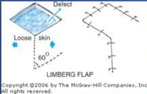
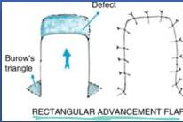

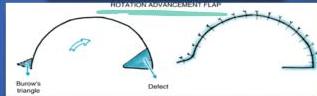
Types based on how its taken geometrically
2/3/2024
Dr. M. Almadani
22
Flaps
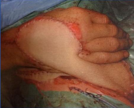
- Distant flaps: Remains attached to its original blood supply until new one developed locally (2-3 weeks), Then pedicle is divided.
- Free flaps: Grafts blood supply is restored locally by microsurgery.
- Composition: Cutaneous, fasciocutaneous, myocutaneous, osteo-myocutaneous Include many things: Skin, fascia, muscle, osteomyocutaneous tissue
2/3/2024
Dr. M. Almadani
23
Pedicle flap
Distant flap
2/3/2024
Dr. M. Almadani
24
Skin, & soft tissue lesions
→ Superficial
-
Skin lesions are attached to skin → eg. sebaceous cyst, epidermal cyst → skin can’t be pinched
-
Epidermal lesions cause surface irregularity
-
Dermal lesions do not
-
Pigmented lesions indicate melanocytic lesions
-
Subcutaneous lesions: → eg. lipoma, Dermoid cyst → below epidermis and dermis
-
skin can be pinched up or moved independently over swelling
2/3/2024
Dr. M. Almadani
25
Skin infections
become Folliculitis (infection of hair follicle)
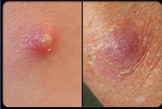
Boil (Fruncle) → involve Skin, surrounding hair follicle and surrounding subcutaneous tissue
Carbuncle → when may Boils join together and there are multiple openings → most severe form → may cause toxemia
Stages of some infection
- Erysipelas
- Impetigo
- Cellulitis
- Necrotizing soft tissue infection (Necrotizing fasciitis)
2/3/2024
Dr. M. Almadani
26
Skin infections
- Folliculitis is a tender pustule that involves the hair follicle
- Furuncle involves both the skin and the subcutaneous tissues in areas with hair follicles
- A carbuncle is an aggregate of connected furuncles and has several pustular openings
2/3/2024
Dr. M. Almadani
27
Skin infections
2 • Boil
2/3/2024
Dr. M. Almasani
28
Skin infections
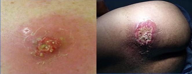
- Carbuncle: is an aggregate of connected furuncles and has several pustular openings
TTT:
incision and drainage and send for culture and sensitivity, do dressing, and give antibiotic
2/3/2024
Dr. M. Almadani
29
Skin infections
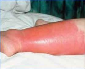
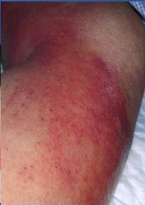
- • Erysipelas: Erysipelas is a skin infection, well demarcated involving the upper dermis extends
into the superficial cutaneous lymphatics.
-
Surgical emergency
-
Swelling, red, hot, edematous appearance over skin
-
usually involve the legs
-
more common in diabetic patients
-
Superficial infection, involve epidermis, and may extend to superficial lymphatics
-
non-necrotizing inflammation
-
well²/³ demarcated
Dr. M. Almadani
30
TTT for both Erysipelas and Cellulitis:
- rest
- antibiotics
- debride pustule if Present
Skin infections
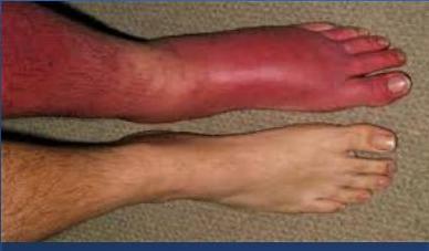
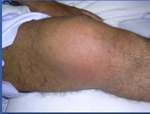
- Cellulitis: Non-necrotizing inflammation of the skin and subcutaneous tissues, usually from acute infection
- acute
- more common in diabetic patients
- non necrotizing inflammation
- deeper than erysipelas
- ill defined margins
- red, hot, tender
2/3/2024
Dr. M. Almadani
31
Skin infections
below skin
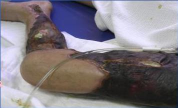
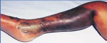
- Necrotizing fasciitis: is a rapidly progressive inflammatory infection of the fascia, with secondary necrosis of the subcutaneous tissues
-
Usually poly microbial →
-
Risk factors
-
DM · Trauma
-
Obesity
-
Malnutrition
-
Immunocompromised
so antibiotics given should cover gram the and -ve. anaerobic (metronidazole)
according to region
- Fournier’s (scrotum; penis perineal)
- Meleney’s (abdominal wall gangrene)
2/3/2024
TTI: aggressive debridment, antibiotic, later repair with graft
32
% of the recosed tissue and normal tissue till it starts to bleed
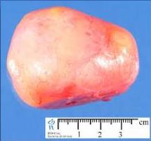
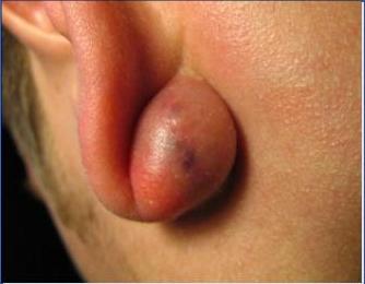
Sebaceous gland produce sebum and gland becomes blocked forming cyst
Sebaceous cyst
- “Epidermal” Cyst
- Dermal swelling covered by epidermis
- Thin wall
- Filled with sebum
- Typical punctum
- Blocked sebaceous glands
- Treatment: excision
2/3/2024
Dr. M. Almadani
33
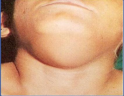
Dermoid cyst
midline swelling
- It is growth of normal tissue enclosed in a pocket called a sac
- Congenital vs acquired
- A cyst is a lump that may contain fluid or other material
- Most often greasy yellow material but may be other
like skin appendages
TTT: excision
2/3/2024
Dr. M. Almadani
34
Tumors of the skin, & subcutaneous
• Benign:
- Papilloma
- Dermatofibroma
- Keratoacanthoma
- Benign pigmented moles
- Haemangioma
- Neurofibroma
- Lipoma
• Premalignant lesions: → if left untreated become malignant
- Actinic (solar) keratosis
- Bowen’s disease
- Marjolin’s ulcer
• Malignant:
- Squamous cell carcinoma
- Basal cell carcinoma
- Malignant melanoma
- Dermatofibrosarcoma protruberans
- Liposarcoma
- Fibrosarcoma
- Kaposi’s sarcoma
2/3/2024
Dr. M. Almadani
35
1. Benign lesions
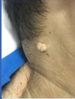
1. Papilloma: → most common one
- Finger like projection from skin.
- Covered by normal skin. usually Pedunculated
- Treatment : Excision looks benign, not hot or red
2/3/2024
Dr. M. Almadani
36
Benign lesions
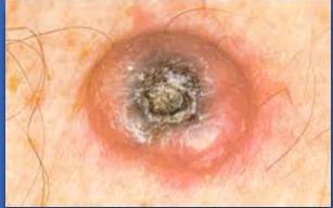
2. Keratoacanthoma:
- Hemispherical nodule → (like BCC or SCC)
- Friable center covered with keratin
- Common site - face
- Age - over 50 → elderly
- Heals spontaneously after shedding central core.
- Confused with squamous cell carcinoma.
2/3/2024
Dr. M. Almadani
37
Benign lesions
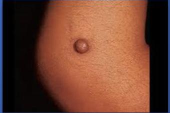
3. Dermatofibroma:
- Brownish, firm to hard, nodular, single/ multiple
- Commonly over hand or feet
- Freely mobile over deeper structures
- Treatment: Excision
2/3/2024
Dr. M. Almadani
38
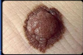
4. Benign pigmented moles
-
A developmental abnormality due to aggregation of melanocytes (naevus or moles)
-
Types: (by histopathology)
-
Junctional moles: Melanocyte activity is at the junction of epidermis and dermis- moles of palm and sole.
-
Dermal naevus: Migration of sheets of melanocytes to dermis.
-
Compound naevus: Migration of sheets of melanocytes to dermis & epidermis.
usually congenital or from childhood, but may appear later on.
2/3/2024
Dr. M. Almadani
39
Benign pigmented moles
- Common moles:
- Flat or raised brown-black lesion covered by normal epidermis.
- May show some activity in childhood.
- Becomes quiescent after puberty.
- Alert: Increased pigmentation, itching, scaliness or bleeding- suggest malignant change.
2/3/2024
Dr. M. Almadani
40
Benign pigmented moles
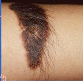
- Giant hairy naevus: → has least malignant potential
- Present at birth,
- Over trunk, face.
- Low risk of malignant change
- Excision: Cosmetic → if its on areas where its cosmetically visible
2/3/2024
Dr. M. Almadani
41
5. Hemangioma
A- Involuting type: → meaning: they regress with time
- True neoplasms arising from endothelial cells.
- Present at birth or appear within weeks
- Predominantly affect head & neck.
- Types:
Superficial lesions (strawberry)
- bright-red, raised, irregular, & bosselated.
Deeper lesion :- blue-black tumor covered by normal skin.
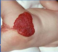
more likely to develop complications of ulceration, bleeding.
- once ulcerated may lead to infections
2/3/2024
Dr. M. Almadani
42
Hemangioma
-
Involuting type:
-
Grows for first 6 months, static until 2-3-years age.
-
Usually disappear before child is 7 years of age.
-
Complications: Ulceration, bleeding, coagulopathy
if complications Treatment: Laser photocoagulation or intra-lesional steroid help early involution
*if no complications → don’t treat, it will resolve on its own
2/3/2024
Dr. M. Almadani
43
Hemangioma
B. Non-Involuting type:
- Hamartoma of abnormal blood vessel formation
- Bright-red patchy lesion on face or scalp.
- Often overlies a peripheral nerve distribution.
- No spontaneous regression or growth.
- Good result with CO above the scan.
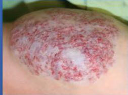
2/3/2024
Dr. M. Almadani
44
Types of noninvoluting:
Hemangioma
-
Cavernous haemangioma: →venous origin
-
“Bluish-purple elevated mass” appearing in early childhood
-
Composed of mature vein like structures.
-
Positive sign of emptying. (compressibility) → (compressed and when release my finger they are filled up again)
-
Treated by excision. → and feeding vessels are ligated
-
Cirsoid aneurysm:
-
Lesion is fed by arterial blood → (arterial origin) → So may bleed alot So embolize it before excision
-
Tortuous, and pulsating
-
Treatment: Angiographic embolization prior to excision.
2/3/2024
Dr. M. Almadani
45
6. Neurofibroma
Commonly seen in Surgical OPDs having nodularity along
-
Hamartoma of peripheral nerve sheath.
-
Neurilemmoma (schwannoma)
-
solitary
-
No malignant potential.
-
Neurofibroma (peripheral nerve tumor)
-
solitary or multiple (von Recklinghausen’s disease)
-
Autosomal disorder, present at birth or early childhood
-
Patches of skin pigmentation (café au lait’ spots)
-
Increase in size or new swelling suggest malignant transformation
become painful
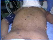
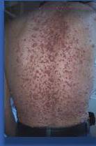
nerve distribution along the body. usually they are non tender and freely mobile
if not complicated → leave it alone don’t treat (because can’t cut all the nerves) 2/3/2024 Dr. M. Almadani
46
2. Premalignant lesions → all are due to UV rays
• Actinic keratosis (solar keratoses):
- Small, single or multiple warty spots on the face, back of neck or hands of fair skinned
- dry scaly patches of skin that have been damaged by the sun
- Elderly
- Biopsy for diagnosis.
- Treated by cryosurgery.
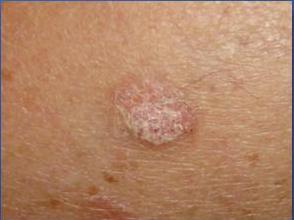
2/3/2024
Dr. M. Almadani
47
Premalignant lesions
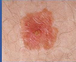
2. Bowen’s disease: (SCC in situ)
-
very early form of skin cancer
-
Cause: long-term exposure to the sun – especially in people with fair skin
-
It affects the squamous cells, which are in the outer layer of skin (epidermis)
-
‘Elevated red scaly plaque’ on the exposed area of body.
-
Gradually enlarge & thickens.
-
Transform into SCC.
-
Biopsy for diagnosis
-
Treatment: excision, cryosurgery or topical 5-fluorouracil.
depend on stage
best one
in early stages
(anti cancer element)
2/3/2024
Dr. M. Almadani
48
Premalignant lesions
3. Marjolin’s ulcer:
- Cutaneous malignancy that arises in the setting of :
- chronic wounds
- longstanding scars
- previously injured skin
- Chronic non-healing burn scars (due to chronic inflammation and cellular proliferation)
- Turn into SCC.
- Grows slowly.
- Lymphatic metastasis unusual- less vascularity & lymphatics in the scar.
- Treatment: excision with wide margin.
2/3/2024
Dr. M. Almadani
49
3. Carcinoma of epidermis
-
Occurs primarily on exposed areas → due to UV rays
-
Albinos, xeroderma pigmentosa (undue sensitivity to sunlight).
-
Chronic irritation: chemicals, chronic ulcer, radiation exposure, exposure to sunlight.
-
Age: > 50 elderly
-
Pathological types: Basal cell (80%) & Squamous cell carcinoma (16%)
- more common
- more aggressive
- less common
2/3/2024
Dr. M. Almadani
50
I - Basal cell cancer (BCC)
- BCC arises from basal layer of epidermis
- Common in Elderly, commonest skin malignancy (80%)
- Slow growing, locally invasive (rodent ulcer).
- Rarely metastasize (malignant cells larger than the size of lymphatics) (<0.1%).
- Site: Middle third of the face- nose, inner canthus of eye, forehead & eyelids.
- Earliest lesion- hard pearly nodule, dimpled in Centre
2/3/2024
Dr. M. Almadani
51
Basal cell cancer (BCC)
- Degeneration makes the lesion translucent and raised.
- Ulcerated lesions - rolled edges. → Typical finding → due to dimpled center
- Repeatedly scales over and breaks down.
- Aggressive tumors burrows deeply and destructive to skin and bone.
- Extremely radiosensitive (when surgery is contraindicated or very difficult e.g. near the eye)
2/3/2024
Dr. M. Almadani
52
Basal cell cancer (BCC)
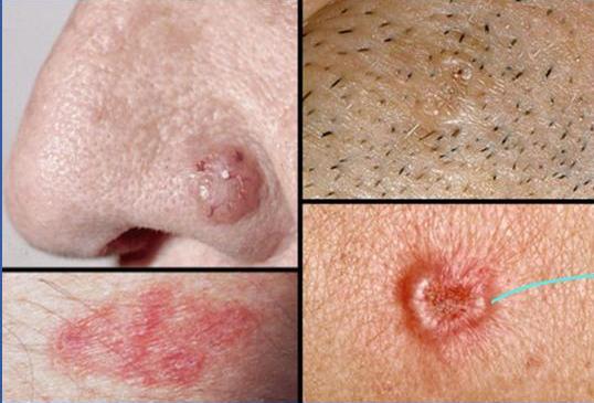
→rolled edges • dimpled center
2/3/2024
Dr. M. Almadani
53
Basal cell cancer (BCC)
- Diagnosis: Biopsy (Bx):
- excisional (small lesions) → Diagnostic and therapeutic → Send Srecimin to histopathology
- incisional or punch bx (large lesions)
- Management:
- Surgical excision with adequate margins or radiotherapy (RT)
- Small lesion-: cryosurgery, curettage and cautery
- Late presentation- reconstructive surgery to restore appearance & function.
- Contraindications of RT: if lesions are close to eye or over the cartilage.
- if Surgery is contraindicated
2/3/2024
Dr. M. Almadani
54
2. Squamous cell cancer (SCC)
- Less common, more aggressive (16%)
- Arise from keratinocytes of the epidermis
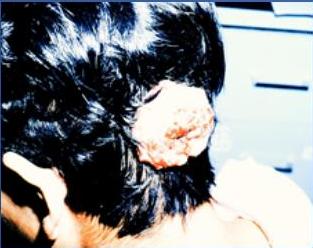
- Common sites: Ear, lips, back of hand. Any other area. back of scalp & common site
- Early lesion - hard erythematous nodule
Cauliflower like ulcer, with raised everted edge. → Typical finding
- Metastasis: Regional lymph nodes (≥ 10 mm thick)
very common to lymphatics and to blood poor prognosis
2/3/2024
Dr. M. Almadani
55
back of scalp
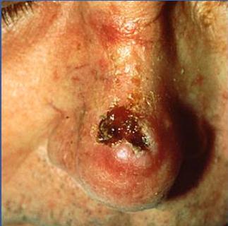
Squamous cell cancer (SCC)
ideal treatment
-
Treatment: Excision with a 1-cm margin.
-
Histologic confirmation of tumor-free margin. → this is necessary
-
Regional lymph node excision- if clinically palpable.
-
Lesions on the face, nose or eye.
-
cosmetically imp area
-
not have much tissue to be excised
Excision by Mohs’ technique
- Serially excise a tumor by taking small increments of tissue until the entire tumor is removed.
- Clear margin confirmed by frozen sections.
(excision with tumor free margins) (normal tissue)
- if metastasize to regional LNs
- if its in foot or leg, it will go to inguinal LN
- if its in arm, it will go to cervical LN
2/3/2024
Dr. M. Almadani
57
other options for treatment: Squamous cell cancer (SCC)
Radiation therapy:
- Small and superficial lesions
- Adjuvant therapy (along with surgery)
- Cure rates are less comparable to surgical excision
- Metastatic disease:
- Poor prognostic sign
- 13% patients surviving after 10 years
2/3/2024
Dr. M. Almadani
58
Malignant melanoma
malignant form of nevus/mole
- Malignant transformation of melanocytes
- Most deadly skin cancer. high mortality and morbidity
- Comprises 4-8% of skin cancers less common than SCC
- Predominantly affect fair-skinned people
- More common in females
- Major cause- exposure to sunlight
- Half of malignant melanoma arises in pre-existing naevi.
- Any new nevi after 40 years old- should be dealt as melanoma
warning sign
2/3/2024
Dr. M. Almadani
59
Malignant melanoma
- Types:
- Superficial spreading melanoma
- Hutchison’s melanotic freckle (lentigo maligna)
- Nodular melanoma
- Amelanotic melanoma
- Acral lentiginous melanoma
- Subungual melanoma
2/3/2024
Dr. M. Almadani
60
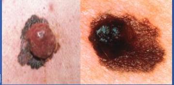
Malignant melanoma
Superficial spreading melanoma
Commonest type.
- Commonly affects middle aged.
- Common site: trunk or exposed areas.
- Premalignant phase:- malignant cells spread outwards, surface slightly raised, pigmentation patchy and outline indistinct.
- Invasive phase:- spreads down into dermis while lesion still small.
- Indurated nodule.
- Soon ulcerates or bleeds.
2/3/2024
Dr. M. Almadani
61
Malignant melanoma
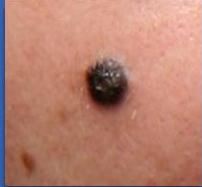
2. Nodular melanoma:
- Elevated, deeply pigmented lesions.
- 2nd most common type & most aggressive form.
- May develop in a pre-existing benign naevus.
- No initial intraepithelial spread.
- Nodule enlarges steadily in all direction.
- Darkens progressively, active area is jet black.
- Typical findings: itching, ulceration, bleeding, crusting, scab formation.
- Appearance of satellite nodules- metastasis
→ (alerting signs in benign conditions)
sign of
2/3/2024
Dr. M. Almadani
62
Malignant melanoma
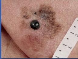
3. Hutchison’s melanotic freckle (lentigo maligna)
light brown colored areas
- Brownish-red patch
- On the face of elderly
- Grows slowly over years.
- Least aggressive
- Serrated margin.
- Premalignant phase- lasts for 10-15 years.
- Malignant change- brownish red papule, develop eccentrically within freckle.
2/3/2024
Dr. M. Almadani
63
Other types of melanoma
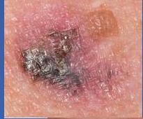
4. Amelanotic melanoma:
- Rare, pale pink lesion (less pigmented)
- Grows rapidly
- Histology demonstrates pigment
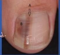
5. Subungual melanoma:
- Nail bed of thumb or big toe
- Pigmentation not visible in early stage
- Misdiagnosed as paronychia (nail infection) or ingrowing toenail
if there is no history of trauma and something appears on nail, be alerted.
2/3/2024
Dr. M. Almadani
64
Other types of melanoma
6. • Acral lentiginous melanoma:
- Site: Over sole and palm.
So may be ignored or misdiagnosed • Thick skin masks some features. • Presents late with nodularity and ulceration.
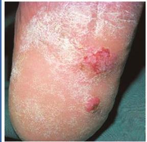
Fig. 18.24 Acral lentiginous melanoma arising on the sole of the foot.
2/3/2024
Dr. M. Almadani
65
Malignant melanoma
-
Spread: → (like SCC)
-
Lymphatics: - regional nodes.
-
In transit metastasis along lymphatics- satellite nodule.
-
Blood-borne: Brain, liver, lungs, skin subcutaneous.
-
Staging:
-
Clinical stage
-
TNM stage
(histological) Mitotic activity & lymphocytic infiltration- influences prognosis
2/3/2024
Dr. M. Almadani
66
Malignant melanoma
| Table 18.6 Prognosis in relation to the stage and depth of malignant melanoma | |
|---|---|
| Clinical stage | 5-year survival rate (%) |
| I Primary lesion only → Breslow depth (mm) | The more thickness of the less survival rate |
| <1.5 | (due to longer duration and more advanced) |
| 1.5–3.5 | about its still curable because its stage 1 |
| >3.5 | |
| II Primary lesion + regional lymph node or satellite deposit | 30 |
| III Metastatic disease | 0 |
| Classification | Thickness (mm) | Ulceration Status/Mitoses |
|---|---|---|
| T | ||
| Tis | NA | NA |
| T1 | ( \leq 1.00 ) | a: Without ulceration and mitosis ( < 1/mm^{2} )b: With ulceration or mitoses ( \geq 1/mm^{2} ) |
| T2 | 1.01-2.00 | a: Without ulcerationb: With ulceration |
| T3 | 2.01-4.00 | a: Without ulcerationb: With ulceration |
| T4 | >4.00 | a: Without ulcerationb: With ulceration |
| N | No. of Metastatic Nodes | Nodal Metastatic Burden |
| N0 | 0 | NA |
| N1 | 1 | a: Micrometastasis* b: Macrometastasis† |
| N2 | 2-3 | a: Micrometastasis* b: Macrometastasis†c: In transit metastases/satellites without metastatic nodes |
| N3 | 4+ metastatic nodes, or matted nodes, or in transit metastases/satellites with metastatic nodes | |
| M | Site | Serum LDH |
| M0 | No distant metastases | NA |
| M1a | Distant skin, subcutaneous, or nodal metastases | Normal |
| M1b | Lung metastases | Normal |
| M1c | All other visceral metastases | Normal |
| Any distant metastasis | Elevated |
Abbreviations: NA, not applicable; LDH, lactate dehydrogenase.
*Micrometastases are diagnosed after sentinel lymph node biopsy.
†Macrometastases are defined as clinically detectable nodal metastases confirmed pathologically.
2/3/2024
Dr. M. Almadani
67
Malignant melanoma
Treatment:
→ excision with tumor free margins → not only surroundings but also in the depth
-
Excision biopsy to determine the depth of tumor.
-
Healthy tissue margin depends on tumor thickness.
-
Small lesion:
-
Excision with a clear margin of 3 mm down to deep fascia.
-
Smaller defects closed by primary suture.
-
Larger defects closed by skin graft or flap.
-
SLNB: Sentinel LN is the 1st node that drain the lesion.
-
LND: Stage II or primary lesion overlies LN
(LN dissection) nodes are enlarged
- Adjuvant therapy:
Immunotherapy, molecular therapy, isolated limb perfusion
To get the tumor free margin of deeper tissues
2/3/2024
Dr. M. Almadani
68
Uncommon malignant lesions
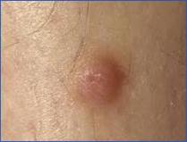
- Dermatofibrosarcoma protruberans:
- Rare invasive tumor of the dermis
- Presenting as red or bluish firm painless mass.
- Rarely metastasize.
- Treatment: wide surgical excision, RT reduce risk of recurrence.
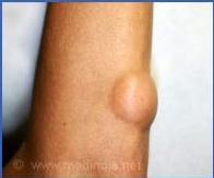
- Liposarcoma:

- Arise from any fatty tissue.
- Treatment: Wide surgical excision.
*malignant transformation from long standing or larger lipomas
2/3/2024
Dr. M. Almadani
69
Uncommon malignant lesions
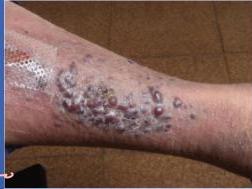
-
Kaposi’s sarcoma:
-
Neoplasm of capillaries and perivascular connective tissue.
-
Multifocal in presentation.
-
May be associated with AIDS or other immunocompromised state.
-
Lesions appear as purplish nodule.
-
Legs most commonly involved.
-
Treatment: unsatisfactory.
-
RT or
-
immunotherapy may help. not sufficient and cure rates are low
2/3/2024
Dr. M. Almadani
70
Thanks
2/3/2024
Dr. M. Almadani
71