جامعة المعرفة ALMAAREFA UNIVERSITY

Leenah Turjoman
نرتقي مع المعرفة
جامعة المعرفة ALMAAREFA UNIVERSITY
Upper GIT
Bleeding
Omar Alaidaroos MSc, MD, FRCS Associate Prof. of General Surgery
نرتقي مع المعرفة
جامعة المعرفة ALMAAREFA UNIVERSITY
Objectives
At the end of this presentation students will be able to:
- Define upper GI haemorrhage.
- Describe the resuscitative measures.
- Enumerate the causes of upper GI bleeding.
- Describe the symptoms & signs of UGI bleeding.
- Describe diagnostic work up.
- Describe the non-surgical management and indications for surgical intervention.
GI bleeding
- Upper(70%) & Lower (30%) Smore Common
- Defined by the location of source of bleeding
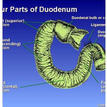
- Either proximal or distal to the ligament of Treitz (junction between duodenum & jejunum)
Forms of GIB
➤ Hematemesis or Coffee ground vomitus (50% of UGIB)
❖ Hematemesis Vs Hemoptysis
bleeding blood
coughing of blood frothy
➤ Melena: black, tarry, sticky, offensive,
black stool
- Blood must be in GI tract for 8 hours to turn black
- 70% from Upper GI tract, 30% from lower GI tract
- 50-100 cc of blood to the intestine.
small amount
➤ Hematochezia (bright red blood in stool)
- Usually from lower GI tract → defend on site
- 10-15 % from brisk upper GI bleeding. These patients are more likely to be older and to have duodenal ulcers.
left side of colon: bright red
right side of colon: marron color
if bleeding is heavy and intestinal motility is rapid
➤ Positive Occult blood in stool
RBCs in stool : no visible bleeding
Severity of GI Haemorrhage
- Mild to moderate haemorrhage is defined as :
✓ Patient < 60 years ✓ No PMH (no past medical history) ✓ No hypovolaemia ✓ Hb > 10 g/dl
- Life-threatening haemorrhage is defined as :
✓ Patient > 60 years ✓ Hypovolaemia (systolic blood pressure <100mmHg or diastolic blood pressure falls on sitting and standing) ✓ Hb < 10 g/dl ✓ Severe disease, e.g. liver, cardiovascular, respiratory
PMH
1. UGIB
- Is defined as bleeding derived from a source proximal to the ligament of Treitz (D J flexure)
- More common in male 2 : 1
- 4 times more common than lower GI bleeding.
- 30% pts are older than 65 years.
- 80% are self-limited.
- 20% of pts who have recurrent bleeding (within 48-72 hrs) have poor prognosis. brequine major intervention
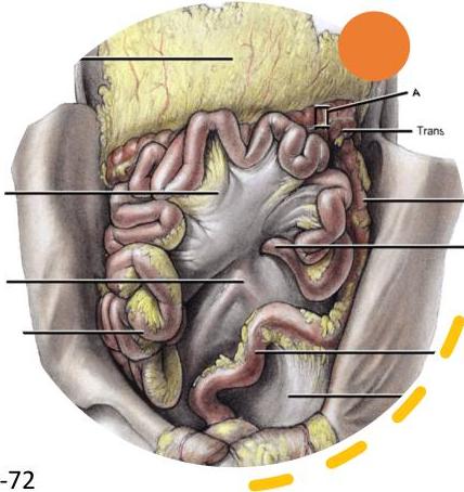
Causes of UGIB
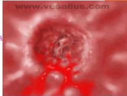
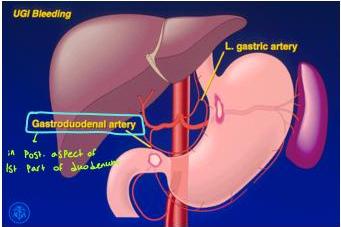
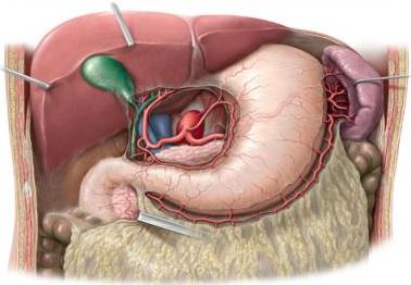
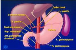
- Peptic ulcer disease (DU 30-37%, GU 19-24%) ↳ most common cause of UGIB → cause bleeding due to penetration of Post. wall, due to presence of gastro duodenal artery in 1st part of duodenum. bleeding will enter duodenum to stomach and vomited.
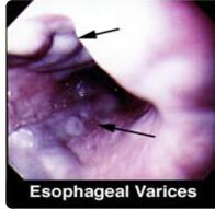
- Oesophageal varices (portal hypertension) (6-10%) ↳ due to
- Erosive gastritis /esophagitis. (5-10%) ↳ due to Stress
- Mallory-Weiss syndrome- mucosal tears of the mucosa of esophagus. (3-7%) ↳ in patients with projectile vomiting, severe vomiting leading to
- Dieulafoy lesion. ↳ spare ↳ submucosal bleeding seen by endoscopy, occur due to malformation. leading to rupture and bleeding, but bleeding is not seen directly.
- Gastric cancer. ↳ spare ↳ usually asymptomatic, unless it is metastasis
- Ulcerated gastric stromal tumor (GIST) = leiomyoma or leiomyosarcoma ↳ arise from stroma/muscle/mesenchyme (could be benign or malignant)
- Aortoenteric fistula- erosion of the aortic graft into the bowel.
- Angiodysplasia- dilated, thin-walled vessels appearing as cherry spots
1. Peptic ulcer disease
- The most common cause of UGIB.
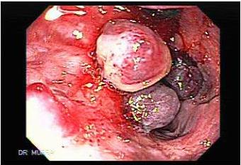
- High-risk for PUD: H pylori, alcohol abuse, chronic renal failure, and/or (NSAID) use. Most common risk (Box)
- Ulcer penetrate the wall then burrows deeper into the mucosa, causes weakening and necrosis of the arterial wall (gastroduodenal artery), leading to a pseudoaneurysm. The weakened wall ruptures, producing hemorrhage.
- Approximately 80% bleeding from PUD stops spontaneously.
2. Oesophageal Varices
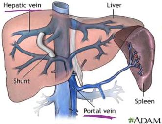
occur due to blood regurg → cause is hepatic or pre or post hepatic. So blood not pass from portal vein to hepatic veins to IVC to Systemic circulation.
instead, the blood regurg to many places including lower veins found in lower part of esophagus and in fundus of stomach (short gastric vessels)
- Portal hypertension leads to portosystemic shunting.
- Development of varices- lower oesophagus and gastric fundus
- Elevated portal pressure transmitted to esophageal / gastric varices resulting in rupture of varices.
- Mucosal ulceration can be a bleeding source.
- Normal portal pressure 5-15 cm of H₂O → >15 → Portal hypertension
- Bleeders- usually > 25 cm of H₂O
- 20 % may have peptic ulcer or gastritis may be associated with
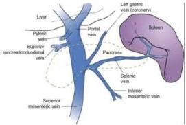
Causes of portal hypertension
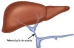
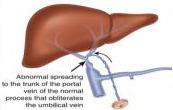
- Pre-hepatic:
Portal vein
Congenital atresia of PV, PV thrombosis,
Compression of PV (tumours)
Splenic vein + SMA + IMA = Portal vein
- Intrahepatic:
Pre-sinusoidal- Schistosomiasis Sinusoidal- Cirrhosis 4 bilhar Ziasis may be due to alcoholic
- Post-hepatic (Post-sinusoidal): Budd-Chiari syndrome, Constrictive pericarditis
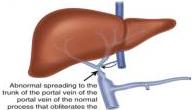
Budd-Chiari syndrome
Definition
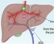
Venous obstruction
from the small hepatic veins to the junction of IVC with the right atrium
sós right-sided cardiac disease
Symptoms
budd Chiari Syndrome
Triad:
ascites
83%
abdominal pain
61%
hepatomegaly
TRIAD
67%
Other:
-
fever
-
pedal edema & truncal hepatic veins
Less common:
-
esophageal bleeding (5%)
-
hepatic encephalopathy (9%)
20% - asymptomatic
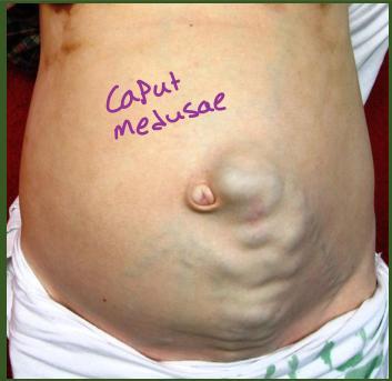
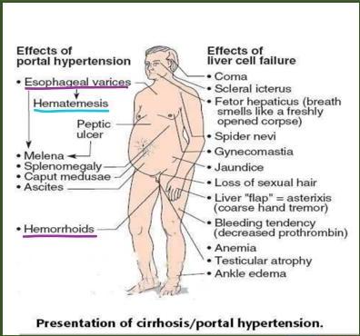
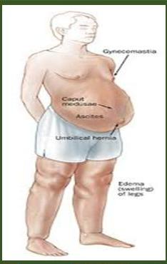
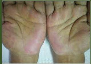
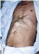
Manifestations of Portal hypertension:
due to increased dilatation of surface capillaries in the hand.
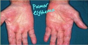
Palmar erythema
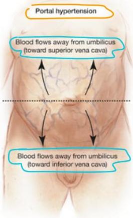
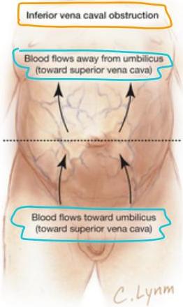
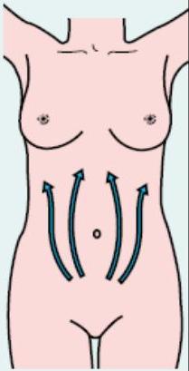
Cavut medusae could be due to: Portal hypertension or IVC obstruction → So to know the cause:
A How to determine blood flow direction in a vein


A segment of vein without blood is now present

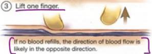
④ Repeat steps 1 and 2, and lift the other finger.

If blood did not refill in Step 3, and blood now flows to fill the empty segment, the direction of flow is confirmed.
B Pattern of blood flow in distended abdominal wall veins
place two fingers at one end of the vein and apply occlusive pressure

move one finger along the vein, thus emptying a section

blood flow direction is indicated by whether or not the vein refills once the finger is removed


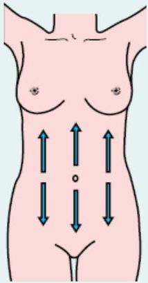
(a)
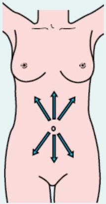
(b)
(c)
Determining the direction of blood flow in abdominal veins. (a) Normal blood flow pattern and those characteristic of (b) portal hypertension and (c) obstruction of the inferior vena cava.
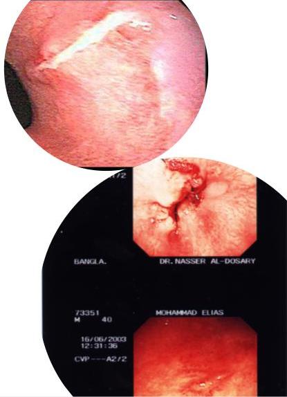
3. Mallory-Weiss syndrome
- Mucosal laceration- result of forceful or long-term vomiting or coughing
- 80-90%- tear along the lesser curve of the stomach just distal to the gastro-esophageal junction
- It occurs more frequently in individuals with alcoholism.
- Affect more males than females.
- The ages varies, with a peak at ages 40 - 60.
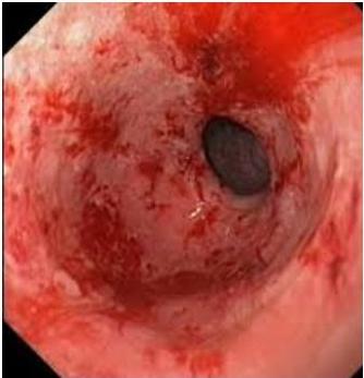
4. Acute stress gastritis
-
Seen in shock, multiple trauma, acute respiratory distress syndrome, acute renal failure, and sepsis patients, burns, Stress, Surgery, MVA
-
The principal mechanisms- decreased splanchnic mucosal blood flow and altered gastric luminal acidity.
Many Patient who will undergo surgery is given PPIs, to avoid gastritis, cuz Patient will be in stress
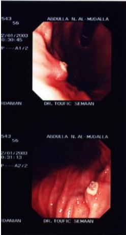
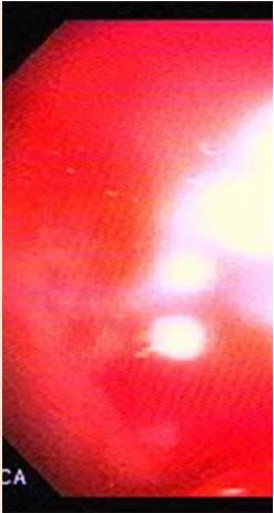
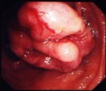
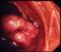
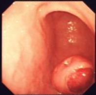
5. Dieulafoy lesion
- A vascular malformation of the proximal stomach.
- 2-5% of acute UGIB episodes.
- Endoscopic appearance: large ulcerated submucosal vessel.
- Bleeding can be massive and brisk.
- Vessel rupture occurs in the setting of chronic gastritis
- Alcohol use is associated with the Dieulafoy lesion.
- Mostly- men in their third to tenth decade.
- Can occur anywhere along the GI tract
DR. NASSER AL-DOSARY
AHMAD F. AL-GHAZALI
DR. NASSER AL-DOSARY
AHMAD F. AL-GHAZALI
MAN
2/2 5 003
MAN
-61/2
26/2003 6:46
70
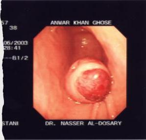
6. GIST (gastrointestinal stromal tumour)
- Mesenchymal tumour, submucosal lesions.
- 50-60%- stomach
- 20-30%- small intestine
- 10%- rectum
- Benign or malignant
Asymptomatic, bleeding or obstruction
38
6/2003
55
-82/2
ANWAR KHAN GHOSE
DR. NASSER AL-DOSARY
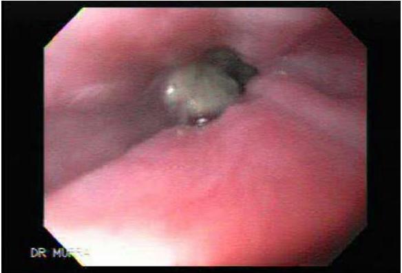
7. Gastric carcinoma
- Common- chronic blood loss (anaemia)
- Haematemesis- uncommon
Symptoms and signs of UGIT Bleeding
- Hematemesis
- Melena
- Hematochezia (15%)
- Syncope
- Dyspepsia
- Epigastric pain
- Heartburn
- Diffuse abdominal pain
- Dysphagia → from obstruction of tumor
- Weight loss → may be due to malignant tumor
- Jaundice → from metastasis to liver
Initial
workup
- Vital signs: Pulse, BP
- CBC: WBC with differential, platelet
- Hemoglobin level
- Coagulation profile (PT, PTT, INR)
- Type and crossmatch blood
- U & E, LFTs
Nasogastric lavage
Diagnosis
❖ Nasogastric lavage
❖ Chest radiography
❖ Gastrin level (Gastrinoma) → can lead to ulceration in many sites for Zollinger-Ellison syndrome
❖ CT scan & ultrasonography:
- Liver disease with cirrhosis
- Pancreatitis with pseudocyst and hemorrhage
- Aortoenteric fistula
❖ Endoscopy → do it once bleeding is less, not while there is severe bleeding → do it once patient is stable → otherwise do CT angio or Technichium scan ❖ Angiography (persistent bleeding, source not identified by endoscopy) (Diagnostic & Therapeutic)
- Intra-arterial vasopressin
- Embolization
❖ Tagged red blood cell (TRBC) scan
- Only diagnostic & usually for occult bleeding
- More sensitive than angiography
- Can detect bleeding rate of 0.1-0.5 mL/min
if bleeding is 0.5-1.5
if bleeding is
1.5
(both will have no value if the bleeding has stopped) (so do them only if there is continuous bleeding)
Management: Resuscitation
ABC
-
Airway + O₂
-
Two peripheral IV lines To restore blood volume of Patient if can’t get a line because Patient is shocked and collapsed vein, do: Venous cut down: Put a line in long Saphenous vein (1cm behind and above medial malleolus)
- open a 2cm opening in skin and dissect with an artery (instrument), till reach clear blue vein and put the cannula there.
-
X-match, CBC, u/e, coagulation profile, LFTs
-
Crystalloid solution normal Saline ringer lactate
-
NG tube: Gastric wash, monitor bleeding, prevent aspiration.
-
Foley catheter- evaluation of urinary output.
*oligouria: <500 cc /day ; <30 cc/hour ; <0.5 cc/min
- Peptic ulcer patients: 80 mg IV PPI.
high dose → as first dose, then can give 10 mg /hour
- Endoscopic hemostatic therapy.
Endoscopic hemostatic therapy
*do the endoscope once patient is stable and bleeding has dec.
- Endoscopy: Diagnose + control of bleeding
- Injection of 1:10,000 adrenaline
- Laser or bipolar electrode coagulation
- Clips or bands
- PUD: epinephrine injection; diathermy (cauterization)
- esophageal varices: banding; sclerotherapy
Complications:
- Perforation
- Necrosis on high dose epinephrine
- Induce acute and delayed hemorrhage
- Prophylaxis antibiotic should be applied
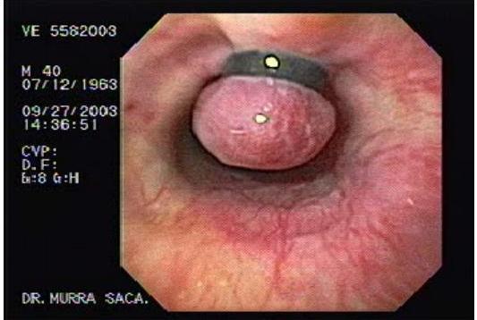
Recurrent bleeding in PUD
- A minority - recurrent bleeding after endoscopic therapy
- Risk factors for rebleeding: Presence of Coagulopathy Age>60 years , Comorbidities
- shock upon admission, Active pulsatile bleeding, Presence of cardiovascular disease.
- H pylori infection- recurrent bleeding is extremely low.
in 20% of cases
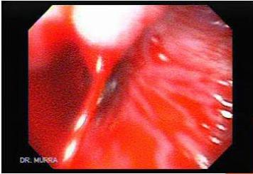
Indications for surgery in bleeding peptic ulcers
- Life-threatening bleeding not responding to resuscitation. (not even able to do endoscope)
- Failure of endoscopic hemostasis or recurrent bleeding.
- Prolonged bleeding, with loss of 50% or more of blood volume
- A second hospitalization for peptic ulcer bleeding. (recurrent)
- A coexisting perforation or obstruction.
- Failure of medical therapy
Management of recurrent bleeding
(in PUD)
both choices have same effect. So the best is Choice A to avoid surgery
- Re-endoscopy to achieve hemostasis.
and do band ligation of the bleeding vessel
- Surgical management Duodenal ulcer:
→ duodenum is retroperitoneal, so open peritoneum, and cut ant. wall of 1st part of duodenum then see post. wall and gastroduodenal artery, and ligate the artery. Then close ant. wall of duodenum.
A Duodenotomy+ under-run with suture + anti-ulcer medications.
→ high dose PPI (80mg) IV bolus, then every hour 8 mg for 3 days, then continue on oral.
B Duodenotomy+ under-run with suture + anti-ulcer surgery-
(pyloroplasty+ bilateral truncal vagotomy)
either this, or do:
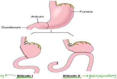
Billroth 1 or 2
if duodenum is healthy
if duodenum is not healthy
→ cut ant. and post. trunk of vagus n.
→ There will be dec motility of stomach so should do a drainage procedure with it.
dec ucl secretion → either pyloroplasty, or Billroth 1 or 2
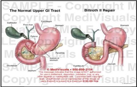
Exhibit# 698053-01X
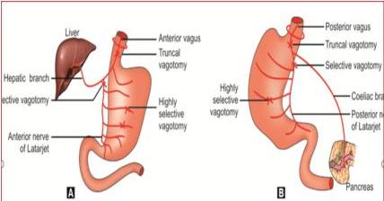
Figs 89.1A and B: Showing different types of vagotomy of (A) Anterior and (B) Posterior vagus nerves
BILLROTH 1
- Partial gastrectomy with gastro-duodenostomy. It is the most physiologic type of gastric resection, since it restores normal continuity.
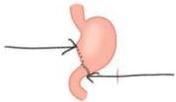
Billroth I
Source: Fauci AS, Kasper DL, Braunwald E, Hauser SL, Longo DL, Jameson JL, Loscalzo JL, Harrison’s Principles of Internal Medicine, 17th Edition: http://www.accessmedicine.com Copyright © The McGraw-Hill Companies, Inc. All rights reserved.
2 Surgical management Gastric ulcer: 4 quadrant biopsy (because could be malignant)
Young & fit- wedge excision of ulcer. 4 biopsies
Old & unfit- Under-run the bleeding point+ biopsy
Benign ulcer: Anti-ulcer medical treatment. PPI
after biopsy
Malignant ulcer: Staging the disease, surgery if indicated.
may need to excise the stomach (either Partial or Complete)
☐ Acid- suppression therapy
- I.V. Proton pump inhibitor is more effective than I.V. H 2 blocker in increasing intragastric pH
- High dose omeprazole significantly reduces the frequency of further bleeding and of surgery in patients with bleeding ulcer, dosage 40 mg i.v. every 12 hrs. for 5 days
- Somatostatin / Octreotide infusion In massive UGIH with Hx of advance liver disease is recommended
PROSTAGLANDIN ANALOQUE - Cytoprotective agent
- Octreotide (Somatostatin analogue) 50 microgram i.v. bolus Highly selective
- Then 50 microgram/ hr for infusion rate for 5 days , it can be discontinued without tapering.
**Vasopressin should not be used due to its systemic side effect
- Somatostatin causes
Splanchnic vasocostriction
Reduces Azygos venous blood flow
Reduces portal collateral circulation and decreases portal pressure
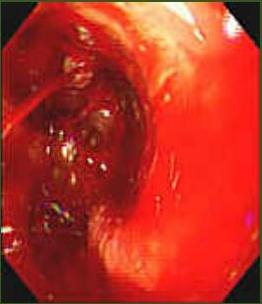
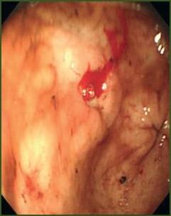
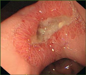
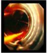
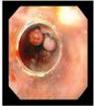
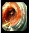
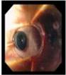
endoscopic classification of peptic ulcer:
Endoscopic features, Stigmata of recent bleeding and risk of re-bleeding
That bleeding happened one of the following:
①
Active bleeding 55 – 90%
②
ulcer with
Clean base 3 – 5%
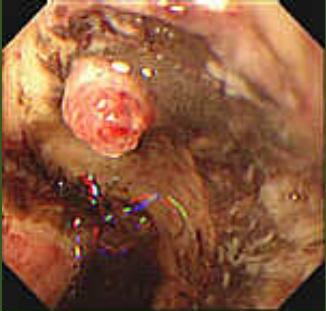
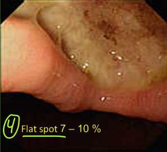
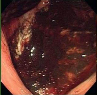
Non bleeding visible vessel 40 – 50 %
Adherent clot 10 – 33%
Forrest Endoscopic Classification
Endoscopic Observation
Rebleeding Chance%
Endoscopic intervention is only required in Forrest Ia, Ib, IIa and probably IIb at first to stop the active bleeding (Ia, Ib) and prevent subsequent rebleeding.

Acute variceal bleeding- management
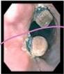
Endoscopy is required
Just temporarily
-
Octreotide infusion- lowers portal pressure
-
Endoscopic banding
-
Endoscopic injection sclerotherapy
Seither intravariceal or paravariceal
-
Balloon tamponade
-
TIPPS (Transjugular intrahepatic portosystemic shunting)
-
SURGERY:
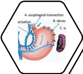
✓ Gastro-oesophageal devascularization + stapled oesophageal transection
✓ Liver transplantation
once Patient is Stable and bleeding doesn’t have to be Stopped but its best if its minimal
if bleeding is still there (20%) do
• Endoscopic Intervention For Esophageal varices
1- Endoscopic sclerosing therapy 1 Effect of Ectoxy sclerol solution 0.5-1cc /point
INTRAVARICEAL INJECTION ( underfluoroscope and venogram)
PARAVARICEAL INJECTION
2- ENDOSCOPIC MUCOSAL VARICEAL BAND LIGATION
3- Ballon Tamponad for temporary control after fail endoscopic intervention
control ( Senstaken Blakemore tube preparation)
9 2 ballons, one for lower part of esophagus to compress on veins and another one in fundus of stomach once placed, inflate gastric balloon first, then pull balloon outside so the inflated balloon compress
- Complication Prophylaxis antibiotics cover gram negative bacteria on the fundal varices, then inflate the other such as ciprofloxacin, levofloxacin, ceftacidime, amoxicillin-culvulanic acid and aztreonam are appropriate choices.
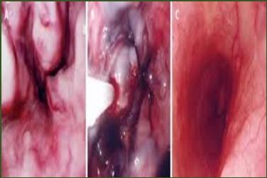
Endoscopic sclerotherapy for OV
→max for 48 hours. but, to avoid pressure necrosis, should deflate every 12 hours, and if there is still bleeding, then inflate it again and if still bleeding after 48 hours, then prepare for surgery.
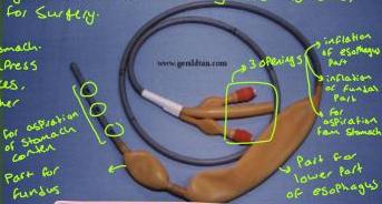
Sengstaken-Blakemore Tube
- Compression of varices for not excess 48 hours
- Deflate the esophageal balloon for about 30 mins every 12 hours
- Major complications — aspiration and esophageal perforation
- Control hemorrhage >90%, but it is temporary If bleeding is unresolved with endoscopy or endoscopy is contraindicated
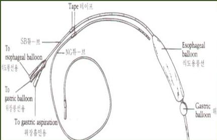
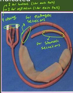
Minnesota tube with four lumens, used in treatment of esophageal varices; having a lumen for aspiration of esophageal secretions
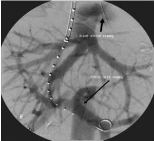
TIPS( Transcutaneous-jugular intrahepatic portosystemic shunts)
*Enter through jugular vein transcutaneously, and reach liver using interventional radiology and make shunt between intrahepatic vein and Portal vein. So blood will go from Portal to intrahepatic vein ■ Non-operative shunt to IVC.
- Use in stage of cirrhosis with liver failure
- Less effective to stop GI bleeding than operative shunt, but less invasive.
- Technique need radio-intervention
SURGERY:
in case of failure of ballon Tamponade
• Gastro-oesophageal devascularization + stapled oesophageal transection
Cut the junction between esophagus and stomach. Then reanastomose & so its cutting all dilated veins Liver transplantation
And should also give definitive treatment for the portal hypertension
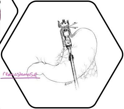
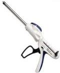
Endo GIA stapler
Cut and anastomose at the same time
Treat accordingly
Management of uncommon causes of UGI bleeding
- Conservative/ endoscopic management:
Mallory-Weiss syndrome-
Erosive gastritis /esophagitis.
Dieulafoy lesion.
- Surgical management after stabilization & diagnosis:
Gastric cancer.
Ulcerated gastric stromal tumor (GIST)
Management Strategies to minimize risk of recurrence
- Proton pump inhibitor (PPI) therapy
Omeprazole 80mg as a bolus IV dose then a continuous IV infusion at 8mg/hr for 72 hours then switch to oral therapy which should continue long term. 5
-
H.Pylori screening and eradication
-
This is often performed at the same time as endoscopy and is known as the Campylobacter-like organism test (CLO). The basis of the test is the ability of H.Pylori to secrete the urease enzyme, which catalyses the conversion of urea to ammonia and carbon dioxide.
-
Medication review
-
Discontinue NSAIDs.
An alternative pain management
Prognosis
Risk factors for: Increased mortality, recurrent bleeding, the need for endoscopic hemostasis, or surgery : Poor Prognosis:
- Age >60 years
- Severe comorbidity
- Active bleeding (witnessed hematemesis, blood in nasogastric tube, fresh blood per rectum)
- Hypotension
- Blood transfusion ≥ 6 units
- Severe coagulopathy