Urology 1
Dr. Mohamed Khaled Zaky
Objectives
- Review of anatomy & physiology of UT
- Commonly used UT investigations
- Common disorders of UT:
- Infection
- Stones
- Tumours
- Congenital
- Functional
- Trauma
- Urologic emergencies
- Surgical disorders of the external genitalia
Anatomy of the Kidney
Right Kidney Sectioned in Several Planes
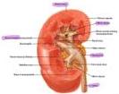
- Left kidney: T12 - L2
- Right kidney: L1-L3
Between Kidney + liver = Morrison pouch
Trauma to the kidney is rare but can be through a trauma to the back, or penetrating to the abdomen, very serious
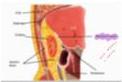

Renal capsule (Glisson’s capsule)
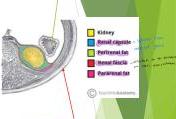
Perirenal fascia (Gerota’s fascia)
Lymph nodes:
- Drainage of Scrotum = Inguinal
- Of testis + kidney = paraaortic
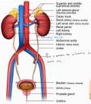
Anatomy of the UT
- Vein
- Artery
- Pelvic
- So to approach the pelvis? Posteriorly to reduce risk of injury
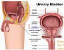
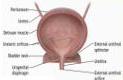
Base of the bladder in between the three orifices (2 ureters, urethra), mucosa is adherent to the muscle unlike the rest
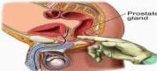
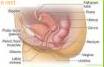
- Females = short urethra = high risk of UTI
- In PR we feel cervix + pouch of Douglas collection
- In males: PR we feel prostate, urethra has 3 parts
Two sphincter:
- Internal sphincter = smooth muscle, opens and give the desire to pass
- External sphincter = skeletal muscle, holds/ voluntary until it fatigues
UT Symptomatology
-
Pain
-
Dysuria
-
Change in urine
-
Change in urination (the process)
-
Swelling
-
Deterioration in kidney function
-
Or systemic manifestation as fever
-
Kidney: dull aching pain at the back, or costophrenic angle — happens due to stretch of capsule
-
Ureter: colicky pain from loin to groin
-
If stone reached the ureterovesical junction: tip of penis
-
Male colic: ureter, intestine
-
Female: … and uterus
-
Pain in perineal? Prostate
-
Pain in suprapubic? Bladder

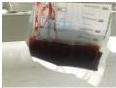
LIGHT YELLOW

YELLOW

AMBER

BROWN

RED
Basic UT investigations
-
Urine analysis — Initial
-
Kidney function tests (KFT)
-
Plain X-ray (KUB) — Starts with
-
Ultrasound
-
Excretory urography
-
CT urography — more common
-
IV urography / pyelography
- Contra: Dehydrated/low GFR, allergy to iodine
-
Ascending urography, cystoscope — if I can’t rely on kidney for excretion
-
+PH, serum electrolytes
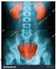


Hilum of kidney at L2 — Radioopaque stones along the transverse process (in the ureter) — Midline = bladder

Calyces lateral, ureters medial
Congenital Disorders of the Kidneys
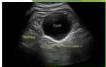
Simple cysts — very common
- Benign lesions of the renal parenchyma, having a thin wall, and being completely fluid filled.
- Rarely require intervention or follow up unless they cause symptoms (due to very large size) or are infected.
- When to worry? Send for cytology:
- More than 5 cm
- Complex cyst — not thin wall — (echoes, thick wall)
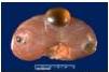
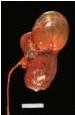
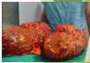
Polycystic kidney disease (PKD)
Infantile or autosomal recessive:
Extremely rare. Symptoms can begin in the earliest months of life, even in the womb. It tends to be very serious, progresses rapidly, and is often fatal in the first few months of life. Will compress the surroundings
Adult or autosomal dominant:
- The most common form. Symptoms usually begin between the ages of 30 and 40, but they can begin earlier, even in childhood.
- Polycystic disease can affect other organs besides the kidney, such as the liver, pancreas, spleen, ovaries, and/or cerebral aneurysm.
- Cystic fibrosis disease (lung + pancreas cysts)
Ttt: renal transplantation
PKD — C/P
- Asymptomatic
- Pain (recurrent)
- Swelling
- Haematuria
- Infection
- HTN — compresses the parenchyma
- RF — compresses the nephrons
First: pain, then swelling, change in urine (hematuria due to cyst rupture)
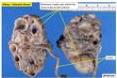
Very characteristic


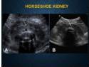
Horseshoe Kidney
The most common fusion anomaly of the kidney — kidneys lie lower than normal and are malrotated
C/P:
- Asymptomatic
- Ureteral kink → back pressure
- Hydronephrosis
- Abdominal mass — at the level of umbilicus
Inferior Mesenteric at L3 prevent kidney from reaching T12 (celiac trunk), so low lying kidney


Calyces medial + ureter lateral

Other Anomalies
Ectopic to the pelvis or crossed ectopia to the other side but ureter still opens at the previous site
Ectopic kidney
May be in the pelvis (dd: nephroptosis — rapid weight loss, loss of perirenal fat, normal in supine, ptosis of kidney downward when stands + kinking of ureters), or on the opposite side
Duplication
Double pelvis and/or double ureter — usually no problem but some will have crossing of the ureters = hydronephrosis
PUJO
May be congenital or acquired. Pelvic ureter junction obstruction (real or spasm) = hydronephrosis due to obstruction = dilatation or we cut the junction + anastomose
-
- unilateral Agenesis

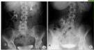


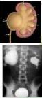
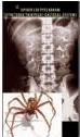
Stones (Urolithiasis) — Triad
Aetiology
Change in urine composition: super saturated solution to crystallise
- Increase in certain products — hypercalcemia due to Hyperparathyroidism, hyperuricemia in gout
- Decreased water content — decreases the dilution
- Decrease in stone-forming inhibitors (eg citrate)
Change in pH: normally acidic
- Found naturally in UT
- Proteus infection → alk. urine
- Stasis — why? Splitting of ammonia
Stones usually produced in kidney except some produced due to infection
- Supersaturation
- Usually with other samples, allopurinol
- Stones between 0.1 and 1.0 g per ml water

- Aggregation — stone formation
- Aggregates retention
Types
- Ca oxalate: most painful as have spikes, hematuria — 80-85%, radiopaque
- Uric acid: filling defect in contrast, plain x ray won’t show — 5-10%, radiolucent, friable
- Struvite (Mg, Ammonium, phosphate): 2-20%, alk urine, radiopaque, staghorn
- Cystine: 1%, genetic, slightly radiopaque, v hard
- Xanthine: extremely rare, genetic, allopurinol ttt
- Ca phosphate: rare, alk urine, hyperpara, radiopaque
Alkaline urine = phosphate accumulates
Pt for long in Foley catheters, phosphate encrustation (can’t pull the catheter out) = give Acetic acid
Calcium — stones are formed due to an excess of a mineral called oxalate — commonly found in some fruit, vegetables, nuts and chocolate. Bladder + spiky → Jackstone calculus
Struvite — a struvite stone is less common and caused by infection in the urinary tract. It can grow quickly and become quite large. Branches in the kidney → Staghorn calculus
Uric acid — stones form due to chronic dehydration. The risk increases in those with gout, a genetic tendency or a diet too high in protein.
Cystine — stones form in people with an inherited disorder that causes the kidneys to excrete an excess of certain amino acids.
Xanthine — stones are caused by an enzyme deficiency that causes the build-up of xanthine deposits.
Silica — stones are rare and caused by certain medications or herbal supplements
Effect
- Obstruction → back pressure, pain
- Stasis → infection
- Deterioration in renal function
- Migration → pain, hematuria
- Ch. irritation → malig. in Urothelium / squamous cell carcinoma
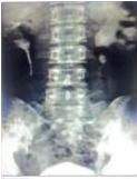

Diagnosis
C/P: may be asymptomatic in Lower calyx of the kidney
- Plain X-ray (90% of stones)
- U/S — stones = back shadow
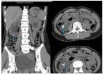
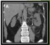
- CT before & after contrast
- Urine analysis:
- RBCs
- WBCs
- Crystals
- pH — alkaline = phosphate stone
- KFTs

Figure 5. Ultrasound Of Kidney Stones Showing Shadowing Effect

Total cell counts in 1st day of study
Treatment
- Conservative: stone < 5 mm — lots of fluids
- NSAID for pain — relax spasm in colic
- MET: Tamsulosin (a-blocker) — relaxation of ureter
- Dissolution: uric a. & cystine stones → Pot. Citrate, or Sod. Bicarb. (alk urine)
- Vitamin c for acidification of urine
- handwritten on slide: → dissolve the acidic stone
- ESWL: for kidney & upper ureter, < 2 cm. Best used away from bone. Can cause hematuria — Gel + shock waves
- Ureteroscopy: for ureteric stone, extraction of fragments, stenting — Lithotriptor for large stones
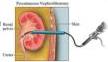
- PCNL: percutaneous nephrolithotomy — opens in the costovertebral angle, used in obstructed - infected urine
- Open surgery: diseased kidney — open to treat + remove the stone w it
- Prevention: plenty of fluids, dietary advice, Lithotripsy
- Push + bag: pushback the stone to the renal pelvis and then fragmentation

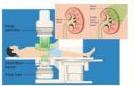
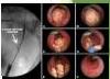
Contraindications to ESWL
Absolute:
- Children < 10 y
- Pregnancy — teratogenic
- Active urinary tract infection / urosepsis
- Decompensated coagulopathy — bc it causes hematuria
- Uncontrolled arrhythmia
- AAA > 4.0 cm.
Relative:
- Renal impairment
PCNL: Stones in upper ureter + pelvis
- For stones > 2 cm, or failed ESWL
- A nephrostomy tube is placed for 48 h after the procedure — after stone removal for drainage
- Advantages:
- Local anaesthesia
- Short hospital stay
- Complications:
- Hge
- Infection
- Injury to other structures
- Residual stones
Open surgery
- For very large stones
- Failure of other modalities
- For associated PUJO
- Pyelolithotomy — if the stone in the renal pelvis
- Nephrolithotomy — stone within the kidney
- Partial nephrectomy — if kidneys are damaged we remove it so doesn’t cause HTN


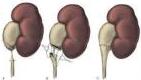
Hydronephrosis
Due to partial or intermittent obstruction of urine flow → gradual dilatation of the pelvicalyceal system → compression of the renal parenchyma and deterioration of kidney function

Dilatation of the renal collecting system


Normal Kidney
Mild hydronephrosis


Moderate hydronephrosis
Severe hydronephrosis
Hydronephrosis = partial or intermittent obstruction Acute / sudden = complete = AKI / AH
Causes
Extramural obstruction:
- Tumour from adjacent structures, e.g. carcinoma of the cervix, prostate, rectum, colon or caecum — Pelvic
- Idiopathic retroperitoneal fibrosis, pregnancy
Intramural obstruction:
- Congenital stenosis, PUJO
- Ureterocele and congenital small ureteric orifice
- Stricture ureter
- Neoplasm of the ureter or bladder cancer involving the ureteric orifice
Intraluminal obstruction:
- Calculus in the pelvis or ureter, blood clot
Functional:
- Vesico-ureteric reflux
Or acute hydronephrosis
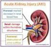
Due to sudden + complete obstruction — severe flank pain — Beware !!
If uni = the RFT might not be elevated
Causes of bilateral hydronephrosis:
- congenital:
- posterior urethral valves;
- urethral atresia;
- acquired:
- benign prostatic enlargement or carcinoma of the prostate;
- postoperative bladder neck scarring;
- urethral stricture;
- phimosis.
Obstruction in bladder and below or Vesicoureter reflux in kids (when bladder contracts the urine goes up) = bilateral hydronephrosis
C/P
- Mild pain or dull aching in the loin
- Palpable kidney
- New onset HTN
- Recurrent UTIs
-
- C/P of the cause
Treatment
- Drainage to relieve pressure
- TTT of the cause
- Nephrectomy in advanced cases
- Double J catheter or stent

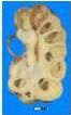
Pyonephrosis
Pathology
- Obstruction + infection → suppurative destruction of the parenchyma of the kidney
- Multilocular cavities filled with pus, lined by necrotic tissue + nephritis
- 2ndy calculi
C/P
- Pain, swelling and fever — might have no pus cells if complete obstruction
TTT
- Urgent drainage (percutaneous nephrostomy) or ureteric catheter if I can reach the area (first)
- Abx
- Nephrectomy — if can’t be controlled, sepsis, renal damage
- Try to save renal function as much as possible


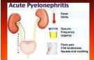
Pyelonephritis
Infection reached the pelvis / kidney / urine is still passing
Routes of infection
- Ascending infection (most common)
- Hematogenous (inf endocarditis, septicemia)
- Lymphatics
Organism
- G -ve: E. coli (70-80%), Proteus, others (intestine) — ascending or through lymphatics (chronic constipation)
- Females > males
C/P
- Remittent high fever, rigors, vomiting, severe loin pain, pyuria
TTT
- Abx (C&S), alkalinization of urine, fluids
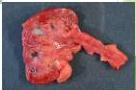
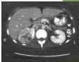
Renal abscess & Perinephric abscess
- Localized collections of purulent material within the renal parenchyma
- A rare but critical complication of urinary tract infections (UTIs) and bacteremia
- May extend to the perinephric space
- Commonly caused by E. coli, or hematogenous seeding from Staph. Aureus
- Fever, flank pain, and leukocytosis — “Urine analysis is normal” — no pus cells
- Flank tenderness is the most consistent finding, with costovertebral angle tenderness
- Abscess (≥ 5 cm) typically requires percutaneous drainage, smaller ones can be treated by antibiotics alone
- US or CT


TB. If skin, intestine, lung, cervical lymph nodes = direct, anywhere else is hematogenous
TB kidney (Rare)
2ry to pulmonary TB (Hematogenous)
- Destroys renal parenchyma, cheesy material may → PUJO & hydronephrosis
- TB toxemia: night fever & sweating, loss of appetite, loss of weight
- Hematuria, pyuria, flank pain, burning micturition & frequency
- U/A: sterile pyuria — why? TB needs special culture to grow in
- Imaging: CT
TTT
- Anti TB
- Nephrectomy may be needed
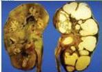
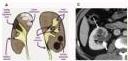
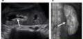
Tumours of the Kidney
Benign tumours
- Papillary renal adenoma: most common. Small, grow slowly and usually don’t cause symptoms
- Oncocytoma: can grow quite large, may be multiple
- Angiomyolipoma: blood vessels + muscles + fat — can grow and destroy surrounding structures. May cause hge. (hemorrhage) hematuria
TTT:
- Active observation
- Embolization (for angiomyolipoma)
- Surgery
Malignant tumours
- Nephroblastoma (Wilms tumour)
- Hypernephroma
- Urothelial tumours
- Secondaries
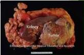
Wilms’ Tumour
- The most common pediatric abdominal cancer, and the 4th most common pediatric cancer.
- May be associated with:
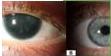
- Paraneoplastic syndrome: No Iris
- Hemihypertrophy, aniridia, cryptorchidism, horseshoe kidney, and hypospadias — undescended testis
- 90% < 6 y, 5-10% bilateral, equal sex distribution
- Usually presents as an asymptomatic abdominal mass + abd. pain, hematuria (late), HTN, fever
- Diagnosis: usually not this presentation — no early invasion to the parenchyma
- U/S, CT, MRI (best)
TTT:
- Nephrectomy, chemotherapy



Suprarenal gland
Neuroblastoma (Symphatoma)
- Associated with opsoclonus-myoclonus syndrome
- May CROSS the midline
- Usually fixed and immobile
- Constitutional symptoms ARE common
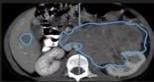
Crosses the midline
Wilms Tumour
- Associated with WAGR syndrome and Beckwith-Wiedemann syndrome
- Usually does NOT cross the midline
- May be DISPLACED
- Constitutional symptoms NOT common
Nephroblastoma (Wilms tumor)
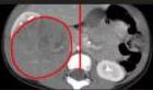
Doesn’t cross the midline
Hypernephroma (Renal Cell Carcinoma, RCC)
Why hyper? Usually from the upper pole
- The most prevalent form of kidney cancer in adults
- Usually diagnosed in patients aged 50-70
- A triad of: gross hematuria (painless, recurrent total), flank pain, and a palpable abdominal mass
- Painless hematuria: hypernephroma or urothelial tumor
- Paraneoplastic syndromes, eg hypercalcemia, polycythemia, HTN
- May spread along the renal vein → 2ry varicocele (left), intracaval extension
- Cross inside the renal vein and pass to the IVC
TTT: If elevate the scrotum = doesn’t disappear
- Partial nephrectomy (tumour < 7 cm, confined to one pole)
- Total nephrectomy


Urothelial Tumours
- Urothelial tumours (also known as transitional cell carcinomas) are a group of cancers that arise in the urothelium, the specialized lining of the urinary tract
- Renal Pelvis and Ureters (Upper Tract): rarer cases (5-10%) that occur in the tubes or collection areas of the kidney
- May be non-invasive (in situ) or invasive, low grade or high grade
- Usually → painless hematuria, may cause obstruction → flank pain
- Spread by surface implantation
Diagnosis:
- Urine cytology, CT urography
TTT (for kidney & ureter):
- Nephroureterectomy with excision of bladder cuff
- High recurrence rate → long term monitoring
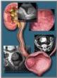

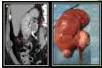
Ureteric Stone
- Migrating from the kidney
- Presents with ureteric colic + hematuria
- Nausea, vomiting
- Loin pain due to hydronephrosis + infection
TTT:
- Medical ttt (tamsulosin + NSAID) for stones < 5 mm
- ESWL
- Ureteroscopy: basket, lithotripsy, Push-bang
- Surgery: ureterolithotomy
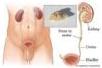


Other ureteric problems
Ureterocele
A cystic swelling at the end of the ureter where it enters the bladder, caused by faulty development during fetal growth
Vesico-ureteric reflux
Urine flows backward from the bladder into the ureters and kidneys, due to a congenital defect in the valve between them → back pressure & rec. UTI


- Narrowing of the lower end of the ureter
- Udder head appearance
- Ttt: Small opening or reimplantation of the ureter in another place
Hematuria
Causes
Within the UT
- Infection
- Stones
- Tumours
- Trauma
General
- Coagulopathies
- Medications
- Pigments (beet root)
Pt on blood thinners came w hematuria? Exclude pathology predisposing to bleeding
- Initial hematuria: urethral injury
- Terminal hematuria: bladder source (bilharziasis)
- Painless + causeless + recurrent = malignancy
Gross: Red/Dark urine
- 1ml blood/liter urine can induce color change
- Gross hematuria + clots → Lower Urinary tract source
- Hematuria: 3 or more RBC/HPF spun urine sediment
Microscopic
Detectable by examination of urine sediment by microscopy (Urinalysis or dipstick)


Glomerular bleeding:
- IgA nephropathy
- Thin basement membrane disease
- Alport syndrome
- Mesangioproliferative GN without IgA deposits
- Post-infectious GN
Glomerular hematuria:
- Immune mediated injury
- Noninflammatory glomerulopathies
Signs of glomerular hematuria:
- RBC casts — diagnostic of GN or vasculitis
- Dysmorphic RBC
- Brown urine
- Proteinuria → suggestive of glomerular disease
Loin pain hematuria syndrome: flank pain + gross or microscopic hematuria
HEMATURIA
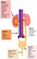
Pseudohematuria:
- GI bleed
- Menstruation/Postpartum
- Drugs: Pyridium, Phenytoin, Rifampin
- Beets
- Pigmenturia
RENAL:
- Pyelonephritis
- Hydronephrosis
- Malignant HTN
- Renal vein thrombosis
- Renal artery embolism
- AVM
- Papillary necrosis (sickle cell)
- Renal mass
URETER:
- Stone
- Stricture
- Malignancy
- Fistula
- Post-procedural
Urethra/Prostatic:
- Urethritis
- Urethral diverticulum
- Traumatic catheterization
- BPH
- Prostate cancer
- TURP/biopsy
BLADDER:
- Malignancy
- Cystitis
- Bladder stones / polyps
- Radiation
- Rapid decompression
- Diverticulum
Nutcracker syndrome:
- Compression of left renal vein between aorta and SMA
- Can cause microscopic and gross hematuria
Renal/Ureter/Bladder
Upper and Lower urinary tract
- Infection (Viral, Bacterial)
- TB
- Schistosomiasis
- Urolithiasis
- Malignancy
- Trauma
- Intervention
- Exercise induced bleeding
- Anticoagulation
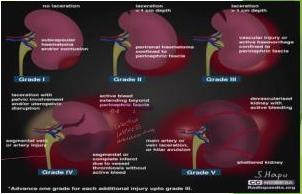
Kidney Injuries
- Generally uncommon
- May cause hematuria, or extravasation of urine or both
- Commonly treated conservatively, but surgery may be needed in severe cases (Grade IV, V)