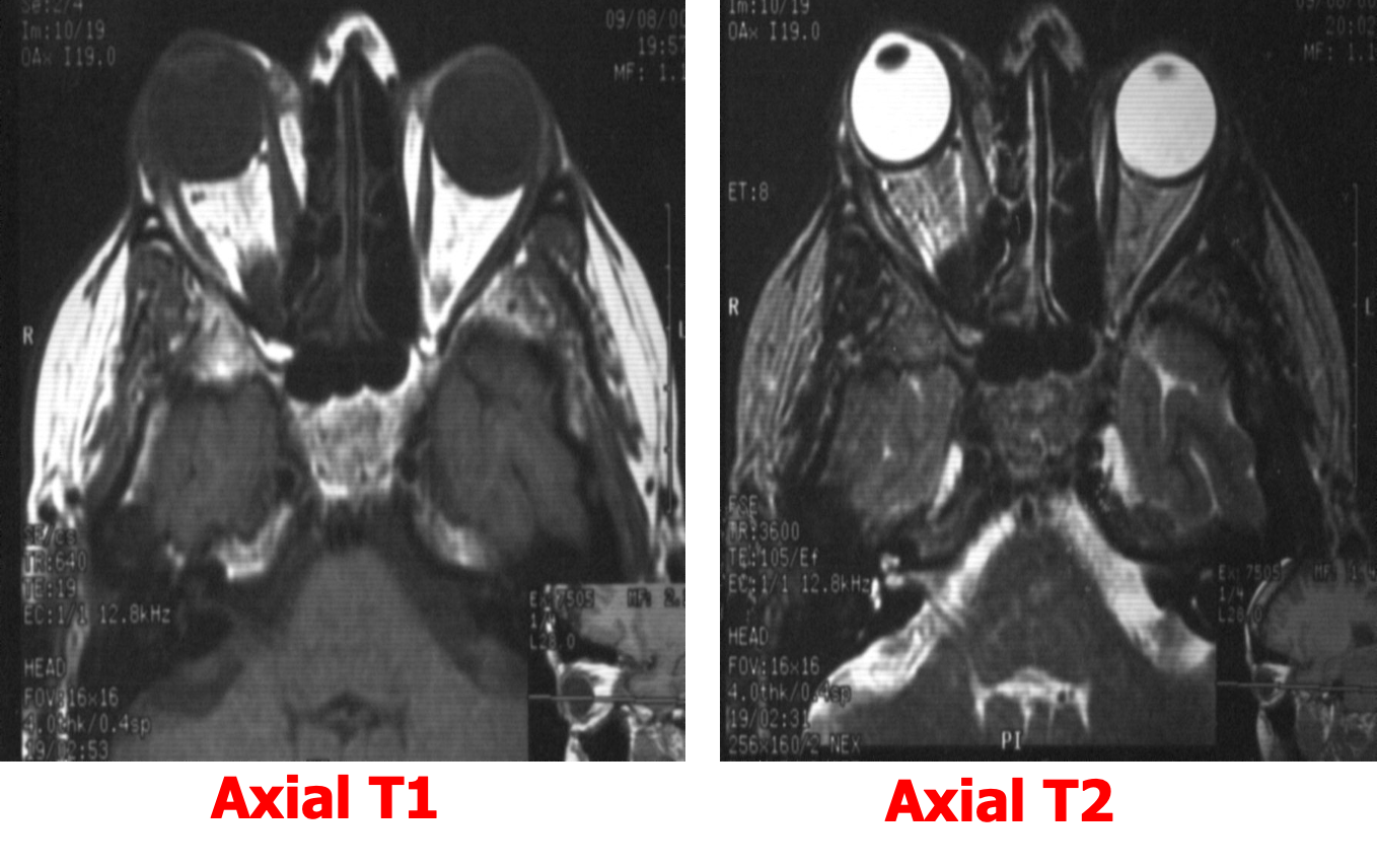
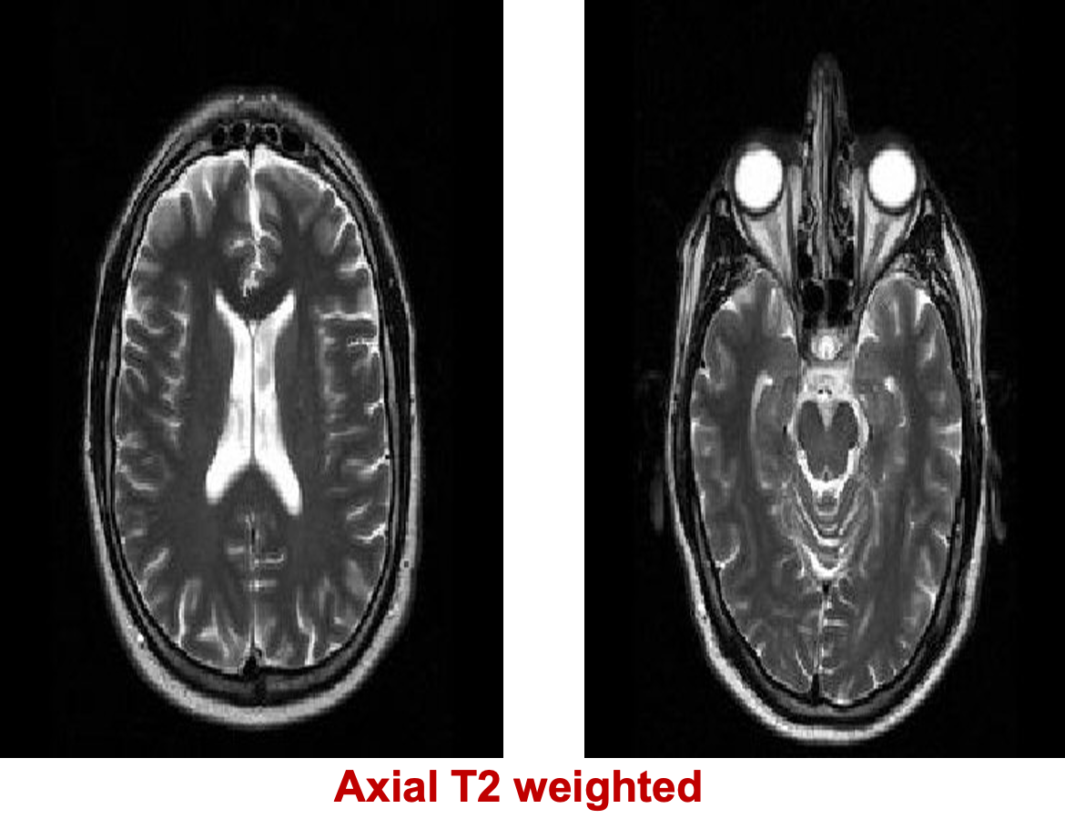
Multiplanar imaging: Axial ,Sagittal & Coronal Any MR examination should include T1 and T2 Weighted images
Signal intensity
- Low signal lesion = Hypointense = Dark
- High signal lesion = Hyperintense = Bright
- Intermediate signal = Isointense = Gray
T1 WIs [CSF BLACK ] T2 WIs [CSF BRIGHT ]
T1-low signal / T2 – low signal [Non mobile protons]
- Cortical bone
- Mature fibrous tissue ( ligaments and tendons)
- Calcifications ( physiological, pathological)
- Air in the sinuses, lungs,…
T1 high signal / T2 high signal
- Subacute blood [met Hb]
T1 high signal/ T2 high signal
- Fat ( subcutaneous fat, dermoid cyst, Melanin ,…)
T1 low signal / T2 high signal
- Fluids ( CSF, urine, pleural effusion, ascites.,…)
- Edema and infarctions
- Most of tumors

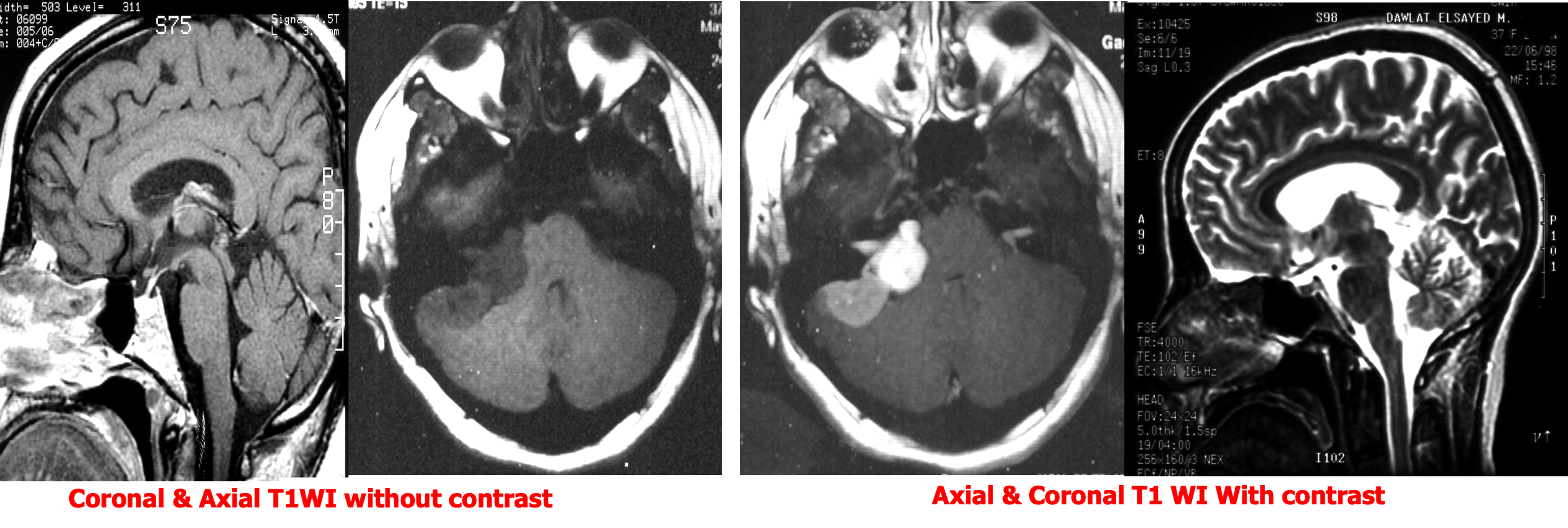
Gadolinium – DTPA
Only T1 weighted images are obtained after Gd- DTPA injection
- Differentiate SOLs
- Assess activity of some lesions like MS
- Assess post operative tumor recurrence
Intracranial enhancement on computed tomography and magnetic resonance imaging
Physiological
- Choroid
- Anterior pituitary gland
- Arteries
- Dural venous sinuses
Pathological
- Metastases
- Some primary gliomas
- Meningiomas
- Abscess
- Acute demyelination
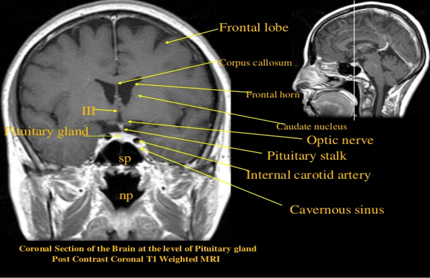
Coronal
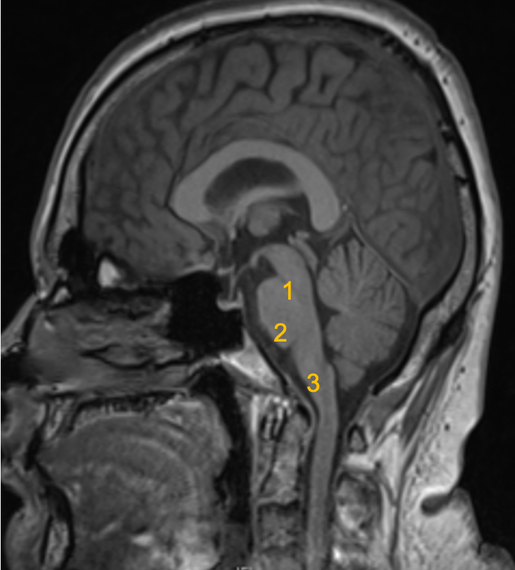
Brain Stem..
Three parts from superior to inferior:
1 Midbrain
2 Pons
3 Medulla oblongata
