Interstitial Lung Diseases
DR MUHAMMAD FAROOQ, FRCP
DEFINITION
These are a group of lung diseases in which there is infiltration of the interstitial tissue by cells, fluid and sometimes connective tissue.
There are almost 200 ILDs which differ in etiology and radiographic appearances. Many features are common to all.
CLASSIFICATION
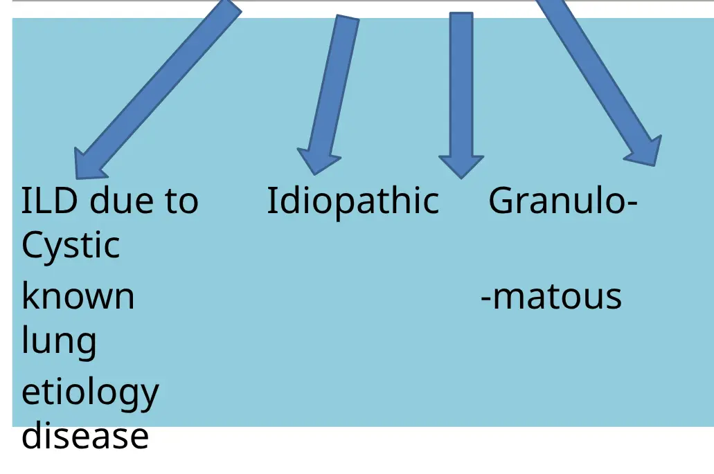
ILD DUE TO KNOWN ETIOLOGY
- Medicines: amiodarone, bleomycin, etc.
- Smoking
- Connective tissue diseases: Rheumatoid arthritis, scleroderma, SLE
- Radiation
- Pneumoconiosis: These are ILDs which occur due to exposure to environmental/occupational substances:
- Coal worker’s pneumoconiosis
- Asbestosis
- Silicosis
- Exposure to bird feathers (even pillows containing bird feathers can be the etiology)
IDIOPATHIC ILDs (No Known Etiology)
- Idiopathic Pulmonary Fibrosis (IPF) - Also called Idiopathic “Usual Interstitial Pneumonia (UIP)”
- Idiopathic Non-Specific Interstitial Pneumonia (NSIP)
- Acute Interstitial Pneumonia
- Cryptogenic Organising Pneumonia (COP)
GRANULOMATOUS ILD
Sarcoidosis can cause changes in the interstitial tissue of the lungs.
REMEMBER
The chest X-ray and CT scan patterns in all these diseases are a little different from each other, as we will see.
SIGNS AND SYMPTOMS (S/S) OF ILDs
1) History
- Dyspnea (worsens gradually)
- Dry cough
2) On Examination
- Clubbing of fingers
- Inspiratory crepts on lung auscultation
Mnemonic: DDCC = Dyspnea, Dry cough, Clubbing, Crepts
3) Features of Other Diseases
If ILD is secondary to another condition, look for features of:
- Rheumatoid arthritis
- Scleroderma
- Other connective tissue diseases
IMPORTANT POINTS IN HISTORY
Ask For:
- Medicine use
- Exposure to:
- Asbestos
- Silica
- Birds (even pillows containing bird feathers can be the etiology)
- Detailed occupational history
INVESTIGATIONS
Imaging and PFTs (Pulmonary Function Tests) have a major role and are the most important investigations.

IMAGING
1) Chest X-ray: Features Common to All ILDs
- Interstitial thickening
- Loss of lung volume (shrinking)
- Cystic changes
- Opacities:
- Reticular
- Nodular
The above pathological changes produce “honeycomb” and “reticular” patterns on X-ray and CT.
Honeycomb Pattern on X-ray


2) High Resolution CT Chest (HRCT) Z
- Must be done if signs/symptoms and X-ray suggest ILD
- Best imaging test for ILDs
Cystic Changes on HRCT


Interstitial Thickening
 Note the white lines
 reticular pattern
Honeycomb Pattern (Additional Images)


Reticular Pattern

PFTs (Spirometry)

- Shows a restrictive pattern
- FEV1 reduced
- FVC reduced
- FEV1/FVC ratio normal
Labs
- No specific labs for ILDs
- Blood tests for specific diseases are done if you suspect them:
- Rheumatoid arthritis
- CREST syndrome
- Other connective tissue diseases
Lung Biopsy
- Only done if diagnosis is uncertain
- Risks: Can sometimes worsen the condition, and the patient can even die, so it should be done only if really needed
SOME SPECIFIC ILDs
1) Idiopathic Pulmonary Fibrosis (IPF)
- Also called idiopathic UIP (Usual Interstitial Pneumonia)
- Commonest ILD in old people
- Slowly progressive disease with poor prognosis
CT Chest Shows:
- Subpleural and basal opacities
- Honeycombing: cystic spaces with thick fibrous tissue walls
Honeycomb Pattern on X-ray (IPF)
Chest Radiograph:


Conglomerating cysts of varying size and wall thickness - “Honeycomb” sign
Basal Opacity (IPF)
BASAL OPACITY


IPF Treatment
Long Term Treatment:
- Pir-feni-done
- Tyrosine kinase inhibitors
- Oxygen (O2) - for some patients
- NO steroids (in long-term management)
- Pulmonary rehabilitation
Short Term Treatment (Acute Exacerbation):
- Oxygen (O2)
- Steroids
2) Smoking-Related ILDs
- Smoking can cause different types of ILDs
- Some have a good prognosis after smoking cessation
- Ground glass opacities in the upper lung zones
Ground Glass Opacity (Upper Lobes)


3) Pneumoconiosis
- These are ILDs which occur due to exposure to inorganic dusts (usually occupational exposure)
We will discuss 2 types:
- Asbestosis
- Silicosis
Asbestosis
- Asbestos is a naturally occurring fibrous material which has been used in making floors, ceilings, and fireproof materials
- It is carcinogenic
Jobs with Chronic Exposure to Asbestos:
- Shipyard workers
- Carpenters
- Asbestos miners
Diseases Caused by Asbestos:
-
ILD (asbestosis)
-
Pleural thickening
-
Mesothelioma
-
Lung cancer
-
Asbestosis is irreversible. Management only prevents further worsening
Asbestos-Related Images


Pneumoconiosis Continued: Silicosis
- Due to exposure to silica dust, found in stone and sand
- Occurs in:
- Construction workers
- Stone blasters
- Foundry workers (metal factory)
X-ray Findings:
- Nodules in the upper and middle lung zones
- Plus hilar adenopathy
Silicosis Images

General Treatment of All ILDs
- Oxygen (O2) therapy is most important, to correct hypoxemia (O2 sat should be 90% at least)
- Stop smoking, avoid any environmental pollutants
- Pulmonary rehabilitation (breathing and exercise training, social support)
- Flu vaccine and pneumovac
- Corticosteroids and immunosuppressive drugs are beneficial in many ILDs but not in IPF
Lung Transplant for ILDs
- Lung transplant improves survival in Idiopathic Pulmonary Fibrosis. However, organ availability is very limited.
- Without transplant, prognosis is very poor (few years)
- There are definite criteria for lung transplant
COMPLICATIONS OF INTERSTITIAL LUNG DISEASES
- Pulmonary hypertension (HTN), which may cause right heart failure (called?)
- Lung cancer (Ca. lung)
SUMMARY OF X-RAY CHANGES
- Idiopathic Pulmonary Fibrosis: Subpleural and basal opacity + honeycombing
- Smoking-related ILD: Upper lung opacity (ground glass type)
- Asbestosis: Pleural thickening + opacities
- Silicosis: Upper and middle lobe nodules