Collected from Haneen H. Alsultan, Faisal Alkharji, Faisal Alturki, Talal M. Ak., Toqa Alsulaim, Rawan Althaqib, Abdul Rhman Abou Al-Shaar, Mohammed Al Khaldi, Basim Alhomida, , Muath Bin Abdullah Muath bin abdullah, Khalid Alanazi.
اللهم يا معلّم موسى علّمني، ويا مفهم سليمان فهّمني، ويا مؤتي لقمان الحكمة وفصل الخطاب آتني الحكمة وفصل الخطاب اللهم اجعل ألستنا عامرة بذكرك، وقلوبنا بخشيتك، وأسرارنا بطاعتك، إنك على كل شيء قدير، حسبنا الله ونعم الوكيل
https://www.youtube.com/playlist?list=PLDIP6wfTrzsE
Master Table of Contents
- 1. Abdominal Pain History & Examination
- 2. Hernia Examination
- 3. Peripheral Vascular Examination
- 4. Diabetic Foot Examination
- 5. Neck Lump Examination
- 6. Thyroid Examination
- 7. Breast Examination
- 8. Laparoscopic & Surgical Instruments
- 9. Laboratory Interpretation
1. Abdominal Pain History & Examination
History-Taking Checklist
- WIPER: Wash hands, Introduce yourself & role, Privacy (chaperone), Explain purpose, Request permission/consent.
- Focused history demographics→CC (exact duration)→HPI (SOCRATES + red flags)→past medical/surgical→drugs/allergies→social→family; ask progression, timing, episodes and fluctuation (first noticed by patient/other, bigger/smaller over time) without leading with character/site.
- Pre-touch summary: demographics, CC, key positives/negatives, background, risk factors, working diagnosis.
Pain analysis (SOCRATES)
- Site: landmark; document migration: periumbilical→RIF (appendicitis), epigastrium→back (pancreas), RUQ→shoulder tip (diaphragmatic irritation).
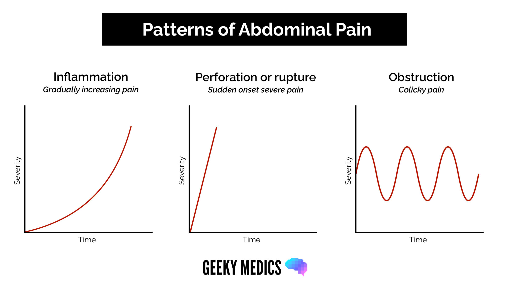
- Onset: sudden/“thunderclap” (perforated viscus, ruptured AAA, torsion) vs. gradual (inflammatory).
- Character: colicky: luminal obstruction (biliary, ureteric, bowel); burning: acid; stabbing: ischaemia; dull: capsular stretch.
- Radiation: back (pancreas, AAA), shoulder tip (diaphragm), groin/genitalia (ureter).
- Associations: nausea/vomiting, fever/rigors, jaundice, urinary symptoms, haematemesis/melaena, altered bowel habit, weight loss.
- Timing/triggers: intermittent vs. continuous; meal relation: duodenal ulcer hunger pain 2–3 h post-prandial, gastric ulcer worse with food; movement, respiration, defecation, sitting forward (relief in pancreatitis); severity 1–10.
- Visceral vs. somatic: foregut→epigastric, midgut→umbilical, hindgut→suprapubic (poorly localised); parietal peritoneum→well-localised somatic pain.
- ICE: explore ideas, concerns, expectations.
Systemic enquiry & red flags
- GI/urinary: dysphagia (solids vs. liquids), haematemesis, melaena, haematochezia, altered bowel habit, bloating, bilious vs. non-bilious vomiting; gross painless haematuria, acute retention, frequency.
- HPB: fever/rigors, RUQ pain and jaundice = Charcot’s triad (ascending cholangitis).
- Systemic/B-symptoms: unintentional weight loss, drenching night sweats, anorexia.
- Pain red flags: severe pain, persistent vomiting, distension with absolute constipation, blood in stool.
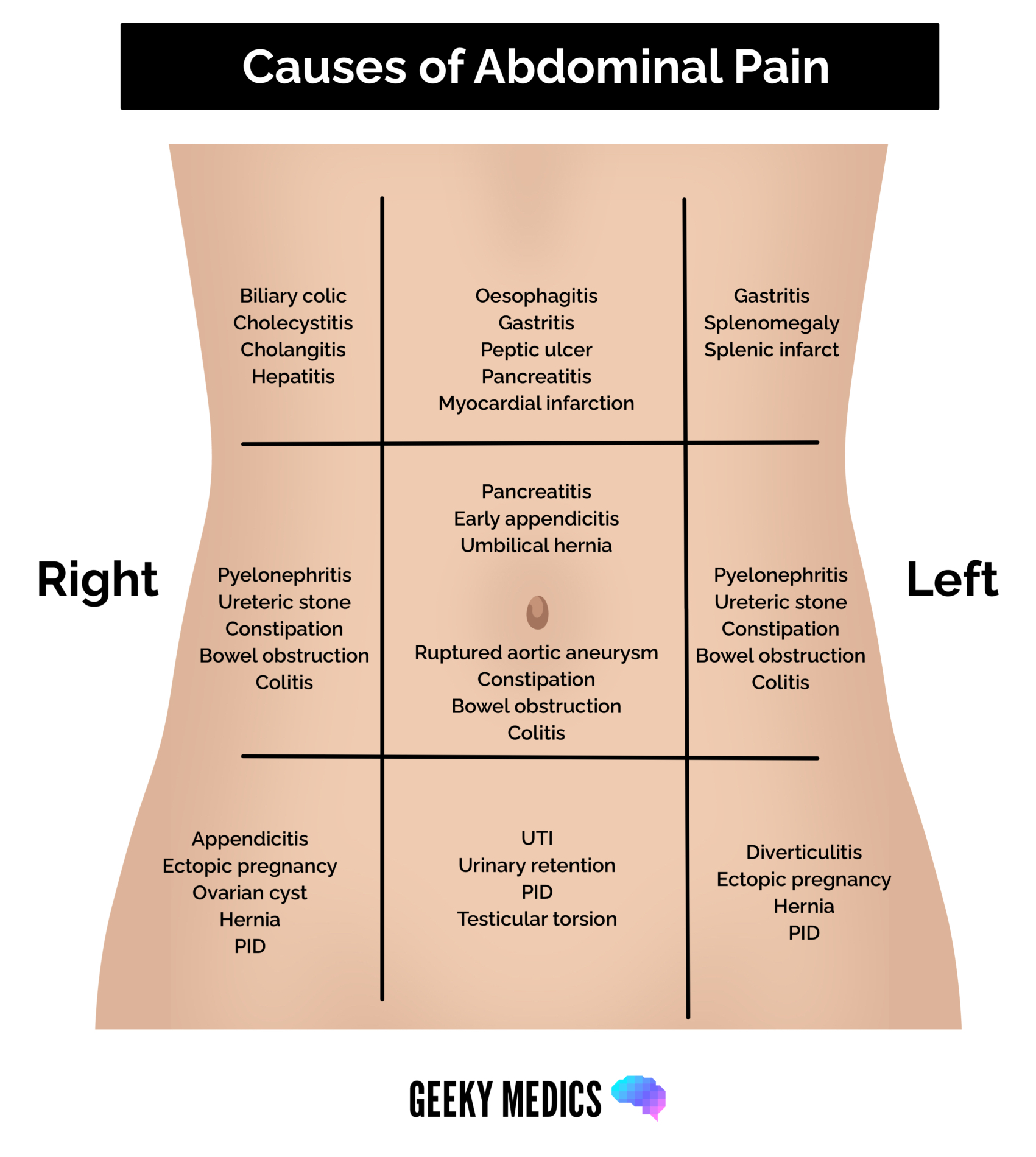
- Pain patterns & location:
- Medical mimics: MI (epigastric), pneumonia, DKA, herpes zoster, acute porphyria, hypercalcaemia.
- Pregnancy screen (MANDATORY): urine β-hCG in ALL females of reproductive age with acute lower abdominal/pelvic pain, before ionising radiation (CT/AXR) or surgery; rules out ruptured ectopic pregnancy.
Past surgical/medical/drug history
- Surgical: previous abdominal/hernia surgery/scars (adhesions→adhesive small bowel obstruction), DVT/PE, anaesthetic complications.
- Medical: DM, HTN, IHD, COPD, CKD, liver disease.
- Drugs/allergies: anticoagulants/antiplatelets (warfarin, DOACs, aspirin, clopidogrel), NSAIDs/steroids (peptic ulceration), immunosuppressants; pain causes: NSAIDs (ulcers), ferrous salts (GI upset), opiates/codeine (constipation), antibiotics (clarithromycin, amoxicillin, co-amoxiclav), DPP-4 inhibitors (saxagliptin, linagliptin→pancreatitis); penicillin, IV contrast, latex.
- Social/family: home adaptations (stairlift), carer input, ADLs; family GI disease with age at onset; sexual history/pregnancy; pack-years: years × packs/day (1 pack = 20).
Jaundice history
- Cholestasis triad: dark tea-coloured urine (conjugated bilirubinuria), pale clay-coloured stools (acholic), generalised pruritus (bile salts); ask onset/duration/progression (intermittent), itching, dark stools, weight loss, fever/malaise, bruising; pre-hepatic→normal urine/stool (unconjugated bilirubin water-insoluble), post-hepatic→pale stool and dark urine (only when conjugated).
- Discriminators: painless progressive jaundice and weight loss: periampullary/head of pancreas malignancy (Courvoisier’s law); colicky RUQ pain: choledocholithiasis; fever/rigors: acute ascending cholangitis.
- Pseudo-jaundice (carotenaemia): yellow-orange skin (palms/soles) with sclera intact; no bilirubinuria — exclude true jaundice.
- Risk screen: alcohol, gallstones, travel/water (Hep A/E), transfusions/tattoos/IV drugs (Hep B/C), hepatotoxic drugs (paracetamol, statins, augmentin, anti-TB).
Upper GI/peptic ulcer history
- Duodenal ulcer: epigastric burning/sharp pain, hunger pain 2–3 h post-prandially, relieved by meals/antacids, waking at 1–2 AM (unbuffered nocturnal acid); gastric ulcer: pain worsened by food→food avoidance, weight loss.
- Complications: haematemesis/melaena (bleeding), sudden board-like peritonitis (perforation), projectile non-bilious vomiting (gastric outlet obstruction).
- Risk factors: NSAIDs/aspirin, H. pylori, smoking; alcohol/gallstones/medications (pancreatitis).
Examination Checklist
Environment & exposure
- WIPER as above; supine, arms at sides, knees slightly flexed; expose nipple to mid-thigh, genitalia covered (hernia stations: stand the patient).
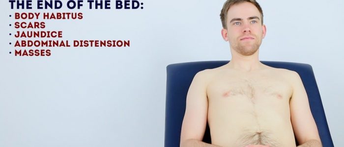
- General inspection: age-look, build, condition (alert, pain/distress), position (comfortable/restless), devices (IV cannula, catheter output), vitals; confusion: hepatic encephalopathy; cachexia: malignancy/advanced liver failure; bronzing hyperpigmentation: haemochromatosis.
Hands/arms/axillae
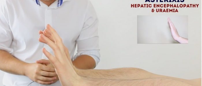
- Asterixis (flapping tremor): arms outstretched, wrists cocked back, hold 30 s→irregular loss of posture; causes hepatic encephalopathy (hyperammonaemia), uraemia, CO₂ retention.
- Dupuytren’s contracture: palmar fascia thickening with cord-like bands→finger contractures; genetics, alcohol excess, increasing age, male sex, diabetes.
- Bruising (clotting defects), excoriations (pruritus/cholestasis), needle-track marks (viral hepatitis), palmar erythema (chronic liver disease; also pregnancy).
- Axillae: acanthosis nigricans (insulin resistance/T2DM; also gastric malignancy), axillary hair loss (iron deficiency/malnutrition).
Face/mouth/neck
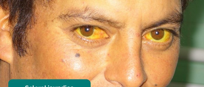
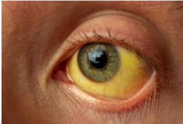
- Scleral jaundice best seen superiorly (look down, lift upper lid); conjunctival pallor.
- Corneal arcus (benign >60 y; <50 y→hypercholesterolaemia), xanthelasma (hypercholesterolaemia), Kayser–Fleischer rings (Wilson’s), perilimbal injection (anterior uveitis, IBD).
- Mouth: angular stomatitis (iron deficiency, malabsorption), glossitis (iron/B12/folate), oral candidiasis (immunosuppression), aphthous ulcers (benign; Crohn’s), hyperpigmented macules: Peutz–Jeghers (AD, GI polyps).
- Virchow’s node: left supraclavicular drains the abdomen (gastric/GI malignancy), right drains the thorax (oesophageal/thoracic).
- Chest: spider naevi (>5→cirrhosis; also pregnancy/COCP); gynaecomastia (cirrhosis, digoxin, spironolactone); chest hair loss (oestrogen).
Abdomen: inspection
- From end of bed: symmetry; contour (scaphoid/normal/distended, 6 Fs: fat, fetus, fluid, flatus, faeces, fibroid); movement with respiration (abdomino-thoracic males, thoraco-abdominal females); from patient’s right: peristalsis (increased “step-ladder”: early obstruction; absent: ileus; left-to-right: pyloric stenosis), pulsations, umbilicus (central/inverted/everted, hernia, discharge), masses.
- Distension, 5 S’s: Skin/fat, Surface fluid (ascites), Swellings (incl. Fetus, Fibroid), Silent flatus (gas), Subcutaneous (bowel loops); normally only mildly convex.
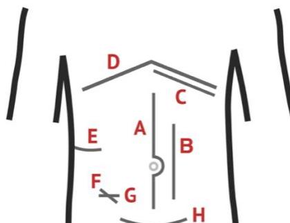
- Scars/incisions: midline (trauma/peritonitis/laparotomy), Kocher’s (open cholecystectomy/biliary), McBurney’s/Lanz (open appendectomy), Pfannenstiel (C-section/pelvic), rooftop/Chevron (Whipple/liver transplant), paramedian (kidney/adrenals/spleen), loin (retroperitoneal kidney); gridiron injury to iliohypogastric/ilioinguinal nerves denervates the conjoint tendon→direct inguinal hernia.
- Skin: striae (Cushing’s/pregnancy); dilated veins: caput medusae (portal hypertension) or IVC obstruction; ecchymoses: Cullen’s periumbilical, Grey Turner’s flank, Fox’s femoral (retroperitoneal haemorrhage; Cullen’s/Grey Turner’s: late haemorrhagic pancreatitis).
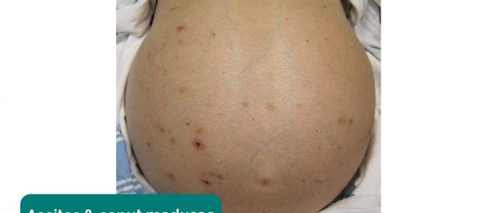
- Objects/drains/stomas: stoma bags (location); drains (site, type open/closed ± suction, fluid serous/serosanguinous/purulent, amount); feeding tubes (NG/nasojejunal); catheters (urine colour/volume); TPN; mobility aids/charts/prescriptions; stomas: location (colostomy LIF, ileostomy/urostomy RIF), output, base attachment, peri-stomal skin, mucosal viability (red healthy, black ischaemic), consistency (liquid ileostomy vs. solid colostomy), spout (ileostomy/urostomy spouted; colostomy flush).
- Hernial orifices: cough/head-lift→expansile cough impulse (→ Inguinal Hernia Examination).
Palpation
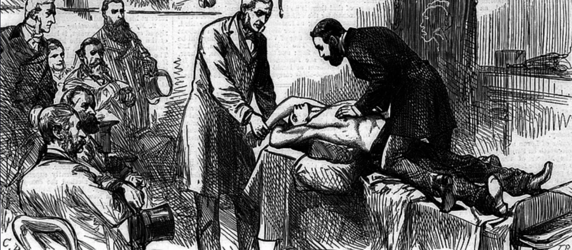
- Warm hands; sit/kneel beside the bed; watch the face; examine painful areas last.
- Superficial (9 regions, anticlockwise): tenderness, voluntary guarding, involuntary rigidity (board-like: generalised peritonitis), rebound tenderness (Blumberg), Rovsing’s sign (LIF pressure→RIF pain, appendicitis), superficial masses, temperature; deep: all quadrants for deep masses/organomegaly.
- Appendix signs: Psoas (RIF pain on passive right hip hyperextension, retrocaecal appendicitis), Obturator (RIF pain on passive internal rotation of flexed right hip, pelvic appendicitis).
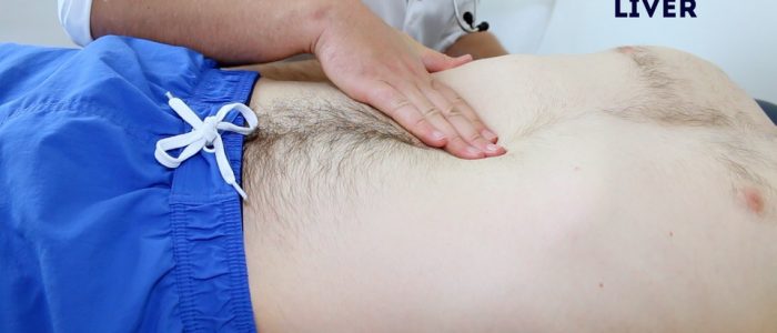
- Liver: start RIF during expiration; feel edge below right costal margin (surface, consistency, edge, tenderness); percussion span mid-clavicular line 6–12 cm; Murphy’s sign: inspiratory arrest (acute cholecystitis). Hepatomegaly: hepatitis, HCC, metastases, Wilson’s, haemochromatosis, leukaemia, myeloma, glandular fever, primary biliary cirrhosis, tricuspid regurgitation, haemolytic anaemia; pulsatile liver: tricuspid regurgitation.
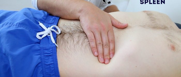
- Spleen: palpate diagonally RIF→left costal margin (below 10th rib tip); not palpable unless enlarged; lift with left hand behind lower left ribcage; versus kidney: spleen dull, not ballotable, notched, may cross midline; kidney ballotable, resonant, moves with respiration. Splenomegaly: portal hypertension, haemolytic anaemia, congestive heart failure, splenic metastases, glandular fever; palpable spleen at costal margin ≈ 3× normal size (look for splenic notch).
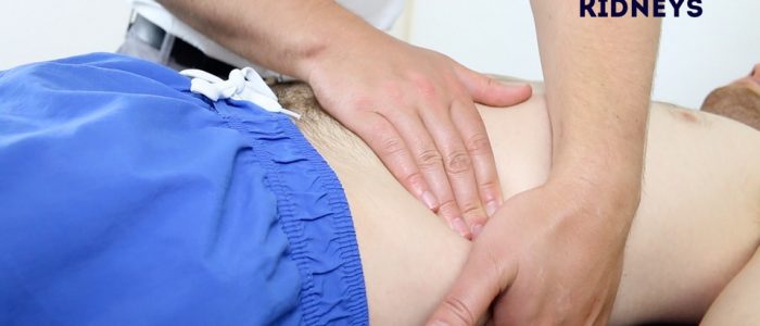
- Kidneys: bimanual ballotment in the loin (between 12th rib and iliac crest), posterior hand lifting the kidney anteriorly while the anterior hand palpates (distinguishes retroperitoneal kidney from spleen/liver); enlarges vertically; largely unrelated to intraperitoneal findings; bilateral ballotable enlargement: polycystic kidney disease, amyloidosis, unilateral: renal tumour.
- Aorta: above the umbilicus, width/character; expansile (outward): aneurysm, transmitted: overlying mass; A: no impulse; B: transmitted; C: expansile. Bladder: ask the patient to void first; distension arises from behind the pubic symphysis.
- Mass? Describe: site/quadrant, size, shape, surface, edge, consistency, temperature, tenderness, mobility/relations (skin vs. deep), fluctuation, fluid thrill, transillumination, compressibility, reducibility, bruit, pulsatility.
- Courvoisier’s law: in jaundice, an enlarged palpable non-tender gallbladder is not gallstones but malignancy (head of pancreas, cholangiocarcinoma); chronic stones cause a fibrotic, shrunken gallbladder. Mucocele: cystic-duct obstruction→distended gallbladder; infected→pyocele.
Percussion
- All 9 regions: resonant: gas; dull: solid/fluid/organ; hyper-resonant: bowel distension. Technique: fix non-dominant middle finger; strike with dominant middle finger two taps, lifting quickly.
- Liver: examine during expiration (inspiration exaggerates hepatomegaly); spleen: rotate the patient, percuss the left 8th–10th intercostal spaces (dull: subclinical splenomegaly; normally resonant due to the gastric bubble); patient fasted ≥2 h.
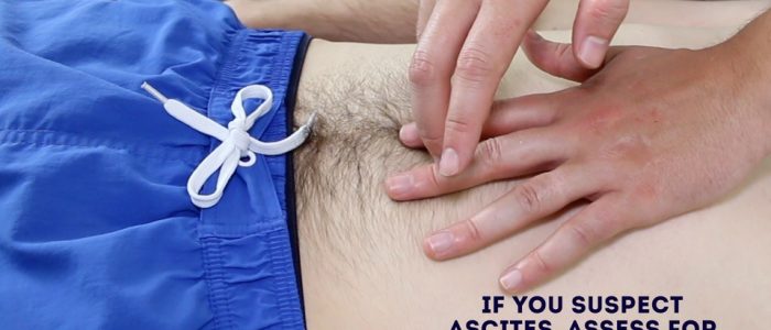
- Ascites: distension, everted umbilicus, bulging flanks; flanks dull, central resonant (floating bowel loops); shifting dullness: percuss umbilicus→flank until dull, roll opposite, wait 15–30 s, percuss back; fluid thrill: patient’s hand vertical on midline, palm on one flank, flick the other.
Auscultation
- Bowel sounds: at McBurney’s point/all 4 quadrants; normal soft gurgles every 5–10 s; silent periods of several minutes normal; borborygmi: exaggerated; absent > 30 s (only heart/breath sounds): paralytic/adynamic ileus (auscultate ≥3 minutes before calling absent); tinkling/high-pitched: mechanical obstruction; hyperactive: gastroenteritis.
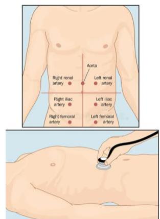
- Bruits: aorta (just above umbilicus), renal arteries (lateral to aorta): systolic, arterial stenosis or AV fistula; hepatic bruit: HCC/vascular hepatic tumours; venous hum over umbilicus/liver: portal hypertension (portosystemic shunting via umbilical/paraumbilical veins).
- Succussion splash (shake/roll the patient): retained gastric fluid (pyloric stenosis/gastric outlet obstruction, advanced bowel obstruction).
Completion & summary
- Finish with genitalia + PR (DRE checklist → Digital Rectal Examination (PR); proctoscopy pearls → 8. Laparoscopic & Surgical Instruments), groin/femoral orifices, back for tenderness/bed sores, femoral pulses; legs for pitting oedema→hypoalbuminaemia (cirrhosis, protein-losing enteropathy). Cover and thank the patient.
- Summary to examiner: positives/pertinent negatives→diagnosis→differentials with justification→investigation→treatment plan.
Differential Diagnosis & Management
Regional pain differentials
- Epigastric/acute abdomen stations: pancreatitis, GERD, peptic ulcer (gastric/duodenal), gastric cancer, oesophagitis, left lobe of liver, inferior MI, ruptured AAA.
- RIF: appendicitis, ureteric colic, pyelonephritis, caecal pathology/IBD.
- LIF: diverticulitis, sigmoid pathology, ulcer.
- Loin: renal (stones, pyelonephritis, mass, ischaemia, trauma).
- Cardiopulmonary (RUQ/epigastric): RLL pneumonia, effusion/empyema, CHF (hepatic congestion), MI, pericarditis, pleuritis.
- Gynae (any lower abdominal pain in a female): ectopic pregnancy, PID, endometriosis, threatened/incomplete abortion, hydrosalpinx/salpingitis, ovarian torsion, haemorrhagic fibroid, tubo-ovarian abscess, gynaecological tumours.
- Upper GI bleeding: peptic ulcer, Mallory-Weiss tear, aortoenteric fistula, angiodysplasia, oesophageal/gastric varices, haemobilia, malignancy, gastritis, oesophagitis.
- Lower GI bleeding: massive upper GI bleed, cancer, polyps, angiodysplasia, diverticular disease, IBD, ischaemic colitis, infective diarrhoea, anal fissure, haemorrhoids, rectal varices.
Appendiceal mass/abscess (phlegmon)
- 10–14 days post-rupture: spiking fever, tachycardia, leucocytosis, tender firm RIF mass; rebound/Rovsing’s/Psoas/Obturator signs. Contrast CT abdomen/pelvis distinguishes phlegmon from walled-off abscess.
- Ochsner–Sherren: acute phase: NPO, IV fluids, IV piperacillin–tazobactam, CT-guided percutaneous drainage for collections ≥3–4 cm (laparotomy if peritonitis/sepsis worsens); elective interval appendectomy at 6–8 weeks (prevents recurrence; excludes caecal adenocarcinoma, carcinoid, Crohn’s).
Jaundice & HPB management
- LFTs: direct bilirubin ↑, GGT ↑, ALP ↑→ultrasound first; MRCP/PTC/ERCP define above/below obstruction.
- Choledocholithiasis: ERCP first (sphincterotomy, stone extraction)→cholecystectomy same admission; if ERCP fails→open/laparoscopic bile-duct exploration.
- Before ERCP: consent, prophylactic antibiotics, correct coagulopathy; ultrasound to confirm stones; renal function, CBC, U&E for contrast.
- Head-of-pancreas carcinoma: resectable→Whipple’s pancreaticoduodenectomy, removing duodenum, head of pancreas and common bile duct with 3 anastomoses: gastrojejunostomy, choledochojejunostomy, pancreaticojejunostomy; unresectable→palliative ERCP/stent (± chemotherapy).
- Biliary colic: RUQ pain radiating to right shoulder/scapula, triggered by fatty food (CCK release), colicky; relieved by analgesia or spontaneously; admit only for persistent/repeated attacks.
Upper GI bleeding & pancreatitis stations
- Upper GI bleed: ABC: patent airway→O₂/pulse oximetry→2 large-bore IV lines (CBC, U&E, coagulation, crossmatch), Ringer’s lactate, NG decompression, Foley for output. History: blood amount/frequency/colour; melaena/PR bleeding, pain, nausea, fatigue, fever, jaundice; bleeding disorder, NSAIDs, anticoagulants; liver disease causes (alcohol, hepatitis, travel, transfusions).
- Perforated peptic ulcer: epigastric pain and guarding; erect CXR: free air under diaphragm; IV fluids, antacids, antibiotics, strict input/output, laparotomy/Graham patch.
- Acute pancreatitis: epigastric pain and vomiting with compromised vitals; risk factors alcohol, gallstones, medications; investigations pancreatic enzymes, CBC, type & crossmatch, electrolytes, LFT, coagulation, ABG, LDH (Ranson’s criteria), erect CXR (rule out perforation), AXR (sentinel loop); stones on imaging→gallstone pancreatitis; 2 large-bore IV lines and analgesia (pethidine, not morphine), NGT (vomiting), Foley catheter, cholecystectomy same admission; complications necrosis, DM, pseudocyst, infection/abscess.
Lower GI & colorectal
- PR bleeding history: blood (type, amount, painful vs. painless); change in bowel habit; obstruction (pain, distension, vomiting, constipation); abdominal mass; family history of colon cancer; malignancy symptoms (weight loss, anorexia, fever, fatigue); metastasis screen (cough/haemoptysis, RUQ pain/jaundice, back pain, headache). Examine abdomen and rectal examination with proctoscopy.
- Right colon cancer: RLQ pain, anaemia (fatigue, palpitations, dyspnoea), RLQ mass, general malignancy symptoms; confirm colonoscopy + biopsy; stage with CT CAP (CEA, CBC, U&E, coagulation).
- Colorectal staging: Stage 0 carcinoma in situ (mucosa only); I submucosa ± muscularis propria; II subserosa/adjacent organs; III lymph-node involvement; IV distant metastasis.
Viva & Exam Spots
- Q: Exception to Courvoisier’s law? A: Double impaction: cystic-duct stone distending the gallbladder plus common bile duct stone causing jaundice.
- Q: Triad of a ruptured AAA? A: Pain, hypotension, expansile mass→urgent OR/EVAR.
- Q: Alvarado score for appendicitis? A: MANTRELS: Migration to RIF 1, Anorexia 1, Nausea/vomiting 1, Tenderness in RIF 2, Rebound 1, Elevated temp ≥37.3°C 1, Leucocytosis >10×10⁹/L 2, Shift of neutrophils left 1; ≥7: high probability→appendectomy.
- Q: Reynolds’ pentad? A: Charcot’s triad, hypotension (septic shock), **altered mental state (confusion)**→acute toxic suppurative ascending cholangitis; emergency resuscitation and urgent biliary decompression via ERCP.
- Q: Complications of ERCP? A: Acute pancreatitis, acute cholangitis, perforation, bleeding; stone extraction→cholecystectomy same admission.
Image spots
 : wall >3 mm, acoustic-shadowing gallstones, pericholecystic fluid; complications pancreatitis, obstructive jaundice, cholangitis, gallstone ileus/gallbladder CA.
 : post-antibiotic C. diff (5–10 d post-op), yellow-white mucosal plaques; Rx oral vancomycin 125 mg 4× daily (or oral fidaxomicin 200 mg 2× daily).
 : flat mucosal lesion; endoscopic mucosal resection (EMR)/snare polypectomy and surveillance.
2. Hernia Examination
Examination Checklist
- Introduction & consent: wash hands, introduce, confirm identity and the groin lump, explain, consent, chaperone, expose nipples to mid-thigh.
- Position: standing first (hernias most prominent), then supine; cough test from behind so both groins are visible — never let the patient cough towards you.
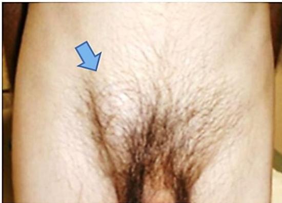
- Standing inspection: inspect both groins — side, shape, size (centimetres or fingerbreadths, or tape/rule) and inguinoscrotal extension; look for scars, visible peristalsis and a cough impulse.
- Landmarks: the first bony prominence is the pubic tubercle. Inguinal hernia lies between pubic tubercle and ASIS, tracking upwards to the deep inguinal ring; the superficial ring is just above and medial to the tubercle; a swelling lateral to the tubercle = femoral hernia (inguinal bulge superior/medial, femoral inferior/lateral at the femoral ring).
- Palpation (supine): expansile cough impulse, reducibility; “can you get above the mass?” — YES (fingers meet above) = pure scrotal swelling (hydrocele); NO (continues into the canal) = inguinoscrotal hernia.
- Reduction: reduce completely and state the direction — inguinal hernias reduce backwards/upwards into the canal; umbilical/paraumbilical hernias reduce backwards. If irreducible, do not attempt surface tests — describe the swelling like any lump.
- Contents tests: “gargling”/object reduction test — difficult to insert a fingertip/object = bowel; easy and doughy = mesentery. Percussion: tympanic = bowel, dull = omentum; auscultate for bowel sounds.
- Deep ring occlusion: reduce the hernia, occlude the deep ring 1.5–2.0 cm above the midinguinal point, ask the patient to stand and cough — controlled = indirect inguinal hernia; bulges medial to the thumb = direct inguinal hernia (Hesselbach’s triangle deficiency).
- Zieman 3-finger test: index over the deep ring → impulse on the fingertip = indirect; middle over the superficial ring/Hesselbach’s triangle → impulse on the pulp = direct; ring finger over the saphenous opening/femoral canal → femoral hernia.
- Scrotal skin invagination: little finger along the spermatic cord into the external ring — cough impulse on the tip = indirect, on the pulp = direct.
- Traction test: downward traction on the ipsilateral testis distinguishes a scrotal hernia from encysted hydrocele of the cord or undescended/ectopic testis.
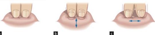
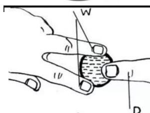
- Fluctuation: sides held with two fingers, centre pressed with another — fluid-filled swellings bulge outwards; repeat in two planes (horizontal and vertical); large cysts may show cross-fluctuation/fluid thrill.
- Transillumination: torch with an improvised folded-paper funnel — hernias usually do not transilluminate; a positive red glow = fluid (e.g. hydrocele).
- Genital/scrotal screen: examine the phallus (urethral stricture, hypospadias); run a finger along the ventral aspect to the median raphe and perineum — hernias track along natural pathways.
- Completion: thank and cover the patient, then summarise findings.
Classification, Causes & Differentials
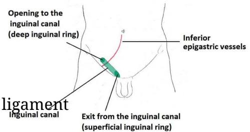
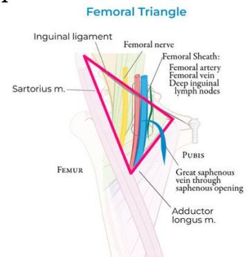
- Anatomy: Hesselbach’s triangle — medial = lateral border of rectus; lateral = inferior epigastric vessels; inferior = inguinal ligament; floor = transversalis fascia (site of direct inguinal hernia).
- Indirect inguinal: patent processus vaginalis; enters the deep ring lateral to the inferior epigastric vessels; controlled by deep ring occlusion; most common; may enter the scrotum.
- Direct inguinal: acquired weakness of transversalis fascia medial to the inferior epigastric vessels; rarely enters the scrotum; not controlled by deep ring occlusion.
- Femoral: through the femoral canal medial to the femoral vein; common in elderly females; mandatory urgent repair.
- Richter: only the anti-mesenteric bowel wall trapped → strangulates without mechanical bowel obstruction.
- Littre: sac containing a Meckel’s diverticulum.
- Maydl: “W”-shaped loop; the intermediate intra-abdominal loop strangulates while herniated loops appear viable.
- Amyand: sac containing the vermiform appendix (± acute appendicitis).
- Pantaloon: co-existing direct and indirect sacs straddling the inferior epigastric vessels (“saddlebag”).
- Incisional: fascial defect along a previous scar; risks = SSI, obesity, malnutrition, cough, steroids.
- Paraumbilical: defect in the linea alba near the umbilicus; complications = incarceration, obstruction, strangulation, skin breakdown.
- Predisposing factors: chronic cough, constipation, straining at micturition (BPH, urethral stricture, prostate cancer), abdominal mass/previous incision, smoking, tuberculosis.
- Differentials: femoral vs inguinal hernia (pubic tubercle rule), hydrocele, saphena varix, inguinal lymphadenopathy, femoral aneurysm, encysted hydrocele of the cord, undescended/ectopic testis.
Investigations & Management


- Children: herniotomy alone (high ligation of the sac).
- Adults: indirect → herniotomy + mesh; young = hernioplasty + herniotomy, elderly = mesh.
- Repair principles: open Lichtenstein tension-free mesh; laparoscopic TAPP/TEP for bilateral, recurrent or female hernias.
- Incisional hernia: sublay/retromuscular mesh repair with >5 cm overlap.
- Paraumbilical hernia: elective mesh repair (open sublay polypropylene mesh hernioplasty or laparoscopic IPOM; Mayo overlap if defect <1 cm).
- Strangulation/obstruction: emergency surgery (laparotomy); never taxis — do not reduce manually.
- Treat the precipitating cause: BPH, chronic cough, constipation, TB; stop smoking.
- Investigations: CBC, bleeding/clotting time, ESR, urine albumin/sugar, blood urea/sugar; blood grouping for irreducible/huge hernia; elderly post-void residual >100 mL is significant → assess for BPH.
- Complications:
- Incarcerated/irreducible: contents cannot be returned (adhesions or narrow neck) but vascularity and bowel lumen are preserved.
- Obstructed: intestinal lumen occluded within the sac → mechanical bowel obstruction (colicky pain, vomiting, distension, absolute constipation) with intact blood supply.
- Strangulated: blood supply compromised → severe continuous pain, exquisite local tenderness, erythema/warmth, tachycardia, fever/vomiting, leukocytosis — surgical emergency.
- Femoral hernia: rigid unyielding canal margins (lacunar ligament medial, inguinal ligament anterior) → highest strangulation rate (~40–45%).
Viva & Exam Spots
- Q: Classic anatomical landmark for the deep inguinal ring? → 1.5–2.0 cm above the midinguinal point (midpoint between the ASIS and pubic symphysis).
- Q: Why do paraumbilical hernias often present as an emergency? → Most present incarcerated early because of the narrow-necked defect in the linea alba.
- Q: Why must a strangulated hernia never be reduced? → Risk of returning non-viable ischaemic bowel to the abdomen — reduction en masse.
3. Peripheral Vascular Examination
Examination Checklist
Peripheral Vascular Examination Checklist
Exposure: consent, wash hands; expose both arms and both legs to mid-thigh; position supine.
General inspection: tar (nicotine) staining, xanthomata (hyperlipidaemia — typically familial hypercholesterolaemia), cyanosis/pallor, gangrene.
Upper limbs:
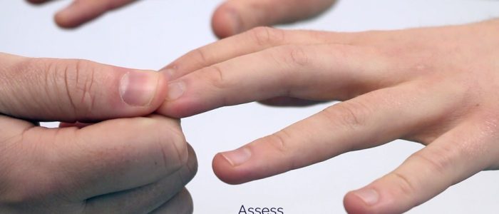
- Capillary refill time (CRT): 5 s pressure; normal refill <2 s.
- Radial pulse, then brachial pulse — support the arm (abducted, elbow partly flexed, forearm externally rotated); palpate medial to the biceps brachii tendon, lateral to the medial epicondyle (deeper than radial).
- Radio-radial delay: loss of synchrony between radial pulses → subclavian stenosis (cervical rib) or aortic dissection.
- BP in both arms: difference >20 mmHg abnormal (aortic dissection); wide pulse pressure >100 mmHg → aortic regurgitation/dissection.
Carotid: auscultate before palpating — a bruit suggests carotid stenosis; palpation may dislodge plaque → stroke; the bruit may be a radiating cardiac murmur (e.g. aortic stenosis). Palpate between larynx and anterior SCM border; assess character (e.g. slow-rising, thready) and volume; warn about reflex bradycardia/syncope. 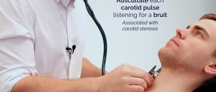
Abdomen: palpate aorta; auscultate bruits over abdominal aorta, iliac and femoral arteries.
Lower limbs:
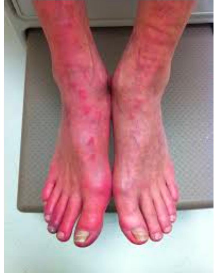
- Inspection: elevation pallor, dependent rubor (sunset foot), ischaemic rubour (dusky-red when dependent — loss of capillary tone in PVD), hair loss, brittle thickened nails, dry skin, muscle atrophy, gangrene.
- Ulcers: venous — large, shallow, irregular, mildly painful, medial ankle; arterial — small, well-defined, deep, very painful, at digits/pressure points.
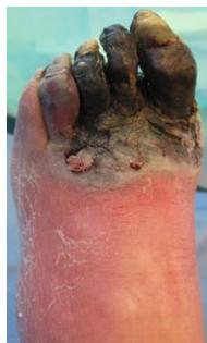
- Dry vs wet gangrene: dry — shrivelled, mummified, well demarcated (may be discharged if uninfected); wet — oedematous, putrefied, discoloured, spreading (admit, urgent debridement).
- Paralysis = critical ischaemia (ask the patient to wiggle the toes).
- Palpation: temperature (dorsum of hand, distal-to-proximal comparison); CRT <2 s normal, >3 s delayed.
- Pulses, proximal → distal:
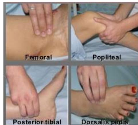
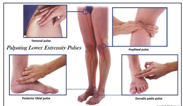
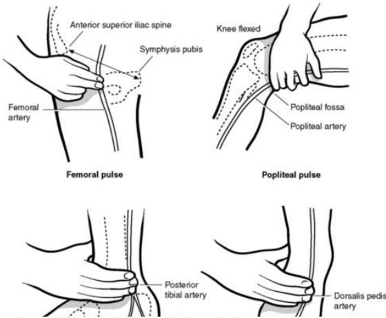
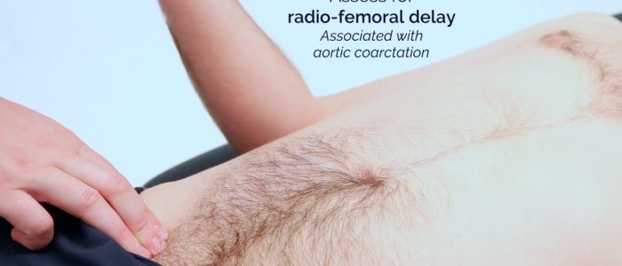
- Femoral: mid-inguinal point (halfway ASIS–pubic symphysis); assess radio-femoral delay; auscultate for a femoral bruit over the femoral canal (most accessible femoral artery point) → femoral/iliac stenosis; also listen over the popliteal artery.
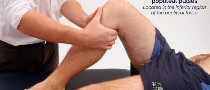
- Popliteal: knee flexed 30°–45°; curl fingers/bimanual thumbs deep into the fossa against the tibial plateau.
- Posterior tibial: 2 cm below/behind medial malleolus.
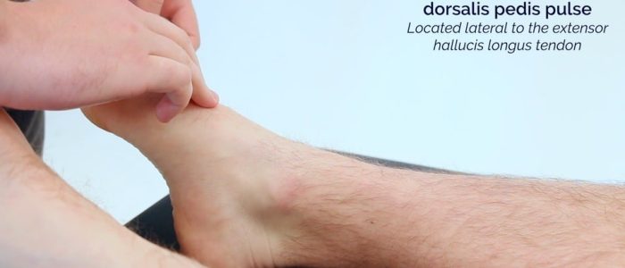
- Dorsalis pedis: lateral to extensor hallucis longus, over 2nd/3rd cuneiform bones; absent in ~10–15% of normal people (congenital variant) — compare sides; use a hand-held Doppler before calling it absent.
- Femoral: mid-inguinal point (halfway ASIS–pubic symphysis); assess radio-femoral delay; auscultate for a femoral bruit over the femoral canal (most accessible femoral artery point) → femoral/iliac stenosis; also listen over the popliteal artery.
- Sensation: chronic neuropathy → glove-and-stocking loss, compare sides, distal → proximal; acute critical ischaemia → rapid paraesthesia.
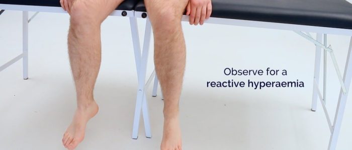
Buerger’s test: elevate supine legs 45°–60° for 1–2 min → pallor. Protocol: elevate in steps of 10° every 10 s from 10° to 45°, watching the plantar surface; normal pallor appears only at about 90°, pallor at 20°–30° = severe/critical ischaemia (Buerger’s angle of circulatory sufficiency; normal >90°). Then sit up and dangle the leg → reactive hyperaemia/dependent rubor (red flushed foot that later settles). 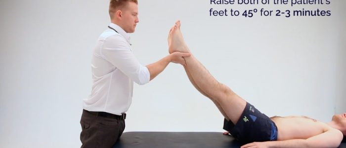
Completion: thank, wash hands, document; check ABPI and hand-held Doppler.
Interpretation & Management
Interpretation, Differentials & Management
Fontaine stages: I (asymptomatic), IIa/IIb (claudication >200 m / <200 m), III (rest pain), IV (ulceration/gangrene = critical limb ischaemia).
ABPI = highest ankle systolic BP (DP or PT) ÷ highest brachial systolic BP.
-
1.30: incompressible, calcified vessels (Mönckeberg sclerosis in DM/CKD; use Toe-Brachial Index).
- 0.90–1.30: normal; 0.70–0.89: mild arterial disease; 0.40–0.69: moderate (claudication); <0.40: severe (critical limb ischaemia; rest pain, necrosis).
- Doppler: normal arterial trace is triphasic; damped/monophasic = stenosis/occlusion.
Acute limb ischaemia — 6 Ps: Pain (sudden, severe), Pallor (chalk-white), Pulselessness, Paresthesia (early nerve ischaemia), Paralysis (irreversible muscle infarction), Perishing Cold (poikilothermia). Action: IV heparin 5000 IU bolus + urgent imaging / Fogarty embolectomy <6 h. 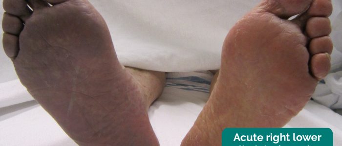
PVD risk factors: smoking (also drives Buerger’s); diabetes and hyperlipidaemia (absent in Buerger’s); CKD (Mönckeberg calcification).
Buerger’s disease (thromboangiitis obliterans): inflammatory occlusive disease of distal small/medium arteries and veins in young male smokers (<45), sparing the proximal vessels, with no diabetes or hyperlipidaemia; presents with distal ischaemia/ulcers and migratory thrombophlebitis.
Ulcer Examination
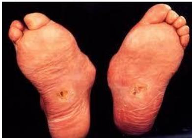
Definition: spontaneous break in epithelial continuity (not direct injury) — exclude direct trauma except repetitive injury.
- 4 S’s + E & F: Site, Size, Shape, Surroundings/Surface + Edge + Floor.
- Floor vs base: the floor is seen, the base is felt — describe floor colour and base consistency separately (a classic mark-loser).
- Edges: sloping — healthy healing/venous ulcer (pale/pink sloping edge = growing epithelium); punched-out — arterial/neuropathic; undermined — TB, syphilis, pressure (sacral/decubitus) ulcers; rolled/beaded — BCC (pearly-white telangiectatic border; locally invasive but almost never metastasises; sun-exposed face/nose, spreading from mouth angle towards ear); everted/heaped-up — SCC/Marjolin’s.
- Floor/base: granulation (red, vascular), slough (yellow/grey “cotton-wool” fibrin), necrosis/eschar (black, dry/wet).
- Discharge: serous (venous), purulent (infected), blood-stained (malignancy).
- Surrounding tissue: lipodermatosclerosis, haemosiderin, venous eczema, shiny hairless skin (ischaemia), cellulitis.
- Vascular & neurologic exam: distal pulses, ABPI, 10 g monofilament, regional lymph nodes.
Ulcer types:
- Venous stasis: gaiter region above medial malleolus; sloping edge, red granulation, haemosiderin/lipodermatosclerosis; moderate exudate, aching relieved by elevation; ABPI >0.8; 4-layer compression + endovenous ablation.
- Arterial (ischaemic): toes, heel, pressure bony points; punched-out, pale/black necrotic base, shiny hairless skin; severe nocturnal pain relieved by dependency; ABPI <0.8 (<0.5 = critical); compression CONTRAINDICATED; revascularisation.
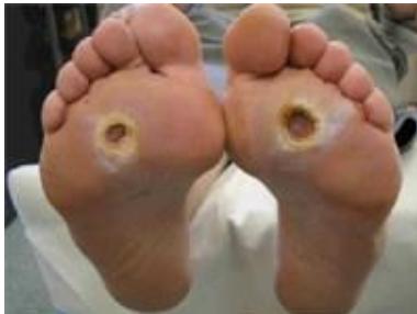
- Neuropathic (diabetic): plantar surface, 1st/5th metatarsal heads; punched-out, surrounded by hyperkeratotic callus, warm foot; variable exudate, painless (loss of protective sensation); offloading (total contact casting) + debridement + glycaemic control.
- Marjolin ulcer: pre-existing burn scar/chronic osteomyelitis sinus; everted, rolled border, vegetating cauliflower floor, indurated base; bloody, foul odour, progressive worsening pain; aggressive SCC — wide excision (≥2 cm) + SLNB.
Venous Disease: Varicose Veins & DVT
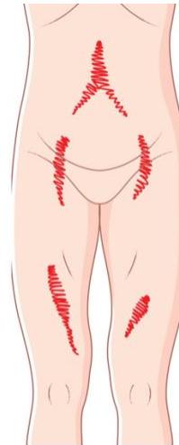
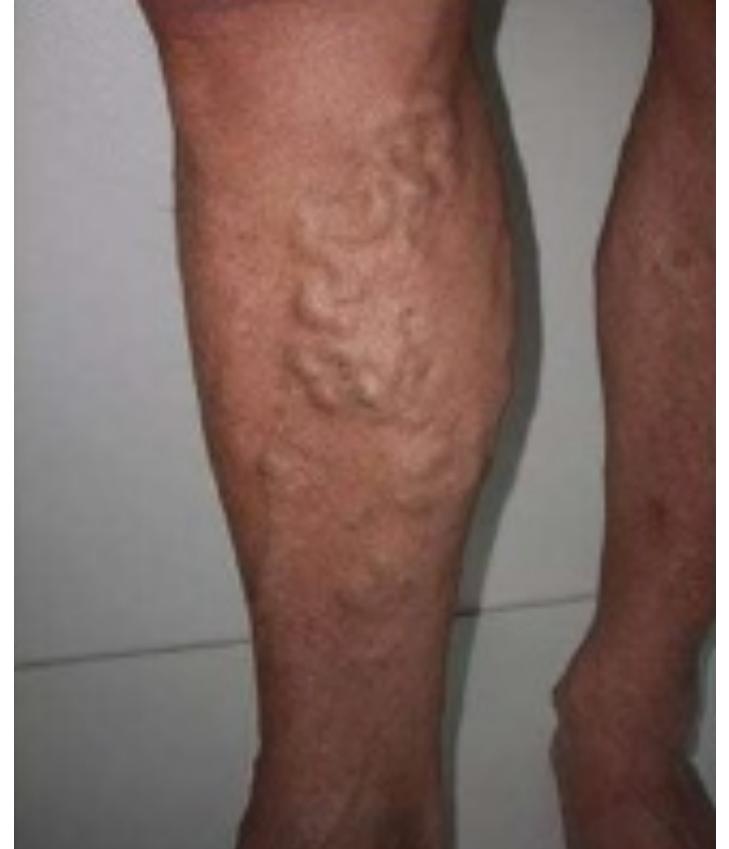

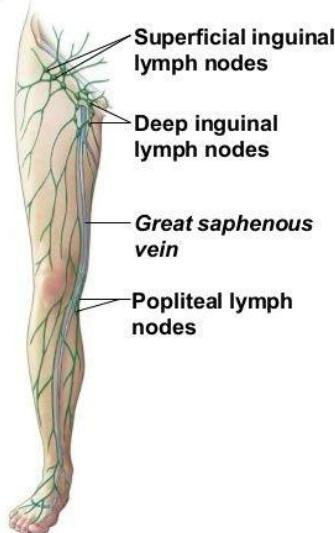
- Inspection: great/small saphenous varicosities, ankle flare, pigmentation, lipodermatosclerosis, medial gaiter venous ulcer. Palpation: pitting oedema (10–15 s medial tibial shaft), fascial defects, thrills/coughs, skin thickening/redness, groin lymph nodes/cough impulse, abdominal masses (pelvic venous obstruction).
- CEAP: C0 none; C1 telangiectasia <3 mm; C2 varicose veins ≥3 mm; C3 oedema; C4a pigmentation/eczema; C4b lipodermatosclerosis/atrophie blanche; C5 healed ulcer; C6 active venous ulcer.
- Trendelenburg test: elevate leg; tourniquet below SFJ; stand. Slow filling from below + rapid engorgement on release = SFJ incompetence; rapid filling despite tourniquet = perforator incompetence.
- Perthes test: tourniquet below knee; walk 5 min. Collapsed veins = patent deep veins; engorged + severe pain = DVT → NEVER STRIP VEINS!
- Schwartz tapping test: tap lower varicosity while palpating the SFJ; proximal impulse confirms an incompetent venous column.
- 4 history questions: prolonged standing; prior DVT/phlebitis; venous symptoms (heaviness, nocturnal calf cramps, evening oedema); multiparity/OCP/HRT. 2 investigations: venous duplex ultrasound (gold standard); MRV/CT venography (pelvic obstruction). 4 complications: superficial thrombophlebitis; variceal haemorrhage; lipodermatosclerosis/eczema; active venous ulcer.
- DVT: unilateral calf swelling (>3 cm asymmetry 10 cm below the tibial tuberosity), erythema, calf tenderness; gold standard compression duplex US; treat with therapeutic anticoagulation (DOAC or LMWH). Homan’s sign: calf pain on passive dorsiflexion — supportive, not diagnostic.
Viva & Exam Spots
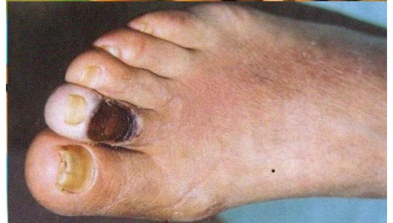
- Q: Non-lab tests for peripheral arterial disease? → ABPI via Doppler; arterial duplex ultrasound; CT angiography (CTA).
4. Diabetic Foot Examination
Examination Checklist
Diabetic Foot Examination Checklist
Exposure: supine, both lower limbs from mid-thigh down. complete the proximal vascular, neurological and musculoskeletal examination first and examine the ulcer last; inspect/palpate the normal side first for baseline; pitting oedema sign — 10–15 s thumb pressure over the medial subcutaneous tibial border (pitting vs non-pitting lymphoedema).
Pathophysiologic triad: neuropathy (sensory, motor, autonomic) + vasculopathy (PAD) + immunopathy/infection; remember microangiopathy alongside retino-, neuro- and nephropathy.
Gait & footwear: reduced speed/broad-based strategy; foot drop → high-stepping gait; turning difficult (patients look at their feet); stay close (falls risk). Footwear: asymmetrical sole wear, correct size, no foreign objects inside.


Inspection: posterior legs and between the toes and behind the heels for hidden ulcers; calluses (abnormal gait/poor footwear); claw toes, fungal nails; venous guttering; Charcot joint — effusion, distortion, erythema, loss of function. 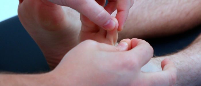
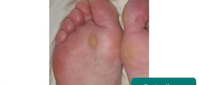
- Plantar trophic ulcer: punched-out circular ulcer over 1st MTP/big toe plantar surface surrounded by hyperkeratotic callus (Mal Perforans); pathogenesis = sensory neuropathy (unnoticed repetitive pressure) + peripheral arterial ischaemia + motor clawing; complications = deep space plantar abscess, osteomyelitis (probe-to-bone), Charcot foot, amputation.
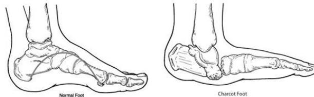
- Charcot neuroarthropathy (rocker-bottom foot): autonomic hyperaemia → osteoclast activation → microfractures → joint dislocations, pathological fractures, debilitating deformities (progressive destruction at weight-bearing joints; one of the most serious diabetic complications). Acute: red, hot, swollen, painless foot.
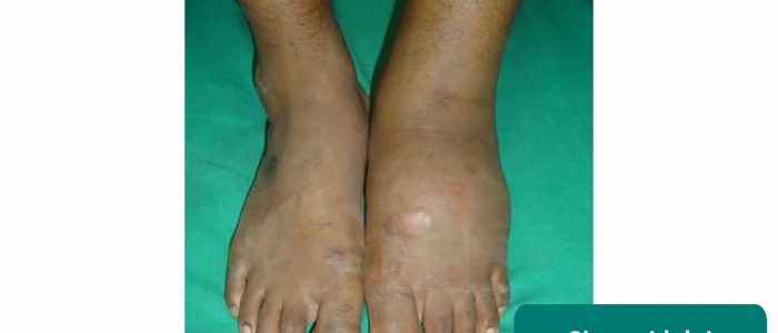
Temperature & pulses: temperature, CRT; palpate dorsalis pedis, posterior tibial, popliteal, femoral pulses; examine the ulcer last for depth/probe-to-bone and calf tenderness (6 palpation items: temperature, pulses, CRT, ulcer depth/probe, calf tenderness, monofilament).
Monofilament (10 g): Semmes-Weinstein; demonstrate on sternum/arm; 10 plantar sites — pulp of the hallux, pulp of the 3rd digit, MTP 1/3/5; bend until it buckles, hold 1–2 s; avoid calluses/scars. ≥2 sites missed = loss of protective sensation (LOPS). 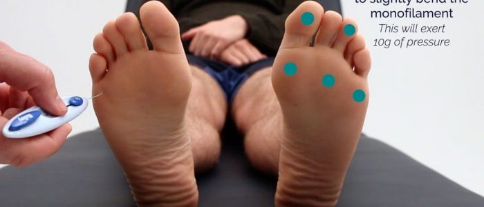
Vibration (dorsal columns): 128 Hz tuning fork — demonstrate on the sternum, then the big-toe IP joint; if impaired progress MTP → ankle → knee; ask when vibration starts/stops.  Proprioception (dorsal columns): hold the sides of the distal phalanx (not nail bed), demonstrate up/down, then 3–4 random movements with eyes closed; progress proximally if impaired.
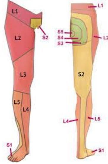
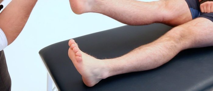
Ankle jerk (S1): leg supported (hip abducted, knee flexed, ankle dorsiflexed) or kneeling on a chair; tap the Achilles tendon → gastrocnemius contraction/plantarflexion; absent in advanced neuropathy.
Proprioception (dorsal columns): hold the sides of the distal phalanx (not nail bed), demonstrate up/down, then 3–4 random movements with eyes closed; progress proximally if impaired.
Ankle jerk (S1): leg supported (hip abducted, knee flexed, ankle dorsiflexed) or kneeling on a chair; tap the Achilles tendon → gastrocnemius contraction/plantarflexion; absent in advanced neuropathy.
Completion: capillary glucose, HbA1c (3-month control); formal neuro/arterial/venous exams; foot-care advice/podiatry. Patient education: supportive footwear; never barefoot; wash feet daily in warm (not hot) soapy water; emollient if dry; check shoes for foreign bodies; no hot-water bottles/radiators; never self-pare callosities or use corn plasters.
Interpretation & Management
Wagner Classification (Diabetic Foot Ulcers)
- 0: intact skin; pre-ulcerative lesion, bony deformity.
- 1: superficial (full skin thickness, no tendon/capsule/bone).
- 2: deep to tendon, joint capsule or bone, without abscess/osteomyelitis.
- 3: deep + cellulitis, deep fascial abscess or osteomyelitis.
- 4: localised gangrene of toe/forefoot.
- 5: extensive foot gangrene requiring major amputation.
Diabetic Foot Infection & Osteomyelitis
- Ranges from cellulitis → complex skin/soft-tissue infection → chronic osteomyelitis; diagnose clinically (cellulitis, purulent discharge, pain, tenderness, gangrene ± systemic toxicity).
- Culture tissue or bone biopsy — surface swabs only show colonising organisms.
- Imaging: plain X-ray (changes are late) and MRI (most sensitive); check kidney function before contrast.
- Management: multidisciplinary; first-line metronidazole (anaerobes) + piperacillin-tazobactam (Tazocin), add vancomycin for Gram-positive cover; surgical debridement/drainage; amputation for chronic osteomyelitis.
- Charcot management: immobilisation/offloading (total contact cast). Diabetic foot risk-score tool guides follow-up/podiatry.
Lower Limb Amputations

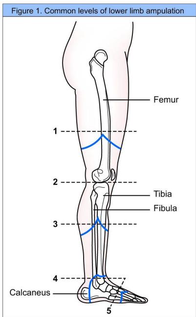
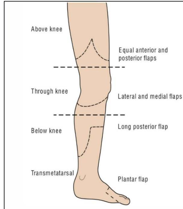
3 major indications: Dead (non-reconstructible critical ischaemia, unsalvageable dry/wet gangrene); Deadly (spreading gas gangrene/necrotising fasciitis with septic shock; osteosarcoma); Damned nuisance (intractable pain, severe flexion contractures, chronic refractory osteomyelitis).
- Above knee (AKA/transfemoral): equal anterior/posterior flaps; non-reconstructible ischaemia, extensive calf necrosis, knee flexion contracture >30°, non-ambulatory.
- Through knee (disarticulation): lateral/medial flaps; full femur lever arm, broad end-bearing stump for wheelchair stability.
- Below knee (BKA/transtibial): long posterior flap (Burgess); sural/posterior tibial vascularity; 80% independent prosthetic ambulation.
- Transmetatarsal/forefoot: plantar flap; thick, durable, weight-bearing glabrous skin; preserves heel pad and ankle joint.
- Ray amputation: removes a toe with its metatarsal (phalanges + metatarsal head) to encourage healing in localised forefoot gangrene/infection; preserve the 1st ray where possible (gait stability).
- Guillotine: open non-closure of all layers; for life-threatening sepsis/gas gangrene; delayed closure 3–5 days.
- Stump examination: stump length, scar position, healing, sinus, redundant tissue (“dog ears”), dog-ear necrosis; palpate temperature, tenderness, neuroma, bone spur, joint contractures; check adjacent hip/knee ROM (flexion contractures) and proximal femoral/popliteal pulses.
Skin grafts: SSG (Thiersch) — epidermis + superficial dermis; high take on suboptimal vascular beds; donor re-epithelialises (7–14 d); secondary contracture; disadvantages = colour mismatch, poor graft sensation, needs general anaesthesia, less cosmetic, trauma-prone. FTSG (Wolfe) — epidermis + entire dermis; superior cosmetic/colour match, minimal secondary contracture; needs a pristine vascular bed; donor closed primarily.
5. Neck Lump Examination
Examination Checklist
- Inspection — scars (previous surgery/radiotherapy), cachexia, hoarse voice (laryngeal compression), dyspnoea/stridor, behaviour (anxious/hyperactive vs low mood), temperature-inappropriate clothing, exophthalmos.
- Midline lump tests — swallow: thyroid masses/goitre and thyroglossal cysts rise; nodes barely move; an invasive thyroid malignancy may be tethered. Tongue protrusion: the thyroglossal cyst rises (persistent thyroglossal duct); thyroid masses and nodes do not.
- Triangle boundaries — anterior: inferior border of mandible, midline, anterior border of SCM; posterior: posterior border of SCM, trapezius, middle 1/3 of the clavicle.
- Lump assessment — site, size, shape, consistency, mobility (turn the head → tethering), fluctuance (hold the sides, press the centre → sides bulge), temperature, overlying skin (erythema, punctum), pulsatility, tenderness; transillumination; auscultate for a bruit.
- Lymph node interpretation — benign: <1 cm, smooth, rounded, non-tender, mobile; reactive: smooth, tender, mobile + infective symptoms; haematological malignancy: widespread rubbery nodes; metastatic: regional, hard/fixed/irregular/tethered.
- Systematic node exam — submental → submandibular → tonsillar → parotid → pre-auricular → post-auricular → superficial cervical → deep cervical → posterior cervical → occipital → supraclavicular; use the pads of the 2nd–4th fingers to press and roll (avoid the “piano-playing” fingertips); examine one side at a time over the anterior chain (avoid carotid compression).
Red Flags & Differential Diagnosis
-
Red flags for malignancy — hard/fixed mass; age >35; mucosal lesion in the head/neck; persistent hoarseness/dysphagia; trismus; unilateral ear pain (referred from the tongue base) → urgent ENT referral.
-
Regional differentials:
- Branchial cyst — remnant of the 2nd branchial cleft; smooth solitary fluctuant swelling in the anterior triangle; appears/swells in young adults after a URTI; conservative if small, else surgical excision.
- Cystic hygroma — congenital lymphatic malformation (prenatal/birth); typically left posterior triangle, transilluminates; benign but disfiguring → drainage + sclerosing agents (prevent reaccumulation).
- Carotid body tumour — pulsatile mass at the carotid bifurcation; moves side-to-side but not vertically; may have a bruit.
- Carotid artery aneurysm — pulsatile mass + audible bruit.
- Pharyngeal pouch (Zenker’s) — reducible midline/left neck mass; regurgitation, halitosis.
- Laryngocele — reducible tense mass enlarging on sneezing/nose-blowing (aerocele).
- Subclavian artery aneurysm — pulsatile mass + bruit above the clavicle.
- Dermoid cyst — formed along lines of embryological fusion, contains keratin; tethered to the epidermis (punctum/dimple); painless; children/young adults.
- Epidermoid cyst — punctum, tethered to the epidermis, contains keratin; painful/erythematous if infected.
- Parotid tail mass — pleomorphic adenoma (benign, most common) vs primary parotid malignancy (pain, rapid growth, facial-nerve palsy).
- Sialolithiasis/sialadenitis — firm duct calculus vs infected gland (pain/tenderness, pus); submandibular gland swellings are usually solitary (whereas lymphadenopathy is multiple).
- Thyroid nodule/goitre/thyroglossal cyst — see 6. Thyroid Examination.
-
Further — thyroid status + TFTs (midline lump), lymphoreticular exam (if lymphoma/leukaemia suspected), oral cavity/oropharynx/nasal cavity exam (mucosal primary), FBC/U&E/CRP, ultrasound ± CT/MRI, FNA for histology, early ENT referral if malignant suspicion.
6. Thyroid Examination
Thyroid Status Examination Checklist
- Prepare & expose (WIPER): Sit upright; expose mid-chest upwards (nipple line and above) to assess a retrosternal goitre (say why if neck-only).
- General inspection: Hyper- vs hypothyroid habitus: agitation vs apathy; hypothyroid eyebrow loss (outer 1/3), macroglossia, generalised myxoedema.
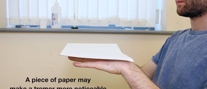
- Hands: thyroid acropachy (clubbing-like periosteal phalangeal bone overgrowth in Graves’), onycholysis (Plummer’s; painless nail detachment), palmar erythema; fine tremor on outstretched hands (paper reveals subtle tremor).
- Pulse: rate over 60 s (30 s ×2 / 15 s ×4; full 60 s if irregular). Bradycardia <60 — athletic, hypothyroidism, AV block, drugs, sick sinus; tachycardia >100 — hyperthyroidism, anxiety, SVT, hypovolaemia; AF (irregularly irregular) associates with hyperthyroidism.
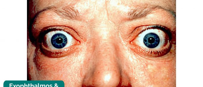
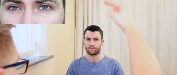
- Face & eyes: lid retraction — sclera visible above the corneal limbus (most common Graves’ sign; also toxic MNG); sympathetic overactivity → superior tarsal + levator palpebrae superioris contraction. Check exophthalmos, chemosis, “H” eye movements (restriction, diplopia, pain), lid lag, Hughes/other lid signs.
- Hands: thyroid acropachy (clubbing-like periosteal phalangeal bone overgrowth in Graves’), onycholysis (Plummer’s; painless nail detachment), palmar erythema; fine tremor on outstretched hands (paper reveals subtle tremor).
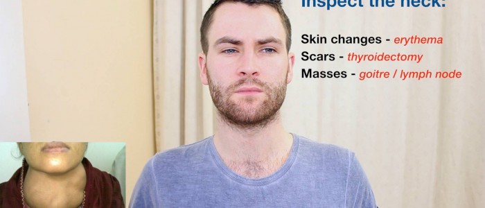
- Neck inspection: Midline anterior swelling, symmetry; scars (Kocher collar incision).
- Deglutition test: swallow water → thyroid mass moves upward (pretracheal fascia attached to cricoid/thyroid cartilages).
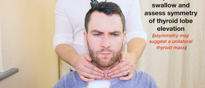
- Tongue protrusion: thyroglossal cyst moves upward (tethered to foramen cecum via hyoid); thyroid does NOT move.
- Deglutition test: swallow water → thyroid mass moves upward (pretracheal fascia attached to cricoid/thyroid cartilages).
- Palpation from behind: Flex neck forward (relaxes SCM), thumb pointing upwards, patient faces forwards (not lift head). Bimanually palpate lobes/isthmus as the patient sips water — 4 S’s + C: Site, Size, Shape, Surface + Consistency (soft = colloid, firm = adenoma/Hashimoto, stony-hard = carcinoma/Riedel), nodularity, tenderness (most exam patients firm). Press one lobe to make the opposite protrude, repeat. Never move both hands at once (nodes may move together). Palpable lower border → no retrosternal extension (no manubrial percussion); unreachable → retrosternal goitre. Berry’s sign: malignant encasement dampens the carotid pulse.
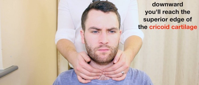
- Percussion: from the sternal notch downwards — manubrial dullness = retrosternal extension. Pemberton’s sign: both arms touching the ears for 60 s → facial plethora, cyanosis, stridor (thoracic inlet obstruction).
- Auscultation: bruit / machinery murmur over the superior thyroid poles (superior thyroid artery) = high vascularity/thyrotoxicosis (Graves’); carotid auscultation is required for the neck, not the thyroid.
- Trachea: 2 fingers on the head of the clavicle, middle finger on the tracheal rings, feel for deviation — before moving behind the patient.
- Lymph nodes: transverse (submental, submandibular, pre-/post-auricular, occipital) and longitudinal (anterior/posterior cervical ± supraclavicular); name groups, not levels.
- Special tests: Reflexes — biceps (thumb over the biceps brachii tendon at the antecubital fossa; tap your thumb), brachioradialis or knee jerk: brisk in hyperthyroidism, delayed relaxation/hyporeflexia in hypothyroidism. Pretibial myxoedema — diffuse mucinosis (excess glycosaminoglycans in dermis/subcutis) → waxy, discoloured, non-pitting shin induration; rare Graves’ complication. Proximal myopathy (MNG, Graves’): proximal wasting, difficulty standing — test by standing with arms crossed (falls risk).
- Completion: thank, wash hands, present findings; suggest TFTs, ECG if irregular pulse, neck ultrasound.
Eye Signs of Graves’ Disease
- Exophthalmos (proptosis): anterior globe displacement (Hertel exophthalmometer) from oedema + lymphocytic infiltration of orbital fat, connective tissue and extraocular muscles; inspect from front, side and above. Consequences: dryness, chemosis, conjunctivitis, severe corneal ulceration.
- Dalrymple’s sign: upper lid retraction (sclera above the superior limbus in primary gaze).
- von Graefe’s sign: lid lag (upper lid lags on downward gaze).
- Stellwag’s sign: infrequent/staring blink; Moebius’s sign: convergence weakness on near vision.
- Ophthalmoplegia: restricted “H” movements with diplopia and pain.
Goitre Types, Causes & Node Levels
- Physiology: ↑T3 raises metabolism (weight loss) and potentiates catecholamines → tachycardia, tremor, anxiety; ↓T3 → weight gain, low mood, constipation, poor memory, hyporeflexia.
- Subtypes: diffuse (hyperplasia), uninodular (single nodule — toxic/autonomously functioning or inactive), multinodular (toxic MNG).
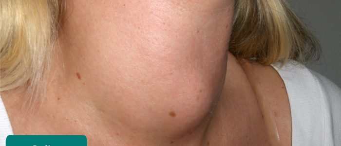
- Thyroglossal cyst: persistent thyroglossal duct; most common congenital neck abnormality.
- Subclinical disease: abnormal TSH, normal T3/T4.
- Symptom review: hyper — heat intolerance, sweating, diarrhoea, weight loss, anxiety/agitation, muscle pain, eye symptoms, palpitations, dyspnoea; hypo — depression/lethargy, cold intolerance, hair loss, periorbital puffiness; constitutional — weight loss, night sweats, fever, anorexia.
- Lymphoma nodes soft and separated; TB nodes matted (scrofula).
- Triangles: shrugging shows the trapezius; within the sternocleidomastoid = anterior triangle, posterior = posterior triangle.
| Level | Contents |
|---|---|
| I | Submental (Ia) and submandibular (Ib). |
| II | Upper internal jugular chain (skull base to hyoid). |
| III | Mid internal jugular chain (hyoid to cricoid cartilage). |
| IV | Lower internal jugular chain (cricoid to clavicle). |
| V | Posterior triangle (spinal accessory nerve chain). |
| VI | Anterior central compartment (pretracheal, paratracheal, Delphian node). |
| VII | Superior mediastinal nodes (below suprasternal notch). |
When describing nodes, name the groups rather than quoting numeric levels — more professional, and examiners will ask what each level contains.
Investigations & Management
- TFTs (TSH, T3, T4); ECG if irregular pulse (AF); neck ultrasound.
| Bethesda Category | Description | Malignancy Risk | Standard Management |
|---|---|---|---|
| I | Non-diagnostic / Unsatisfactory. | 1–4% | Repeat US-guided FNA. |
| II | Benign (Colloid, Hashimoto’s). | 0–3% | Clinical and ultrasound follow-up. |
| III | Atypia / FLUS. | 5–15% | Repeat FNA / molecular testing. |
| IV | Follicular Neoplasm / Suspicious. | 15–30% | Diagnostic hemithyroidectomy (assess capsular/vascular invasion). |
| V | Suspicious for Malignancy. | 60–75% | Near-total or total thyroidectomy. |
| VI | Malignant (Papillary, Medullary, Anaplastic). | 97–99% | Total thyroidectomy ± neck dissection. |
- RLN injury: unilateral — hoarseness, bovine cough; bilateral — stridor, adducted cords, acute airway obstruction → emergency re-intubation/tracheostomy.
- EBSLN injury: loss of high-pitched singing voice, vocal fatigue (cricothyroid paralysis).
- Post-op hypocalcaemia: parathyroid devascularisation/excision; at 24–48 h, perioral tingling, Chvostek/Trousseau signs. Rx 10 mL 10% IV calcium gluconate over 10 min + oral calcium (1–2 g/d) and calcitriol (0.25–0.5 µg/d).
- Tension neck haematoma: bleeding under strap muscles → venous congestion, laryngeal oedema. Remove clips/sutures at the bedside, evacuate the clot with a finger; transfer to OR.
- Thyroid storm: severe tachycardia, fever >38.5°C, agitation. Rx IV fluids, propranolol, PTU, Lugol’s iodine, IV hydrocortisone.
Viva & Exam Spots
- Q: 4 DDx of an anterior neck mass? → MNG; solitary nodule (adenoma/colloid); carcinoma; thyroglossal cyst; Hashimoto’s.
7. Breast Examination
Breast Examination Checklist
- Introduction, chaperone & consent: WIPER; always offer a chaperone; patient-friendly explanation (“inspect, then palpate the breast tissue, then the glands of the neck and armpit”); privacy/dignity and a blanket. Sit upright, expose neck to waist.
- Inspection — 4 positions:
- Arms by sides, hands on thighs: relaxes pectorals; symmetry, contour, skin dimpling, erythema, peau d’orange.
- Hands pressed into hips: contracts pectoralis major; tethered mass moves, puckering accentuated (deep carcinoma tethering).
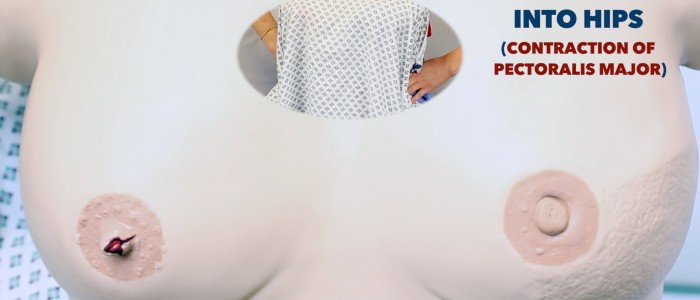
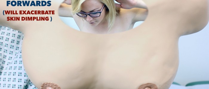
- Arms above head, leaning forward: stretches Cooper’s ligaments; exaggerates asymmetry, dimpling, skin tethering and nipple inversion; assesses pendulous breasts for chest-wall fixation.
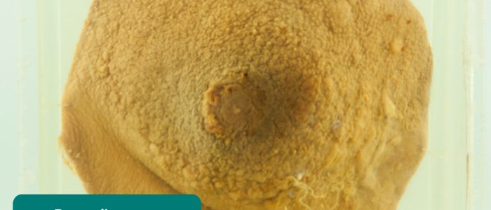
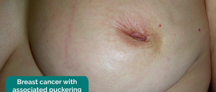
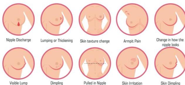
- Skin signs: puckering = suspensory (Cooper’s) ligament invasion drawing skin inwards; peau d’orange = cutaneous lymphatic oedema — tumour emboli obstruct subdermal lymphatics, swollen skin tethering to hair follicles and sweat glands (inflammatory breast cancer); scaling + erythema + pruritus of the nipple/areola = Paget’s disease.
- Nipple inversion: congenital/weight loss (normal) vs pathological — breast cancer, breast abscess, mammary duct ectasia, mastitis.
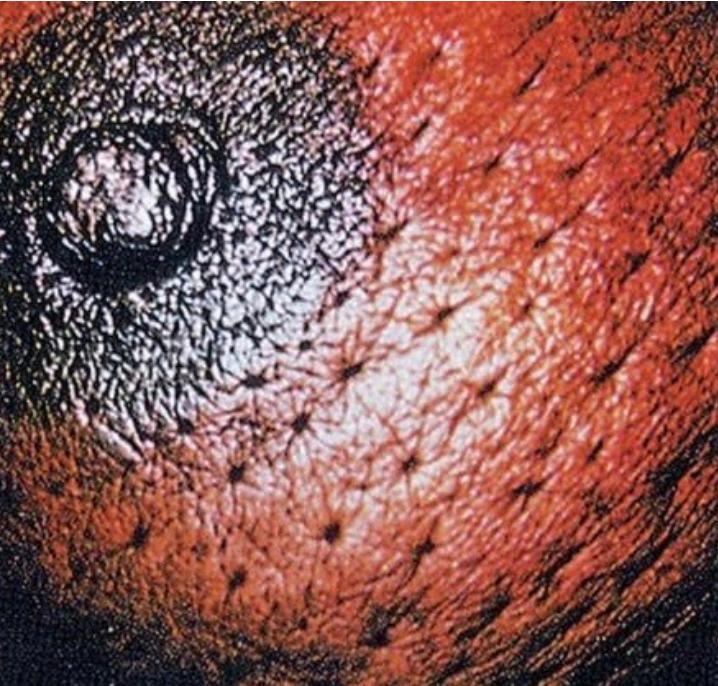
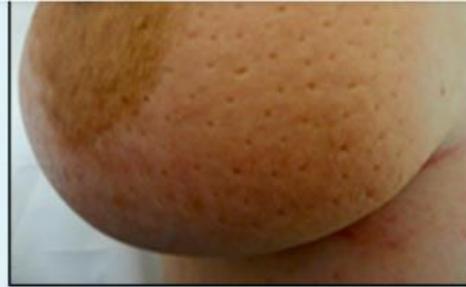
- Palpation (supine at 45°):
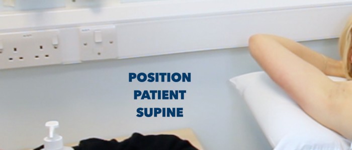
- Ipsilateral hand behind head flattens breast against ribs.
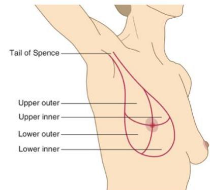
- Palmar surfaces of middle 3 fingers, rotary circular motion (clock/spiral/quadrant technique) over all 4 quadrants (UOQ, UIQ, LOQ, LIQ) + axillary tail of Spence — most cancers arise in the upper outer quadrant. Examine the other (normal) breast first, then the affected side, starting furthest from the lump, painful area last; note discomfort, reassure and be compassionate.
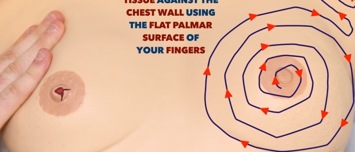
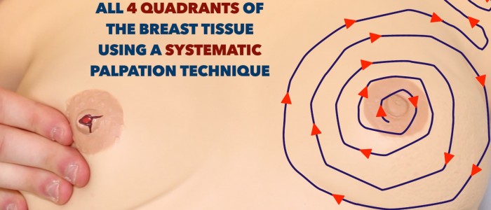
- Lump: pinch skin for tethering, check mobility against contracted pectoralis, test fluctuance; consistency (smooth/firm/stony/rubbery); ask about cyclical change with the menstrual cycle.
- Nipple-areolar complex: elevate the breast to inspect the retro-areolar region; gentle subareolar compression for discharge.
- Describe by clock-face: position and distance from nipple (e.g. “a firm, craggy 3 × 3 cm mass at 02:00”).
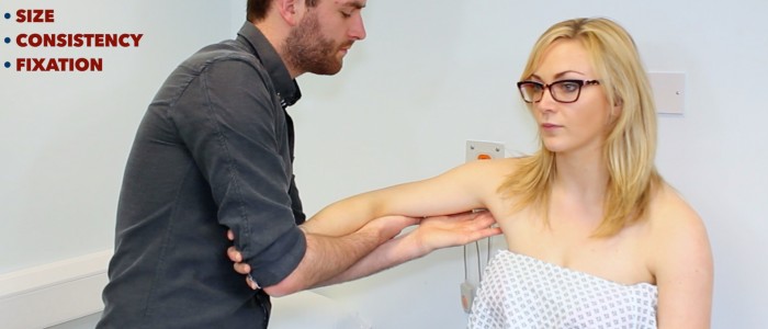
- Lymph nodes (sitting) & handshake maneuver: support the forearm with a “handshake maneuver” (completely relaxes pectoralis major and latissimus dorsi), then examine the 5 axillary groups:
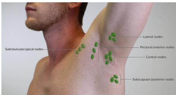
- Anterior (pectoral) — lateral pectoralis border; Posterior (subscapular) — anterior latissimus dorsi border; Lateral (humeral) — upper medial humerus; Central — centre of axilla against ribs; Apical (subclavicular) — apex behind clavicle.
- Also palpate supraclavicular fossa, cervical, infraclavicular and parasternal nodes.
- Completion: summarise findings and state the need for triple assessment.
Interpretation, Assessment & Management
- Nipple discharge: watery/blood-stained → ductal papilloma, carcinoma or DCIS (key to exclude); yellow/green → fibrocystic disease/duct ectasia; white/milky → pregnancy/lactation or galactorrhoea (nonpuerperal — prolactinoma); purulent/offensive → mastitis/central abscess.
- Triple assessment: 1. Clinical — history + bilateral breast and regional nodal exam; 2. Imaging — mammography (bilateral CC/MLO views) first-line ≥35–40 y; ultrasound first-line <35 y (dense breasts) and differentiates cystic vs solid; 3. Histopathology — core needle biopsy (CNB) gold standard, distinguishes in-situ from invasive carcinoma (grade and lymphovascular invasion), tests ER, PR, HER2; FNAC cytology only, cannot assess invasiveness.
- Sentinel lymph node biopsy: sentinel node drains the whole breast via the axilla; inject blue dye or radioactive isotope, identify with a gamma probe (± frozen section); used when axillary imaging is negative but cancer is confirmed; reduces lymphoedema.
- Systemic therapy: chemotherapy if receptors are hormone-unresponsive; targeted therapy for HER2 (antibody + chemotherapy); hormonal therapy (Tamoxifen) for hormone-receptor-positive disease. Nuance: adrenal androgens convert peripherally to oestrogens (relevant in obesity).
- TNM (breast) essentials:
| Component | Categories |
|---|---|
| T | T1 <2 cm; T2 2–5 cm; T3 >5 cm; T4 any size with skin/chest-wall invasion (oedema, ulceration, peau d’orange). |
| N | N0 non-palpable; N1 palpable & mobile; N2 fixed; N3 supra-/infraclavicular spread. |
| M | M0 none; M1 distant metastasis. |
- Risk factors: ≥3 years oral-contraceptive/HRT use; early menarche & late menopause; nulliparity; obesity/saturated-fat diet; family history of breast/ovarian/genitourinary cancers in first- and second-degree relatives; BRCA1/2; prior cancer; first child after 40. Protective: pregnancy and lactation.
- Metastasis red flags: back pain/bone mets; dyspnoea/haemoptysis (lung); RUQ pain/jaundice (liver); seizures/headache/LOC (brain).
- Tethering & fixation: Cooper’s ligament infiltration → dimpling when the lump moves or the arm is raised; completely immobile (superfixed) from the outset, no movement in any direction even with muscle relaxed → deep chest-wall involvement.
- 45° rationale: sitting makes the breast hang down, supine makes it fall laterally; semi-recumbent balances both for palpation and axillary access.
- Puerperal mastitis: lactating woman, tender wedge-shaped erythematous breast sector + fever; Staphylococcus aureus; Rx continue frequent lactation/milk expression, oral flucloxacillin (or clindamycin), analgesia.
- Breast abscess: fluctuant, warm, erythematous collection; Rx US-guided needle aspiration (first-line) or surgical I&D + antibiotics; continue nursing from the unaffected breast.
- Paget’s disease of the nipple: unilateral, persistent erythematous, scaly or ulcerated nipple-areolar lesion mimicking eczema; intraepidermal invasion of malignant ductal cells from underlying DCIS/invasive carcinoma; full-thickness nipple punch/core biopsy mandatory.
- Axillary matted lymphadenopathy pearl: diffuse, rubbery, matted axillary or cervical nodes in a young patient without a breast mass → tuberculous lymphadenitis (scrofula/cold abscess); diagnose via AFB stain, GeneXpert PCR and mycobacterial culture.
- BI-RADS:
| BI-RADS Category | Malignancy Risk | Clinical Management |
|---|---|---|
| 0 | Incomplete. | Additional imaging (views, US, MRI). |
| 1 | Negative (0%). | Routine screening. |
| 2 | Benign (0%) (simple cyst, calcified fibroadenoma). | Routine screening. |
| 3 | Probably benign (<2%). | Short-interval follow-up (repeat 6 months). |
| 4 | Suspicious (2–95%). | Tissue core biopsy recommended. |
| 5 | Highly suggestive (>95%). | Biopsy and surgical oncologic planning. |
| 6 | Biopsy-proven malignancy. | Definitive surgical resection / neoadjuvant therapy. |


Viva & Exam Spots
- Station 23 (SURG 1 Females / Batch 15): Q: Diagnosis? → right breast carcinoma (invasive) — asymmetric enlargement, peau d’orange, nipple retraction. Q: Confirm? → US-guided CNB (histology, grade, ER/PR/HER2/Ki-67).
8. Laparoscopic & Surgical Instruments
Instrument Spotter Atlas
Laparoscopic instruments
Laparoscopic Instruments
| Instrument | Image… | Description / Key Features / Notes | Uses, Indications, Contraindications & Complications |
|---|---|---|---|
| Laparoscope (Telescope) |    | 3 structural variables: 6–18 rod-lens systems; angles 0–120° (0° commonly used); diameters 1.5–15 mm; 3D imaging available. | Uses: visualization of the peritoneal cavity after gas insufflation in minimally invasive surgery. |
| Veress Needle (spring-loaded; closed-technique needle) |   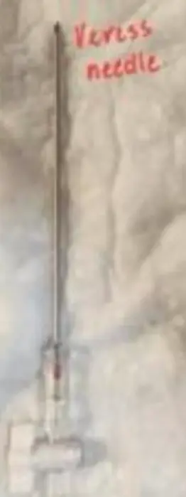 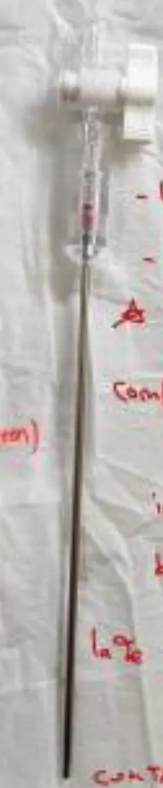   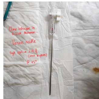 | Sharp outer cannula + inner spring-loaded blunt stylet. Closed technique to access the abdomen. Safe spot: LUQ (left hypochondrium, next to the spleen) at 45° — Palmer’s point (3 cm below left costal margin in MCL); safe in patients with midline scars. Safety checks: saline drop test, aspiration test, initial pressure < 8–10 mmHg. Abdominal access: examine the abdomen for scars (expect adhesions — avoid the area), organomegaly and intra-abdominal masses. Safe spots: lower fold of umbilicus; lateral border of rectus (any of the 4 quadrants). | Uses: closed access technique to create pneumoperitoneum (obese patients; patients with no previous surgery). Contraindications: active bleeding. Complications: injury at port site (“vessel” injury); intra-abdominal injury — bowel, spleen, stomach; late — infection, incisional hernia. |
| Hasson (Open) Cannula | — | Cut-down at the umbilicus under direct vision; incise every layer (fascia and peritoneum); blunt cannula secured with stay sutures. | Uses: open technique for abdominal access — the safest technique; ideal for patients who had previous surgeries. |
| Trocars & Cannulas (laparoscopic ports; optical entry / Hasson balloon / bladeless-dilating trocars) |   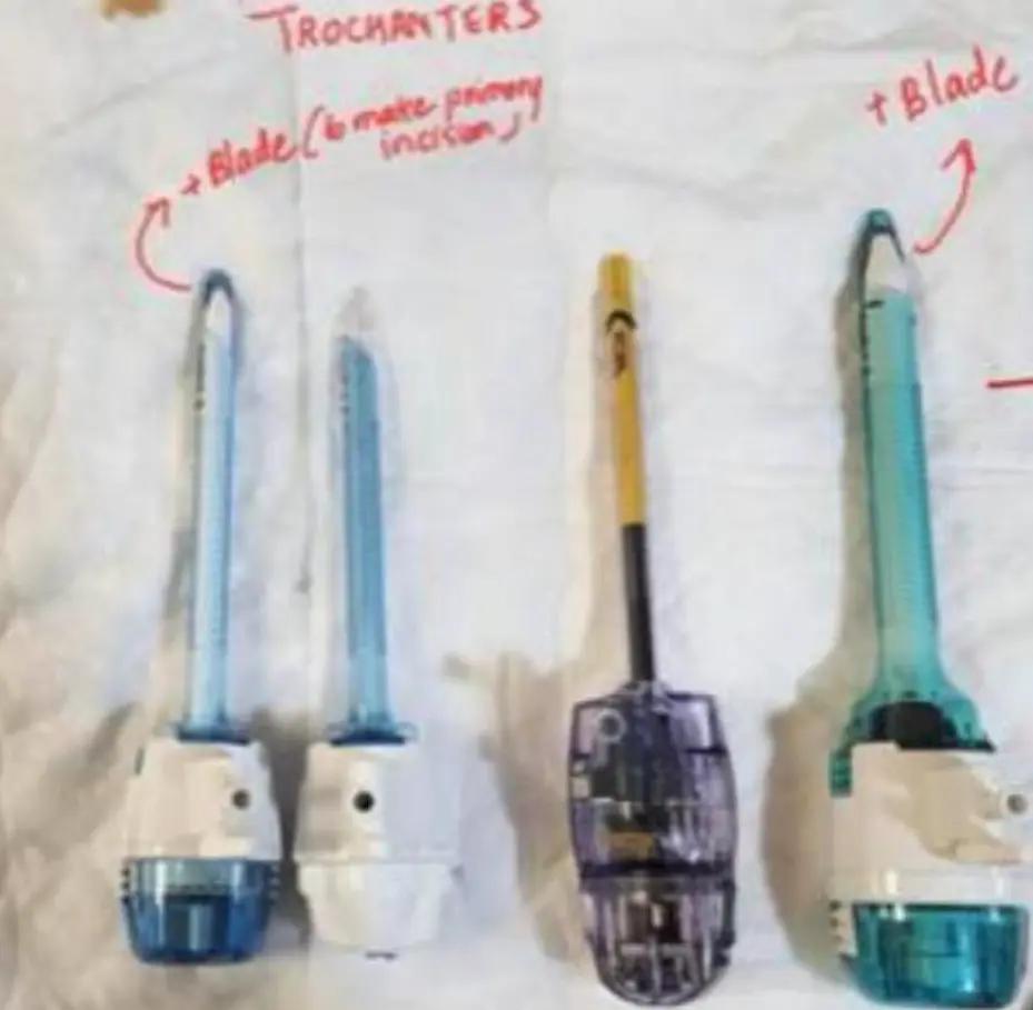 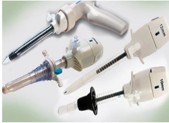 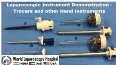 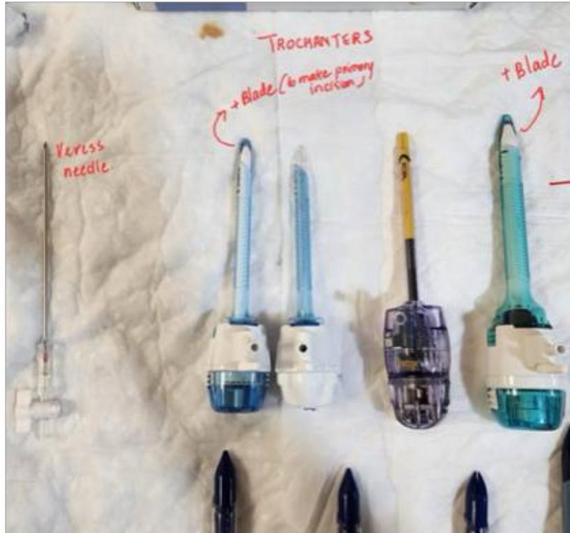 | Made of a blade with a shaft and body; the pointed tip makes the initial incision in the abdominal wall; diameters range 2–30 mm; most common trocar is 5 mm & 10 mm; two parts — obturator + guard; valves to connect the gas; remove the obturator then introduce the laparoscope; reusable. Port position: primary port — umbilicus (central location, hides scars, weak area due to absence of all layers); secondary ports — under direct vision, skin incision, drilling motion to insert. Basic Diamond Concept: 3 ports typical; laparoscope through the umbilical port (10 mm); clip applicator 10 mm port is essential; additional 3–4 ports (5 or 10 mm) depending on the procedure; diagnostic purposes = 2 ports, add a third to make it therapeutic. For gallbladder — a smaller port won’t retrieve it. | Uses: instrument access for laparoscopy. Complications: insertion-related — major vascular injury, GI injury, bladder injury, CO₂ embolism, abdominal wall haemorrhage; post-insertional — GI perforations, laceration & bleeding from solid organs, abdominal wall hernia. |
| Laparoscopic Graspers & Dissectors (Mangeshikar grasping forceps; Kelly atraumatic; long fine dissectors) |    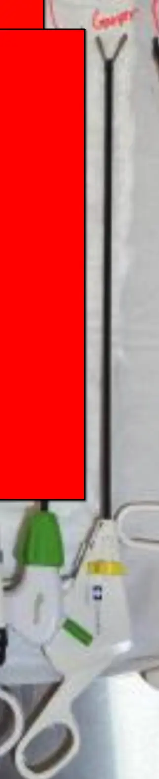 | Atraumatic; Kelly atraumatic; atraumatic with hollow jaws; Mangeshikar grasping forceps — serrated. General instruments: reusable three-piece design (disposables commonly used); sizes 2, 3, 3.5, 5, 10 mm; lengths 20, 30, 36, 43 cm; choice of handle styles; fully rotating 360° sheath; no hidden spaces that trap blood/tissue debris. | Uses: grasping and dissecting tissue in laparoscopic surgery. |
| Maryland Dissector (Maryland forceps) | 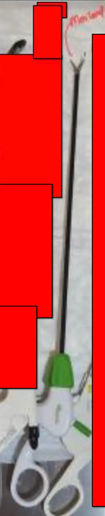  | Curved dissecting jaws (laparoscopic forceps). | Uses: dissection and grasping in laparoscopic surgery. |
| L-Hook (Laparoscopic Monopolar Electrosurgical Hook) (Bovie hook; hooks and spatulas with cautery) | 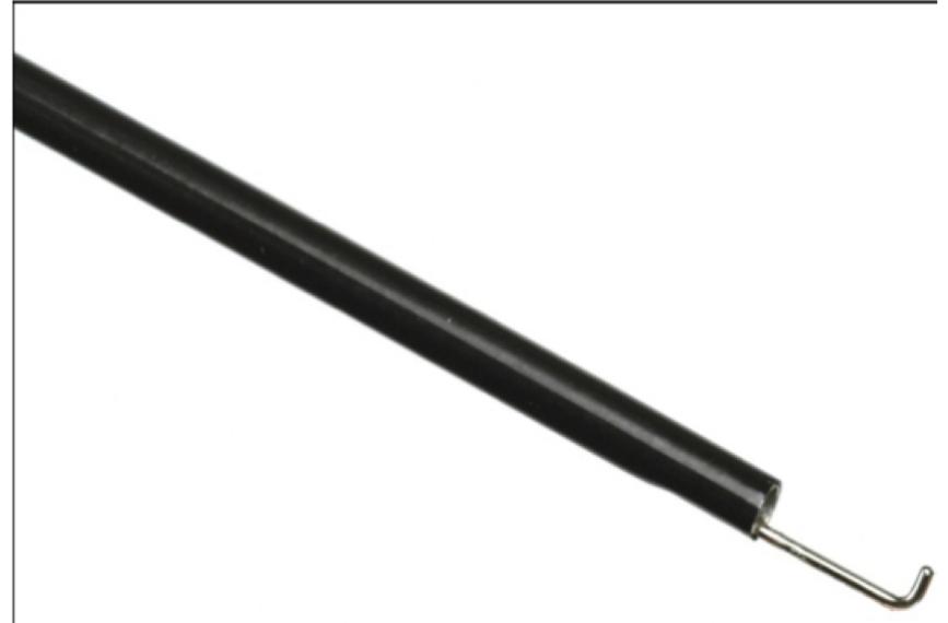 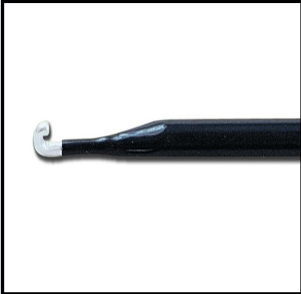   | Monopolar electrosurgical hook electrode; used in laparoscopic surgery — cholecystectomy, appendectomy, hernia. | Uses: electrosurgical cutting and coagulation; adhesiolysis (separate adhesions); blunt tissue mobilization; diathermy; traction to any organ; cauterize a small area of bleeding. Complications: do not use near the bowel — risk of diathermal (thermal) injury. |
| Harmonic Scalpel (ultrasonic scalpel; harmonic scalpel and ligature) |    | Laparoscopic ultrasonic devices (source images show the “Harmonic” jaw and shears); generates heat via ultrasound. | Uses: coagulation and cutting (hemostasis method 7 — ultrasound). |
| LigaSure Vessel-Sealing Forceps |  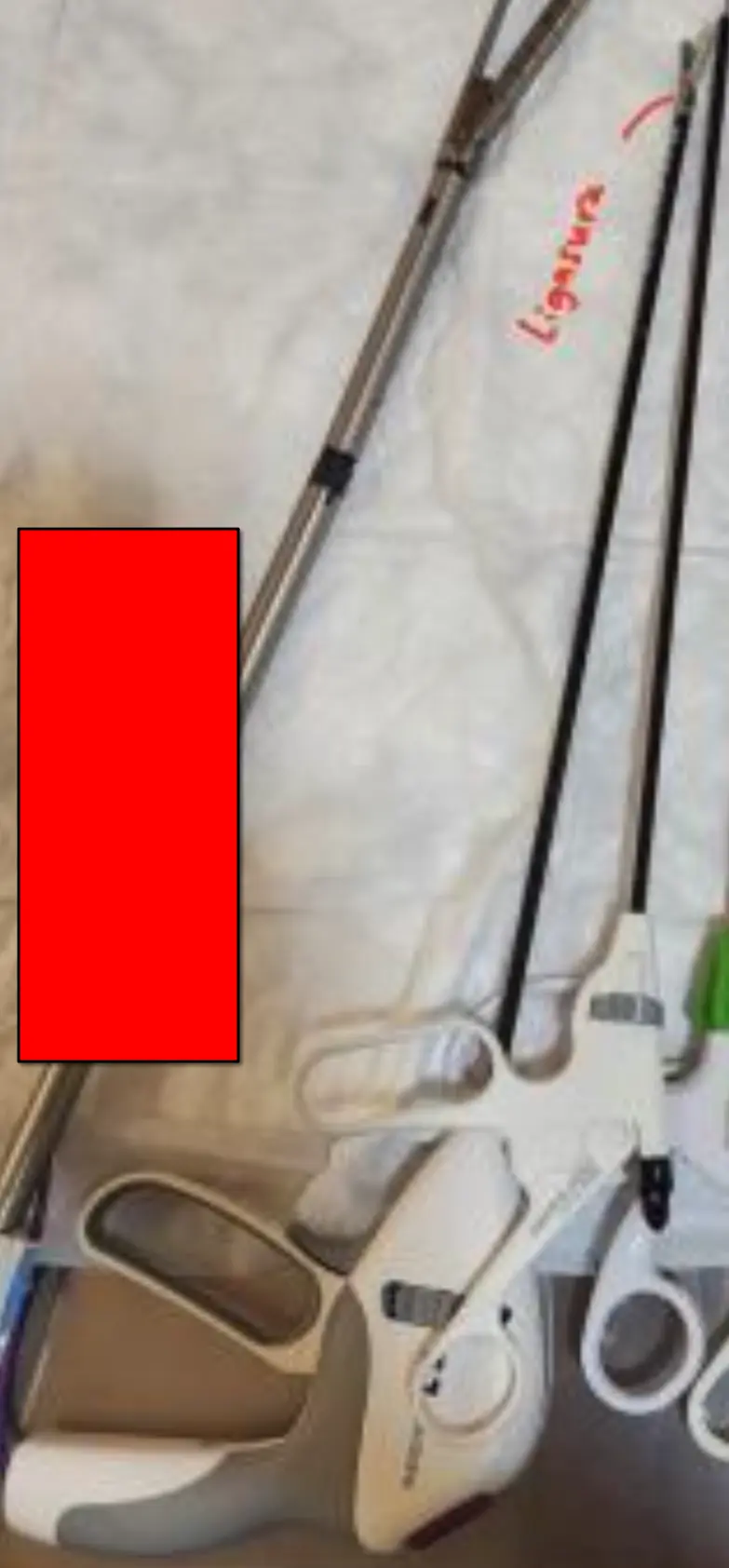 | Electrosurgical vessel-sealing forceps. | Uses: cautery-based vessel ligation/sealing. |
| Electrocautery / Electrosurgical (Diathermy) Pencil (unipolar & bipolar diathermy) |      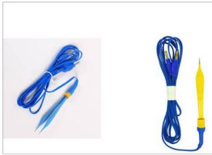 | High-frequency electric current; types: unipolar and bipolar. Unipolar current passes through the patient’s body (generator → patient → back to generator); “cutting diathermy” can be cutting or blunt. | Uses: electrosurgical cutting and coagulation; most commonly used method of hemostasis (thermal coagulation). |
| Laparoscopic Scissors (hook / Metzenbaum / straight) |     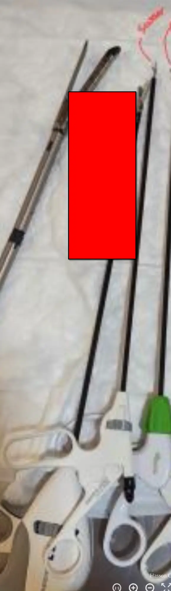 | Hook scissors — single-action jaws; Metzenbaum — curved; straight scissors — only one moving jaw, giving controlled depth of cutting. | Uses: mechanical dissection and cutting in laparoscopic surgery. |
| Endocatch Bag (specimen retrieval bag) |   | — | Uses: bagging organs + specimen for retrieval. |
| Endo Clip Applier (laparoscopic titanium clip applier) | 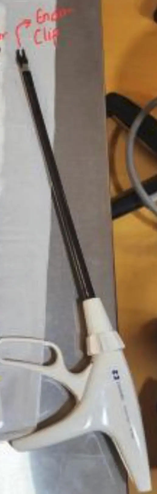 | Applies surgical clips. | Uses: clipping/occlusion (vessels, ducts); a 10 mm port clip applicator is essential. |
| Hem-o-lok Clips (Polymer) | — | Polymer locking clips. | Uses: vessel/duct occlusion during laparoscopy. |
| Endo-GIA Linear Cutting Stapler | 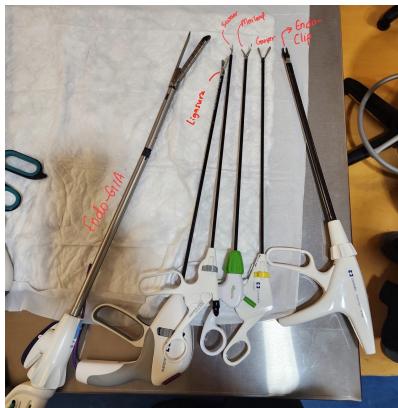 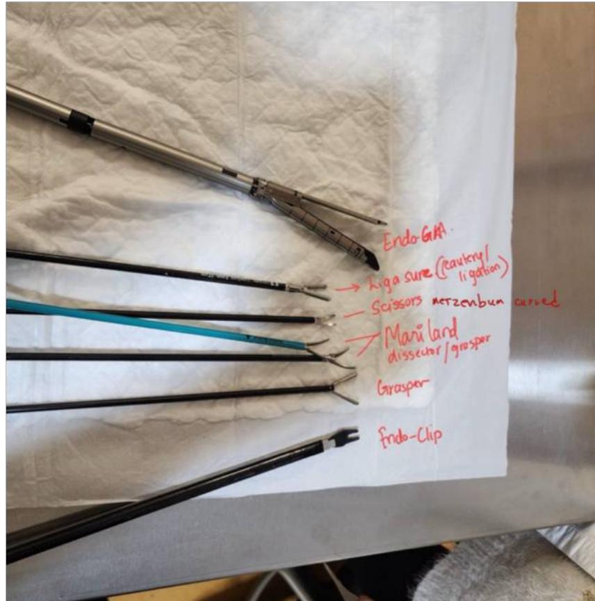   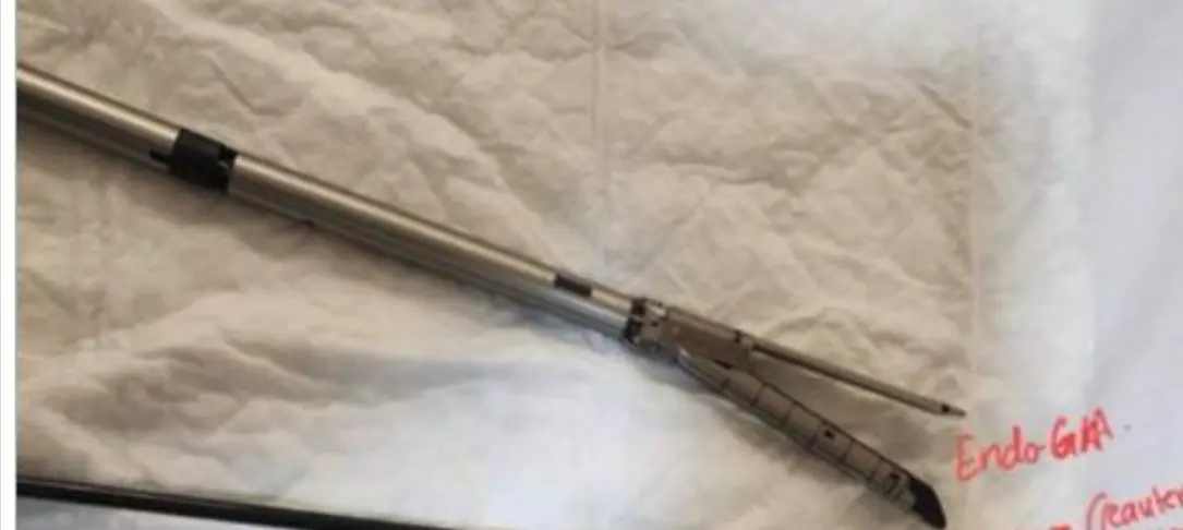 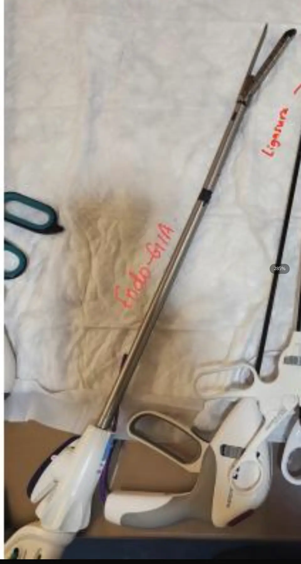 | Deploys 6 staggered rows of titanium staples and divides tissue simultaneously. | Uses: bowel and vascular pedicle transection (“to cut around safely” in laparoscopy). |
| Laparoscopic Suction-Irrigation Apparatus (suction-irrigation cannula) | 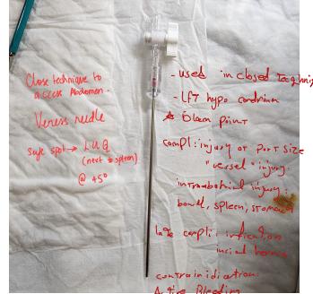 | — | Uses: suction and irrigation (continuous washing during surgery). |
| Reducers | — | — | Uses: negotiate smaller instruments through larger ports. |
Dissecting & Grasping Forceps
| Instrument | Image… | Description / Key Features / Notes | Uses, Indications, Contraindications & Complications |
|---|---|---|---|
| Plain Tissue Forceps |  | Non-toothed dissecting forceps. | Uses: grasping tissue; dressing application. |
| Toothed Tissue Forceps | (plate: Plain Tissue Forceps row) | Forceps with teeth. | Uses: grasping moderate to heavy tissue; used during wound closure. |
| Adson (Toothed) Forceps | 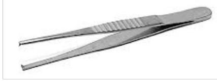 | Fine rat-toothed interlocking tips. | Uses: secure grip on skin and tough dermis without crushing. |
| DeBakey Forceps (DeBakey tissue forceps; vascular tissue forceps) |  | Atraumatic; elongated narrowed blunt tip; longitudinal micro-serrations. | Uses: grasps numerous tissue types; cardiac, vascular and GI procedures; handling blood vessels, bowel and delicate peritoneum; secure grip without tissue damage. |
| Russian Forceps | — | Broad, rounded, spoon-shaped serrated tips. | Uses: handling friable parenchymal tissues, dense fascia and bladder. |
| Ovum Forceps |  | — | Uses: 1. removal of placenta fragments; 2. holding gauze and cotton. |
Clamps & Hemostats
| Instrument | Image… | Description / Key Features / Notes | Uses, Indications, Contraindications & Complications |
|---|---|---|---|
| Mixter Forceps (right-angle forceps; Lahey’s forceps; obtuse clamp; ureter clamp) |  | 45-degree angle clamp with horizontal serrations; used to clamp, dissect and occlude tissue. | Uses: clamping, dissecting and occluding tissue; laparoscopic clipping (in open surgery we usually use this); placing a tie or vessel loop under and around a tubular structure (vessel, duct). |
| Mosquito (Halsted) Forceps | — | Delicate fine jaws with full-length serrations. | Uses: hemostasis of fine subdermal bleeding vessels. |
| Kelly / Crile Clamp (Kelly clamp; Crile clamp) | 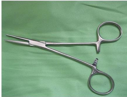 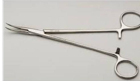 | Medium serrated jaws — Kelly: serrations on half the jaw; Crile: full-length serrations. | Uses: hemostasis of subcutaneous vessels; blunt tissue tunneling. |
| Allis Forceps (Allis tissue clamp) |  | Multiple interlocking fine teeth at the tip; very traumatic. | Uses: lifting, holding and retracting slippery dense tissue that is being removed; upper + lower flaps; tonsils; vaginal, breast and thyroid tissues; grasping bowel during a resection — only for the portion to be removed (very traumatic); most commonly used. |
| Babcock Forceps (Babcock tissue clamp) |  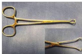 | Atraumatic, non-crushing forceps with a flared, rounded, hollow end and smooth, flattened (fenestrated) tips. | Uses: grasping and encircling delicate tubular structures — ureters, fallopian tubes, bowel, ovaries and appendix. |
| Kocher (Ochsner) Forceps |  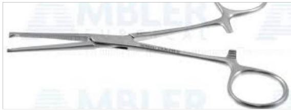 | Heavy transverse serrations with 1×2 teeth. “Very traumatic — use only with tissues to be removed (e.g., nephrectomy)“.  | Uses: grasping tough, fibrous, slippery tissues (muscle, fascia); strong traction on fascia, rectus sheath and bone. |
| Towel Clip |  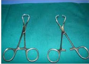 | Spring/interlocking clamp. | Uses: 1. holding towels in place when draping; 2. grasping tough tissue; 3. reduction of small bone fractures. |
| Vascular Clamps & Bulldog Clamp |  | Small vascular occlusion clamps. | Uses: temporary occlusion of the blood supply (source: “To cut BS temporary”). |
| Glassman Intestinal Clamp |  | Note: “Harder plates”. | Uses: clamping bowel during a resection. |
Scissors
| Instrument | Image… | Description / Key Features / Notes | Uses, Indications, Contraindications & Complications |
|---|---|---|---|
| Lister Bandage Scissors |  | Angled blunt scissors in which the lower blade has a smooth flattened tip. | Cutting dressings, drapes and other items; in cesarean section — opening the uterus without harm to the baby. |
| Mayo Scissors |  | Heavy, sturdy blades (curved or straight). | Straight = cutting sutures/drains; curved = cutting dense fascia. |
| Curved Scissors |  | Curved-blade scissors. | Uses: dissection. |
| Metzenbaum Scissors |  | Long slender shanks with thin blunt blades; laparoscopic version: curved, blade length 12–17 mm. | Delicate blunt and sharp deep soft-tissue dissection; laparoscopic version — mechanical dissection in laparoscopic surgery. |
Retractors & Hooks
| Instrument | Image… | Description / Key Features / Notes | Uses, Indications, Contraindications & Complications |
|---|---|---|---|
| Skin Hook (Joseph hook; Gillies hook) |  | Small sharp retraction hook. | Uses: retraction of the skin edges. |
| Parker Retractor (nested right-angle retractor; double round retractor) |  | — | Uses: retraction and exposure of a small or shallow wound. Indications: superficial surgeries (e.g., lipoma). |
| Kelly Retractor |  | Rounded; permits strong retraction. | Uses: retraction of wound edges. |
| Deaver Retractor |  | — | Uses: deep retraction of organs and viscera. Indications: laparotomy. |
| Weitlaner Retractor |  | Self-retaining. | Uses: holds wound edges open. |
| Langenbeck Retractor | — | Right-angled flat L-shaped blade. | Uses: retracting subcutaneous fat, thyroid and hernia incisions. |
| Senn-Miller Retractor | — | Handheld retractor. | Uses: handheld retraction. |
| Richardson Retractor | — | Handheld retractor. | Uses: handheld retraction. |
| Balfour Retractor | — | Self-retaining 3-blade frame with bladder blade. | Uses: sustained lateral and inferior exposure during laparotomy. |
| Morris Retractor |  | — | Uses: deep retraction, especially pelvic. Indications: rectum / rectal exposure. |
| Joll Thyroid Retractor (self-retaining) |  | — | Uses: retracts the upper and lower flaps. Indications: thyroid surgery. |
Needle Holders
| Instrument | Image… | Description / Key Features / Notes | Uses, Indications, Contraindications & Complications |
|---|---|---|---|
| Mayo-Hegar Needle Holder | — | Short cross-hatched tungsten carbide jaws. | Uses: holding heavy curved surgical needles without twisting. |
| Crile-Wood Needle Holder (fine needle holder) |  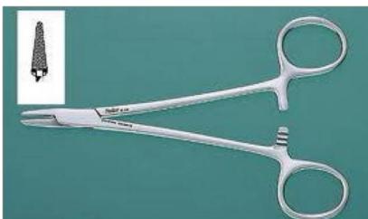 | Narrow rounded tip with a crisscross gripping pattern in the inner jaws. | Uses: holding delicate to intermediate-sized needles when suturing. |
| Laparoscopic Needle Holder | — | — | Uses: suturing during laparoscopic surgery. |
Scalpels, Bone & Special Instruments
| Instrument | Image… | Description / Key Features / Notes | Uses, Indications, Contraindications & Complications |
|---|---|---|---|
| Scalpel Blades No. 10 / 11 / 15 | — | No. 10: large curved cutting belly; No. 11: straight triangular pointed tip; No. 15: small delicate curved blade. | Uses: No. 10 — skin incisions (laparotomy, thoracic); No. 11 — stab incisions (abscess I&D, chest tube, trocar puncture); No. 15 — precision minor surgery, pediatric, plastic surgery. |
| Bone Instruments — Bone Nibbler / Rongeur / Bone Hook | — | — | Uses: bone work. |
| Curette |  | — | Uses: scraping or debriding biological tissue or debris — in a biopsy, excision, or cleaning procedure. |
Electrosurgery & Hemostasis Devices
| Instrument | Image… | Description / Key Features / Notes | Uses, Indications, Contraindications & Complications |
|---|---|---|---|
| Argon Beam Coagulator (ABC) | — | Unipolar coagulation; non-touch technique; less depth of penetration (2–3 mm). | Uses: coagulation of parenchymatous organs (non-touch technique). |
| Surgical Lasers | — | Source marks lasers as rarely used. | Uses: Argon — ophthalmology, vascular anastomosis; CO₂ — cutting tissue; Nd:YAG — paranasal sinus and tracheobronchial tree; Er:YAG — vaporizes cartilage, fibrous tissue and bone. |
Suturing & Stapling Devices
| Instrument | Image… | Description / Key Features / Notes | Uses, Indications, Contraindications & Complications |
|---|---|---|---|
| Surgical Needles (open French-eye; eyeless/swaged; express needle) |        | Open French-eye needle — not used any more; eyeless needles — ready-made with thread already attached; straight or curved (mostly curved used); cross-section: round, triangular or flattened; points: cutting (skin) or tapered/blunt (soft tissue/fascia). Reverse cutting — sharp cutting edge on the outside convex curvature; eliminates cut-through; preferred for tough skin. Round-bodied (tapered) — conical smooth tip; separates tissue without cutting. Figure 1: Eyeless (Swaged) Needle (A) and Eyed Needle (B). | Uses: skin suturing (cutting/reverse-cutting); viscera, bowel, peritoneum and vessels (round-bodied). |
| TA / GIA / EEA Staplers |     | TA: linear everting double line; lengths 30, 55, 90 mm; staple sizes 3.5 and 4.8 mm; 3.2 mm for vessel closure. GIA: two double rows of staples — divide and anastomose. EEA: end-to-end or end-to-side circular staplers. | Uses: TA — linear stapling and vessel closure; GIA — intestinal anastomosis; EEA — circular end-to-end / end-to-side anastomoses. |
| Skin Stapler |  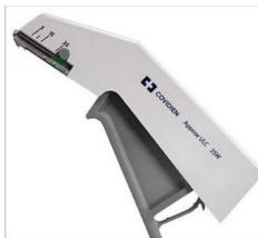 | — | Uses: skin approximation during wound closure. |
| Staple Remover |  | — | Uses: removal of skin staples from the wound. |
Tubes, Catheters & Drains
| Instrument | Image… | Description / Key Features / Notes | Uses, Indications, Contraindications & Complications |
|---|---|---|---|
| Foley Catheter (3-way irrigation catheter) |     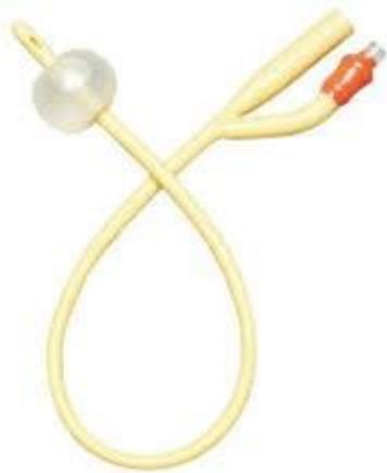 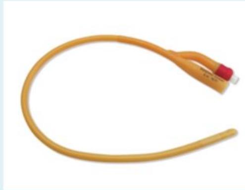 | Retaining (balloon) mechanism to stay in place. Sizing: adults 14–16 Fr (females), 16–18 Fr (males); hematuria with clots — 20–24 Fr 3-way irrigation catheter. Insertion: sterile aqueous chlorhexidine prep + fenestrated drape; males — hold penis vertical at 90°, instill 10 mL 2% lidocaine gel, wait 3–5 min; advance to the bifurcation (“hub”) so the balloon is fully within the bladder; inflate with 10 mL sterile water (never saline — crystals jam the channel; never air — it floats); pull back until resistance at the bladder neck; connect to drainage bag. | Indications: diagnostic — monitoring urine output (strict hourly in shock/ICU; target ≥0.5 mL/kg/h); therapeutic — acute urinary retention (decompression/relief); intraoperative bladder decompression during laparotomy/pelvic surgery. Contraindications: acute urethritis; blood at meatus + history of trauma + ruptured urethra. Complications: UTI; urethral trauma. |
| Sengstaken-Blakemore Tube |   | Balloons: one for the esophagus, second for the esophagogastric junction. | Indications: bleeding oesophageal varices (if endoscopy fails to control the bleeding). Contraindications: history of esophageal stricture; recent esophageal or gastric surgery. Complications: airway obstruction; esophageal rupture; ischemic necrosis. |
| Flatus Tube |   | — | Uses: per rectally; left in place until the anastomosis heals. Indications: post sigmo-anal anastomosis. |
| Nasogastric (NG) Tube |  | — | Indications: therapeutic — aspiration of gastric content after recent ingestion of toxic material; administration of medication; feeding; bowel irrigation. Diagnostic — evaluation of upper GI bleeding (presence, volume); administration of radiographic contrast to the GI tract. |
| Stoma Bag (ileostomy / colostomy bag) |    | Ileostomy — right side, more liquid output, more protruding; colostomy — left side, stool, flat to the skin. | Indications: feeding; colorectal carcinoma; obstruction; fistulae. |
| Chest Tube (ICT) |    | Holes to drain; cm markings for how deep it is. | Uses: drains blood, fluid and air from around the lungs, heart or esophagus. Indications: tension pneumothorax — needle decompression, severe acute, ICT. |
| T-Tube |   | T-shaped tube; temporary — removed once the anastomosis heals, following a T-tube cholangiogram (source: after 72 days). | Uses: biliary drainage after T-tube cholecystectomy. Indications: stone in CBD. |
| Surgical Drains (general) | — | Passive (open or closed) vs active (closed + suction). History: dates back to Hippocrates (metal/glass tubes, bone, gauze, rubber); air-vent suction by Heaton (1889). | Uses: prevents collection of serum, blood, pus/drainage. Indications: prophylactic and therapeutic. |
| Closed Active Suction Drain (Hemovac; Redivac; vacuum evacuator) | 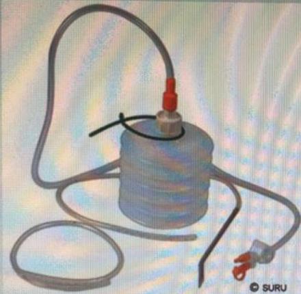 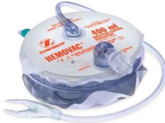    | Multi-holed, polyvinyl chloride or silicone; active closed suction. | Uses: evacuate postoperative blood/seroma under negative pressure; obliterate dead space; prevent infection; effective for draining soft tissue under large skin flaps; lower infection rate. Complications: retrograde infection; clot blockage (clogs and ceases to function); visceral/anastomotic erosion; drain fracture. |
| Penrose Drain |   | Soft, flexible latex rubber wick; non-active open drain; obsolete. | Uses: draining pus, blood or serum; brought out through a separate wound; anchored to the skin with a suture. Complications: risk of infection (efficient but infection risk). |
| Corrugated Drain | — | Corrugated drain (no bag connected). | Uses: leg drains (obsolete — not used any more). |
| Sump Drain |  | Tube within a tube — suction via the inner tube, air enters via the outer tube to aid suction; large and bulky; double/triple lumen allows irrigation and aspiration; relies on continuous flow of air from outside; less likely to be occluded by tissue. | Uses: suction-irrigation drainage (inner suction tube + outer air vent). Indications: pancreatic necrosis; severe abdominal infections; high-volume enteric fistula; rarely used. Complications: predisposed to secondary infection. |
| Therapeutic Percutaneous Drainage Catheter |   | CT- or US-guided. | Uses: draining an abscess — inserted by radiologists to drain accessible localized collections. |
Endoscopy & Examination
| Instrument | Image… | Description / Key Features / Notes | Uses, Indications, Contraindications & Complications |
|---|---|---|---|
| Proctoscope (rigid anoscope; proctoscope with central obturator) |    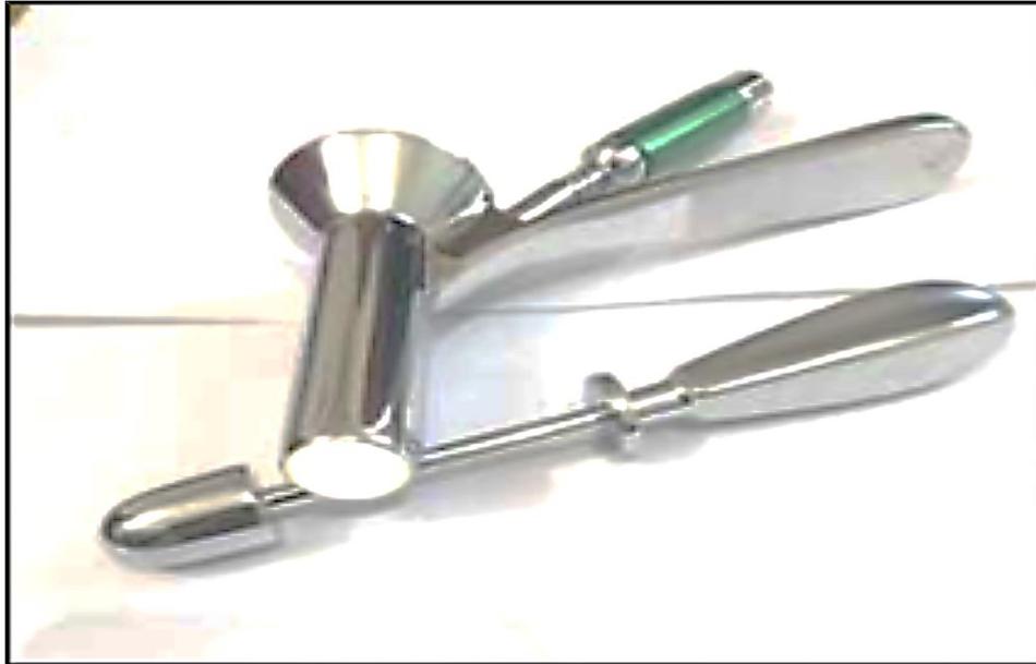 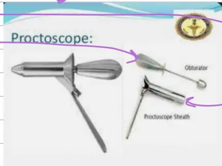 | Components: hollow cylindrical barrel + central bullet-tipped obturator. Insertion: lubricate; insert with obturator fully in place, directed toward the umbilicus for 3–4 cm, then posteriorly into the rectal ampulla; remove the obturator before mucosal inspection. | Indications: hemorrhoids and rectal polyps (only seen with proctoscopy as they are not palpable); need for biopsy, cytology and culture specimens; polypectomy. Uses: diagnostic — direct inspection for internal hemorrhoids, anal fissures, fistula (internal) openings, proctitis; punch biopsy of suspicious lesions/polyps; therapeutic — rubber band ligation (Barron banding) of 1st–3rd degree internal hemorrhoids; sclerotherapy injection; snare polypectomy. Contraindications: large aortic aneurysm; acute myocardial infarction; abnormal coagulation studies. Complications: severe pain; bleeding. |
| Pratt Rectal Speculum |  | Bivalve speculum. | Uses: provides exposure for visualization of the anus and rectum. |
Laparoscopic Equipment, Imaging & Robotic
| Instrument | Image… | Description / Key Features / Notes | Uses, Indications, Contraindications & Complications |
|---|---|---|---|
| Cold Light Source (halogen / xenon lamp; LED) | — | Halogen and xenon lamps; LED light so it doesn’t burn the inside; no shadows. | Uses: illumination for laparoscopy, delivered through the optic cable. |
| Optic Cable (fibre-optic cable) |   | Bundle of optical fibers (glass threads) swaged at both ends; fiber size usually 10–25 mm in diameter; very high quality of optical transmission, but fragile. | Uses: light transmission; remains conducting light even when coiled. |
| 3-Chip Camera / Video Monitor / Laparoscopic Tower |  | 3-chip camera (commonly used, high resolution); video monitor to display images; tower carries the system. | Uses: visualization during laparoscopic procedures. |
| CO₂ Insufflator (gas insufflator) |     | Creates pneumoperitoneum at 12–15 mmHg, distending the abdominal cavity for proper visualization. CO₂ is most commonly used: readily available, cheaper, easily absorbed by tissues, quickly released via respiration; non-combustible and highly soluble in blood → low gas embolism risk. Physiological changes: increased intra-abdominal pressure → vasovagal attack; if the patient becomes hypotensive on inflation, first step = deflate; IVC compression → decreased venous return → decreased cardiac output; increased arterial pressure; diaphragm compression impairs pulmonary compliance; CO₂ → hypercarbia, respiratory acidosis and hypoxia. | Uses: creating the pneumoperitoneum for laparoscopy. Complications: pneumoperitoneum-related — CO₂ embolism, hypercarbia, respiratory acidosis, subcutaneous emphysema, renal failure, venous thrombosis, pneumothorax; pressure should not exceed 15 mmHg (above 20 mmHg very dangerous — reduces venous return/cardiac output). |
| Robotic Surgical System (robotic surgery; consoles) |     | Console screens for the surgeon; can operate from the same room or from other continents; needs an operating surgeon and assistants; a second surgeon must be onsite (source: console — one is learning, one is a surgeon — to exchange arms). | Uses: remote/robotic minimally invasive surgery. Complications / Drawbacks: network connection; another surgeon must be onsite. |
| Incentive Spirometer |  | Sustained maximal inspiration device. | Uses: prevents and treats postoperative atelectasis and pneumonia. |
Sterilization & OR Equipment
| Instrument | Image… | Description / Key Features / Notes | Uses, Indications, Contraindications & Complications |
|---|---|---|---|
| Autoclave (heat sterilization — steam under pressure / dry heat) |  | Machine producing high temperature. Steam under pressure: increasing the atmospheric pressure raises the water boiling point — instruments and pre-packed drapes: 134 °C & 30 lb/in² for 3 min, or 121 °C & 15 lb/in² for 15 min. Dry heat: 160 °C for 2 hrs — suitable for airtight containers and instruments at risk of corrosion. | Uses: sterilization — complete destruction of all micro-organisms including bacterial spores. |
| Gamma Irradiation | — | Gamma rays. | Uses: syringes, catheters, gauze, sutures. |
| Ethylene Oxide | — | Highly penetrative gas. | Uses: delicate items — endoscopes, electrical equipment, single-use plastic items. |
| Glutaraldehyde (Cidex) | — | Liquid chemical. | Uses: cleaning lensed instruments. |
| Bair Hugger & Patient-Warming Devices (heating mattress; warm-air heated blankets; fluid & blood warmers) | — | Plastic sheet covering the patient to control temperature; humidification of inspired anaesthetic gas; warming devices for fluids and blood. Unconscious patients lose the ability to control body temperature; hypothermia risk if ambient temperature < 21 °C for 1–2 hours. | Uses: preventing and managing hypothermia during (long) surgery. |
Sutures, Needles & Local Anaesthesia
Suture materials (synthetic sutures used more often than natural):
| Suture / Material | Structure & Absorption | Tensile Strength | Common Uses |
|---|---|---|---|
| Vicryl (Polyglactin 910) | Braided absorbable (hydrolysis 56–70 days); polyglactic acid — synthetic, braided, very high tensile, absorbed 60 days. | 50% at 3 weeks; minimal residual strength by 4 weeks (fully absorbed 56–70 days). | Subcutaneous tissue; GI anastomoses; soft-tissue anastomosis. |
| Monocryl (Poliglecaprone 25) | Monofilament absorbable (90–120 days). | 50% at 1 week; 0% at 3 weeks. | Subcuticular skin closure (cosmetic); urology. |
| PDS II (Polydioxanone) | Monofilament absorbable (180–210 days). | 50% at 4–6 weeks. | Abdominal fascia (linea alba); biliary; soft-tissue anastomosis. |
| Ethilon (Nylon) | Monofilament non-absorbable; nylon = synthetic polyamide, mono/multifilament. | Permanent (loses ~15%/year). | Interrupted cutaneous skin sutures. |
| Prolene (Polypropylene) | Monofilament non-absorbable; minimal tissue reaction. | Indefinite strength; inert. | Vascular anastomoses; hernia mesh fixation. |
| Silk | Braided natural non-absorbable; silkworm protein filament — dyed, polybutylene-treated, braided. | Loses 50% at 1 year; good tensile. | Vessel ligation; securing drains/chest tubes; natural — not used most of the time. |
| Plain catgut | Cattle/sheep intestine. | Absorption ~10 days. | — |
| Chromic catgut | Chromium salt-treated (source photo: chromic gut 3-0 box). | Lasts up to 20 days. | — |
| Dexon (Polyglycolic acid) | Synthetic, braided, absorbable. | Higher tensile; reabsorption 60–90 days. | Soft-tissue anastomosis (intestinal, gastric). |
| Maxon (Polyglyconate) | Synthetic monofilament. | — | — |
| Dacron (Polyester) | Non-absorbable. | Superior strength and durability. | — |
| Stainless steel | Low-carbon iron alloy; monofilament/multifilament; non-absorbable. | — | Bone suturing. |
| MONOMID® / POLYPRO® / POLYBOND® / UHMWPE | Non-absorbable — monofilament nylon / monofilament polypropylene / braided polyester / ultra-high molecular weight braided polyethylene. | — | — |
- Surgical needles (open French-eye; eyeless/swaged; express needle): open French-eye — no longer used; eyeless — thread pre-attached; straight/curved (mostly curved); cross-section round, triangular or flattened; points cutting (skin) or tapered/blunt (soft tissue/fascia). Reverse cutting — cutting edge on the outer convex curvature; eliminates cut-through; preferred for tough skin. Round-bodied (tapered) — conical smooth tip; separates without cutting. Figure 1: Eyeless (Swaged) Needle (A) and Eyed Needle (B). Uses: skin suturing (cutting/reverse-cutting); viscera, bowel, peritoneum and vessels (round-bodied).
- TA / GIA / EEA staplers: TA — linear everting double line; lengths 30, 55, 90 mm; staple sizes 3.5/4.8 mm; 3.2 mm for vessel closure. GIA — two double rows; divide and anastomose. EEA — end-to-end/end-to-side circular. Uses: TA — linear stapling and vessel closure; GIA — intestinal anastomosis; EEA — circular anastomoses.
- Skin stapler — skin approximation during wound closure; staple remover — removal of skin staples.
- Wound closure: primary closure (clean wounds); delayed primary closure (contaminated wound — left open for dressing, closed after 3–5 days). Suturing methods: simple interrupted, mattress, subcuticular.
- Topical haemostatic agents: oxidised regenerated cellulose, thrombin, gel foam, bone wax — coagulation factors applied to a bleeding area.
- Haemostasis methods: compression (at least 5 min pressure); packing (short/long duration, e.g. liver laceration); ligation (absorbable/non-absorbable sutures, clips); vessel repair (sutures for big vessels); topical agents (above); thermal coagulation (diathermy — most commonly used); ultrasound (harmonic scalpel); laser.
Local anaesthetics
| Agent | Max Safe Dose | Notes |
|---|---|---|
| Lidocaine Plain (1% or 2%) | 3 mg/kg (≈ 200 mg in a 70 kg adult = 20 mL of 1%). | — |
| Lidocaine with Epinephrine (1:200,000) | 7 mg/kg (≈ 500 mg in a 70 kg adult = 50 mL of 1%). | Contraindication: NEVER use epinephrine on end-arteries (fingers, toes, penis, nose, ear pinna) → ischaemic necrosis! |
Drains, Tubes, Catheters & Stomas
| Instrument | Description / Key Features | Uses / Indications / Complications |
|---|---|---|
| Foley Catheter (3-way irrigation catheter) | Retaining balloon. Sizing: adults 12–14 Fr (females), 14–16 Fr (males); haematuria with clots — 20–24 Fr 3-way irrigation catheter. Insertion: sterile aqueous chlorhexidine prep + fenestrated drape; males — penis vertical at 90°, instil 10 mL lidocaine gel (1–2%), wait 3–5 min; advance to the bifurcation (hub) so the balloon is fully in the bladder; inflate with 10 mL sterile water (never saline — crystals jam the channel; never air — it floats); pull back to resistance at bladder neck; connect bag. | Indications: diagnostic — urine output monitoring (strict hourly in shock/ICU; target ≥0.5 mL/kg/h); therapeutic — acute retention; intraoperative bladder decompression (laparotomy/pelvic surgery). Contraindications: acute urethritis; blood at meatus + trauma history + ruptured urethra. Complications: UTI; urethral trauma. |
| Sengstaken-Blakemore Tube | Balloons: one oesophageal, second oesophagogastric junction. | Indications: bleeding oesophageal varices (if endoscopy fails). Contraindications: oesophageal stricture; recent oesophageal/gastric surgery. Complications: airway obstruction; oesophageal rupture; ischaemic necrosis. |
| Flatus Tube | — | Per rectum; left until anastomosis heals. Indications: post sigmo-anal anastomosis. |
| Nasogastric (NG) Tube | — | Therapeutic — aspiration of gastric content after recent toxic ingestion; medication; feeding; bowel irrigation. Diagnostic — upper GI bleeding evaluation (presence, volume); radiographic contrast administration. |
| Chest Tube (ICT) | Holes to drain; cm markings for depth. | Drains blood, fluid and air from around the lungs, heart or oesophagus. Indications: tension pneumothorax — needle decompression, severe acute, ICT. |
| T-Tube (Kehr Tube) | T-shaped; placed in the common bile duct (CBD) after open choledochotomy; external biliary stent, decompresses the biliary tree, allows postoperative cholangiogram. Temporary — removed once the anastomosis heals, after a T-tube cholangiogram; kept ≥14 days (fibrous tract forms, preventing biliary peritonitis); source: after 72 days. 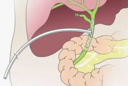   | Biliary drainage after T-tube cholecystectomy. Indications: stone in CBD. |
| Surgical Drains (general) | Passive (open/closed) vs active (closed + suction). History: Hippocrates (metal/glass tubes, bone, gauze, rubber); air-vent suction by Heaton (1889). | Prevents collection of serum, blood, pus/drainage. Indications: prophylactic and therapeutic. |
| Closed Active Suction Drain (Hemovac; Redivac; vacuum evacuator; Jackson-Pratt) | Multi-holed PVC/silicone; spring or bulb continuous negative pressure; active closed suction. Remove when <20–30 mL/24 h. | Evacuate postoperative blood/seroma under negative pressure; obliterate dead space; prevent seroma/haematoma (mastectomy, neck, arthroplasty); prevent infection; effective under large skin flaps; lower infection rate. Complications: retrograde infection; clot blockage; visceral/anastomotic erosion; drain fracture. |
| Penrose Drain | Soft flexible latex rubber wick; non-active open drain; obsolete. | Draining pus, blood or serum; separate wound; anchored to skin with suture. Complications: infection risk. |
| Corrugated Drain | No bag connected. | Leg drains (obsolete). |
| Sump Drain | Tube within a tube — suction via inner, air via outer; large/bulky; double/triple lumen allows irrigation and aspiration; continuous outside air flow; less likely occluded by tissue. | Suction-irrigation drainage. Indications: pancreatic necrosis; severe abdominal infections; high-volume enteric fistula; rarely used. Complications: secondary infection. |
| Therapeutic Percutaneous Drainage Catheter | CT- or US-guided. | Draining an abscess — inserted by radiologists for accessible localised collections. |
| Stoma Bag (ileostomy / colostomy bag) | Ileostomy — right, liquid output, protruding; colostomy — left, stool, flat to skin. 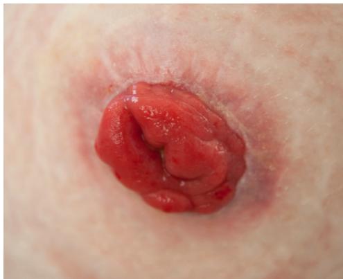 | Feeding; colorectal carcinoma; obstruction; fistulae. |
Colostomy vs ileostomy
| Feature | Colostomy | Ileostomy |
|---|---|---|
| Location | Left iliac fossa (LIF). | Right iliac fossa (RIF). |
| Profile | Flush with skin. | Spouted (2–3 cm). |
| Effluent | Solid/formed; non-corrosive. | Liquid/enzymatic; proteolytic enzymes burn skin. |
| Indications | Hartmann’s (diverticulitis); APR (rectal cancer); obstructing rectosigmoid tumour. | Defunctioning loop (protects low anastomosis); total colectomy/proctocolectomy (UC/FAP). |
| Complications | Ischaemia/necrosis, retraction, parastomal hernia, prolapse, peristomal dermatitis. | High-output fluid/electrolyte loss (>1200 mL/d), peristomal enzymatic excoriation, retraction, ischaemia, food bolus obstruction. |
Laparoscopy Concepts & MIS
- Definition: procedures through small incisions allowing endoscopic access to the peritoneal cavity after gas insufflation creates space between the abdominal wall and viscera, using specialised instruments and visualisation tools.
- Types: intraperitoneal; extraperitoneal; abdominal-wall retraction (gasless); hand-assisted. Stomach, biliary tract, transverse colon intraperitoneal; ascending/descending colon retroperitoneal; hernia either approach; incision extended for large-organ retrieval (e.g., kidney). Also thoracoscopic access; NOTES (natural-orifice transluminal surgery — e.g., gallbladder via the mouth/transgastric route); single-port surgery.
- Why laparoscopy / advantages: Patient — less post-operative pain (smaller incisions); less bowel handling → little/no disturbance of normal function; no abdominal-wall injury → rapid return to normal activity (driving, lifting, sport); shorter hospital stay. Hospital — initial capital cost 250,000, offset by shorter stays (cholecystectomy 5 → 1 day; hiatus hernia repair 7 → 3 days). Surgeon — magnified view, larger operative field, diagnostic ability, operating time. Also: faster recovery/return to work; smaller scars (cosmesis); less internal scarring; less wound infection/incisional hernia; better visualisation; less tissue trauma; reduced wound complications (infection, dehiscence); less patient-blood contact.
- Disadvantages: slower; special expertise necessary (different technique); tactile feedback lost/altered; bleeding control harder; organ extraction sometimes difficult; two small flat-screen views.
- Selection pearls: gold standard for appendicectomy, cholecystectomy and bariatric surgery; first-time inguinal hernia = laparotomy, recurrent/bilateral hernia = laparoscopic; avoid/convert when a previous laparoscopy was difficult, in frozen abdomen or dense adhesions. If an open appendicectomy reveals a normal appendix, it is still removed (prevents future diagnostic confusion).
- Contraindications: absolute — inadequate experience of the surgeon; uncorrected coagulopathy/DIC; shock. relative — severe COPD (CO₂ worsens acidosis); recent MI; ventriculoperitoneal shunt; extensive organomegaly; CHF; trauma; internal bleeding; acidosis.
- Common laparoscopic procedures: cholecystectomy, appendicectomy, hernia repair, adhesiolysis, perforation closure, bowel resection, prolapse repair, nephrectomy, bypass, splenectomy, anterior resection/APR, right hemicolectomy, left/sigmoid colectomy, gastrectomy, oesophagogastrectomy, hiatus hernia repair.
- Future of MIS: AI and augmented reality in surgery; robotic-system advances; single-incision and scarless techniques; personalised and precision surgery.
Laparoscopic equipment, imaging & robotic
| Equipment | Description / Key Features | Uses / Complications |
|---|---|---|
| Cold Light Source (halogen / xenon lamp; LED) | Halogen/xenon lamps; LED so it doesn’t burn the inside; no shadows. | Illumination for laparoscopy via the optic cable. |
| Optic Cable (fibre-optic cable) | Optical fibre bundle (glass threads) swaged at both ends; fibre size usually 10–25 µm; high optical transmission but fragile. | Light transmission; conducts light even when coiled. |
| 3-Chip Camera / Video Monitor / Laparoscopic Tower | 3-chip camera (commonly used, high resolution); monitor displays images; tower carries the system. | Visualisation during laparoscopic procedures. |
| CO₂ Insufflator (gas insufflator) | Pneumoperitoneum at 12–15 mmHg, distending the abdomen for visualisation. CO₂ most used: cheap, readily available, easily absorbed, quickly released via respiration; non-combustible, highly blood-soluble → low gas embolism risk. Physiological changes: ↑ intra-abdominal pressure → vasovagal attack; if hypotensive on inflation, first step = deflate; IVC compression → ↓ venous return → ↓ cardiac output; ↑ arterial pressure; diaphragm compression impairs compliance; CO₂ → hypercarbia, respiratory acidosis, hypoxia. | Complications: CO₂ embolism, hypercarbia, respiratory acidosis, subcutaneous emphysema, renal failure, venous thrombosis, pneumothorax; pressure must not exceed 15 mmHg (above 20 mmHg very dangerous — reduces venous return/cardiac output). |
| Robotic Surgical System (robotic surgery; consoles) | Console screens; surgeon can operate from the same room or other continents; needs an operating surgeon + assistants; a second surgeon onsite (source: one console learning, one surgeon — to exchange arms). | Remote/robotic MIS. Drawbacks: network connection; another surgeon onsite. |
| Incentive Spirometer | Sustained maximal inspiration device. | Prevents and treats postoperative atelectasis and pneumonia. |
- Insulation & capacitance: laparoscopic instruments are black-coated insulated shafts with only the tip exposed → monopolar current acts only at the tip. If the insulation is broken, current leaks into a metallic trocar touching the skin → abdominal-wall burns (capacitance/capacitive coupling). Prevention: check insulation integrity; use a fully plastic trocar.
- SILS (Single-Incision Laparoscopic Surgery): all instruments through one umbilical port; better cosmesis, harder triangulation.
Sterility, OR & wound essentials
- Infection-control definitions: Sterilization — complete destruction of all micro-organisms including bacterial spores. Disinfection — reduces viable organisms; does not inactivate viruses or bacterial spores; non-living surfaces (bleach, alcohol). Antisepsis — destroys organisms at the wound/external-environment interface; living tissues (chlorhexidine, povidone-iodine). Asepsis — keeping as few organisms as possible near the operating field (gloves, gowns, masks).
- Sterilization methods: Heat — autoclave steam under pressure: 134 °C & 30 lb/in² for 3 min, or 121 °C & 15 lb/in² for 15 min (instruments + pre-packed drapes); dry heat 160 °C for 2 h (airtight containers, corrosion-risk instruments). Cold — gamma irradiation (syringes, catheters, gauze, sutures); ethylene oxide (endoscopes, electrical equipment, single-use plastics); glutaraldehyde/Cidex (lensed instruments).
- Patient preparation: shower; shaving vs clipping in the OR (clipping lowers wound-infection risk); wide skin prep with povidone-iodine and alcohol; sterile drapes covering the patient; prophylactic antibiotic before skin incision — as close as possible.
- Surgical-team preparation: scrub hands/forearms 3–5 min with chlorhexidine (Hibiscrub); jewellery removed; dry with a sterile towel; gowns and gloves; double-gloving for high-risk patients (HIV, hepatitis B & C).
- Patient positions (aim: adequate exposure): supine — most abdominal surgery; Trendelenburg — supine, 30° head down; reverse Trendelenburg — head up; lithotomy — knees and hips fully flexed, feet in stirrups (gynaecology, rectal); Lloyd-Davies — Trendelenburg with legs abducted, hips/knees slightly flexed (AP resection for lower-rectal cancer); lateral — upper arm raised above and in front of the head (right thoracotomy → left side down; laparoscopic splenectomy → right side down so the spleen falls for exposure).
- Temperature control: unconscious patients lose temperature regulation; hypothermia risk if ambient <21 °C for 1–2 h. Prevent: minimise uncovered time; heating mattress/warm-air blankets (Bair hugger — plastic sheet over the patient); humidify anaesthetic gas; warm fluids, blood and IV fluids (large volumes can cause hypothermia).
- Principles of surgical technique: adequate incisions along normal skin lines (Langer’s), avoiding joint creases; midline is the commonest abdominal incision (fascia only, xiphisternum → umbilicus or below); atraumatic handling; dissection in natural planes; good haemostasis; debridement of contaminated wounds; continuous irrigation.
- Post-operative wound care: sterile dressing protects from mechanical trauma and bacterial invasion (applied before drapes removed; infected wounds need exudate-absorbing dressings); immobilisation reduces lymphatic flow and spread of wound flora; elevation reduces interstitial oedema; correct timing of suture removal.
- Prophylactic antibiotics & wound classification: prophylaxis = antimicrobials before surgery (before GA) reducing microbes entering tissue/body cavity; chosen by likely site organisms; duration controversial (single dose / 24 h / longer). Classes: I clean — thyroid surgery, breast biopsy (~1–5%; no antibiotics) · ID — graft or mesh (give antibiotic) · II clean-contaminated — biliary/urinary/GI (3–11%; 1–2 doses) · III contaminated — gross contamination, e.g. bowel surgery (10–17%; full 5-day course) · IV dirty — established infection, e.g. peritonitis (~27%+; 5 days). Prophylaxis applies to classes ID–IV.
Viva & Exam Spots
- Stoma spotter (Batch 427 Female / Batch 15): Image 60 = end colostomy (LIF, flush); Image 67 = end loop ileostomy (RIF, 2–3 cm spout).
- Q: Why is an ileostomy spouted while a colostomy is flush? — Ileostomy effluent is liquid, rich in proteolytic enzymes and alkaline bile salts causing severe chemical dermatitis; the 2–3 cm spout directs fluid into the bag away from skin. Colostomy stool is solid and non-corrosive, so a flush stoma prevents leakage.
- Q: Why are uncomplicated internal haemorrhoids NOT palpable on DRE? — They are soft, low-pressure vascular cushions U&E, faecal immunochemical test (FIT), flexible sigmoidoscopy/colonoscopy, CT abdomen/pelvis.
9. Laboratory Interpretation
Reference Ranges & Interpretation
| Test | Normal | Interpretation pearls |
|---|---|---|
| FBC | WBC 4–11 ×10⁹/L · Hb 13–18 g/dL (M) / 12–16 (F) · platelets 150–350 ×10⁹/L | Leukocytosis → infection/inflammation (cholangitis, appendicitis, diverticulitis, abscess); anaemia → bleeding, malignancy, chronic disease; thrombocytopenia → bleeding risk (check before surgery) |
| Coagulation | PT 11–14 s · aPTT 25–35 s · INR 0.8–1.2 | Liver disease/obstructive jaundice → PT, PTT, INR all ↑ (vitamin K–dependent factors II, VII, IX, X); correct with vitamin K, FFP, platelets/cryoprecipitate, RBC (± heparin in selected cases) |
| U&E / KFT | Urea 2.5–7.8 mmol/L · creatinine 60–120 µmol/L · eGFR >90 | Dehydration/renal impairment; check before contrast (CT/ERCP); Na↓ K↑ glucose↓ → adrenal crisis |
| Glucose | 70–100 mg/dL (3.9–5.6 mmol/L) | Hypoglycaemia → adrenal crisis/sepsis; blood glucose + urine ketones in DKA |
| LFTs | Total bilirubin <17 µmol/L · direct <5 µmol/L · ALT 5–30 · AST 10–40 · ALP 25–120 U/L · albumin 35–50 g/L | Obstructive: direct >50% of total, ALP ↑↑ (most sensitive — excreted by hepatocytes/biliary epithelium), ALT/AST normal-mild ↑; hepatocellular: ALT/AST ↑↑; isolated ALP → bone (metastases, Paget’s) or cholestasis (confirm with GGT) |
| Amylase / lipase | Amylase <100 U/L · lipase <60 U/L | >3× ULN = diagnostic of acute pancreatitis; <3× → perforated viscus, ectopic pregnancy, DKA |
| Calcium / albumin | Ca 8.9–10.3 mg/dL (2.2–2.6 mmol/L) · albumin 35–50 g/L | Low in severe pancreatitis (fat saponification); high in malignancy/hyperparathyroidism; low albumin lowers total Ca → interpret corrected calcium |
| CRP / ESR | CRP <5 mg/L · ESR <15 (M) / <20 (F) mm/h | Non-specific inflammation/infection; CRP tracks severity; procalcitonin in bacterial sepsis |
| ABG | pH 7.35–7.45 · PaCO₂ 35–45 mmHg · HCO₃⁻ 22–26 mEq/L · lactate <2 mmol/L | pH → primary disorder → compensation → anion gap → Winter’s formula (method: ABG & acid–base); ↑lactate = hypoperfusion/shock |
| Urine / cultures | — | Dipstick: nitrites/leucocytes (UTI), blood (stones, tumour, trauma), glucose/ketones (DKA); β-hCG in every woman of reproductive age with abdominal pain; blood cultures before antibiotics; deep tissue/bone culture (not surface swab) in diabetic foot |
| Blood bank | — | Group & save/cross-match before surgery or in bleeding; 1 unit PRBC ≈ ↑Hb 1 g/dL (Hct 3%); adverse effects fever, infection, chest pain, allergy (details: transfusion reactions) |
| Tumour markers | — | CEA colorectal — baseline then 3-monthly ×2 y, 6-monthly ×3 y; CA19-9 pancreas · AFP liver · PSA prostate · β-hCG gestational trophoblastic disease · calcitonin medullary thyroid cancer · thyroglobulin differentiated thyroid cancer |
| D-dimer | <500 ng/mL | Fibrin degradation product — used to assess thrombotic disorders; ↑ with PE/DVT (confirm with CTPA/Doppler) |
Lab Order Sets by Scenario
| Scenario | Key labs |
|---|---|
| Acute abdomen | FBC, U&E, LFT, CRP, amylase (± calcium), lactate, troponin, blood cultures, urine dipstick ± M,C&S, β-hCG, ABG; coagulation + cross-match if surgery likely |
| Biliary colic | CBC, LFT, serum amylase — all normal (diagnosis is ultrasound: gallstones + acoustic shadowing) |
| Jaundice | CBC, LFT (ALT/AST/ALP/GGT/amylase), coagulation (PT ↑ in obstruction — vitamin K deficiency), lipids, viral screen (Hep B/C, HIV) |
| Upper GI bleed | CBC, U&E, coagulation, cross-match, LFT; resuscitate then endoscopy |
| Lower GI bleed / colorectal cancer | CBC, U/E, coagulation, CEA; colonoscopy + biopsy; CT CAP staging |
| Breast (early stage) | Bone profile + LFTs (metastases); ER/PR/HER2 (± Ki-67) on core biopsy |
| Thyroid | TSH (most important), T3/T4; ultrasound ± FNA (Bethesda); calcitonin if medullary suspected |
| Diabetic foot | CBC (Hb, platelets, WBC), glucose, HbA1c (3-month control), U&E + creatinine, LFT, blood culture (if unwell) + deep tissue culture, cross-match, urine ketones |
| Pre-operative | CBC, electrolytes, creatinine, glucose, INR/PTT, group & save; CXR/ECG if cardiac or pulmonary disease |
Case Panels — Interpret & Act
| Case | Key results | Interpretation | Next step |
|---|---|---|---|
| Obstructive jaundice (choledocholithiasis) — 50 F, jaundice, RUQ pain, dark urine | Total bili 6.5 mg/dL · direct 5.5 · ALP 450 U/L · AST 150 · ALT 180 | Obstructive pattern (ALP most pathognomonic) | US dilated CBD (initial, not conclusive) → stone: ERCP; head-of-pancreas tumour: CT (MRCP if unclear); ERCP for stone removal |
| Ascending cholangitis (archive) — jaundice + fever 39 °C | WBC 20 · total bili 76 · direct 60 · ALP 215 (µmol/L) | Obstructive pattern + leukocytosis = infection | ABC, IV fluids, analgesia, IV antibiotics, urgent biliary decompression (US ± ERCP) |
| Acute cholecystitis — 40 F, RUQ pain → right shoulder, fever, 2 d | WBC 14,000 · ALP 250 · AST 120 · total bili 1.8 mg/dL | Biliary inflammation | US: wall thickening, pericholecystic fluid, sonographic Murphy +; IV antibiotics + lap chole within 72 h if stable; if sick → conservative then interval lap chole at 6 weeks |
| Acute pancreatitis — severe epigastric pain → back, alcohol | Amylase 1920 (<100) or lipase ↑↑ · WBC 15 · Ca 7.6 ↓ | Amylase >3× ULN; hypocalcaemia = fat saponification; commonest causes gallstones + alcohol | NPO, IV fluids, analgesia, monitor; US + contrast CT (necrosis); surgery if necrotising, haemorrhagic, or pseudocyst >6 cm / pressure symptoms / abscess; pseudocyst = no epithelial lining — follow closely (gastric outlet obstruction, haemorrhage, abscess) |
| Perforated duodenal ulcer (peritonitis) — 50 M, sudden epigastric pain + fever | WBC 18 · lactate 6 mmol/L | Perforation with sepsis | CXR pneumoperitoneum; resuscitate, antibiotics, omental patch repair |
| Pyogenic liver abscess — 65 M, fever/RUQ pain/jaundice, diabetic | WBC 16 · ALP 400 · total bili 2.5 mg/dL · blood culture Klebsiella + | Infection; positive cultures confirm organism | US/CT → antibiotics + percutaneous drainage; DDx: amoebic (Entamoeba histolytica), hydatid (Echinococcus granulosus — daughter cysts; lung/brain/liver) |
| Acute limb ischaemia — 55 M, sudden leg pain, cold, pale | WBC 14 · lactate 7 · CK 4500 U/L (creatine kinase — muscle necrosis/rhabdomyolysis) | Muscle ischaemia with potential necrosis | Doppler no distal pulses → CT angiography (if pulse present → partial occlusion/spasm); well hydrated (rhabdomyolysis → AKI risk) + heparinised; embolectomy (Fogarty — femoral/popliteal) ± thrombolysis |
| Necrotising soft tissue infection — 55 M diabetic, rapidly spreading redness/pain/swelling | WBC 22 · creatinine 2.0 · lactate 6.5 · glucose 350 mg/dL | Severe infection + AKI = sepsis | MRI gas in subcutaneous tissue; extensive surgical debridement + broad-spectrum antibiotics; DDx cellulitis (staph/strep, superficial) vs NSTI (Clostridium perfringens, crepitus) |
| Small bowel obstruction — 65 M, distension, vomiting, cramps 2 d | WBC 13 · Na 129 · K 3.0 · lactate 3.8 · BUN 28 | Possible bowel compromise + electrolyte loss | AXR dilated loops + air-fluid levels → CT double contrast (triple IV/oral/enema if malignancy); mechanical (tumour) vs adynamic (ileus); ischaemia → CT angio |
| Adrenal crisis (post-op) — 60 M, POD2 colectomy, hypotensive/confused; RA on long-term steroids | Na 126 · K 5.8 · glucose 60 · cortisol 2 (<10 in stress) | Adrenal insufficiency (hyponatraemia, hyperkalaemia, hypoglycaemia) | IV hydrocortisone + saline + dextrose; increase stress steroids (rebound phenomenon) |
| Upper GI bleed (archive) — haematemesis ×2 | WBC 9.2 · Hb 7.2 g/dL · platelets 156 | Anaemia, no infection | Resuscitate; target Hb ≥10 g/dL → ~3 units PRBC; endoscopy |
| Liver-disease coagulopathy (archive) | PT ↑ · PTT ↑ · INR ↑ | Deranged synthetic function / vitamin K deficiency | Vitamin K, platelets/FFP/cryoprecipitate, RBC (± heparin in selected cases) |
| DKA ABG (archive) — diabetic | Low pH · low HCO₃⁻ · low PaCO₂ | Metabolic acidosis with respiratory compensation | Check glucose + urine ketones; treat DKA |
| Basic metabolic panel (archive) | Na 142 · K 3.9 · Cl 107 · CO₂ 27 · BUN 10 · creatinine 0.80 · glucose 100 · Ca 8.5 ↓ (8.9–10.3 mg/dL) | Mild hypocalcaemia | Check serum albumin (corrected calcium); IV calcium (≤50 mg/min = 2.5 mEq/min), oral calcium + calcitriol |
| Hypercalcaemia (archive) | Ca ↑ | Malignancy / primary hyperparathyroidism | Saline diuresis + KCl 20–30 mEq/L + furosemide; treat cause: HPT → surgery; metastases → bisphosphonates/calcitonin/steroids (electrolyte emergencies) |
| Anastomotic leak — 55 M, POD5 bowel resection, pain/distension/fever | WBC 19 · albumin 2.5 g/dL · lactate 5 · CRP 45 | Intra-abdominal infection + poor nutrition | CT contrast: extravasation near anastomosis or collection; drain in situ → follow output (↓ = good); no drain → surgery (seal + bypass or stoma) |
| Post-operative ileus — 50 M, POD4, nausea, distension, no flatus/stool | WBC 10 · Na 133 · K 3.2 · Cl 96 | Electrolyte imbalance contributing to ileus (commonest cause of delayed discharge) | AXR diffuse gaseous distension without air-fluid levels (manipulation — commonest cause; peritonitis; electrolytes); NPO, IV fluids + KCl, NGT decompression, mobilise; feed when passes |
| Gallstone ileus — 70 F, distension, cramps, known cholelithiasis | WBC 11 · Na 132 · Cl 96 · lactate 2.5 | Bowel obstruction | AXR/CT: pneumobilia + SBO + ectopic gallstone in terminal ileum (Rigler triad) via cholecystenteric fistula; enterotomy + stone removal; interval lap chole + fistula closure at 8 weeks–3 months |
| SMA occlusion — 70 F, pain out of proportion, atrial fibrillation + hypertension | WBC 20 · lactate 7 · pH 7.25 | Bowel ischaemia + acidosis | CTA: SMA occlusion; laparotomy revascularisation/resection, Fogarty embolectomy, anastomosis + second-look within 48 h |
| Ischaemic colitis — 75 F, abdominal pain, bloody diarrhoea, low-grade fever | WBC 12.5 · Hb 11 · lactate 4.5 | Ischaemia (mild) | CT: well-defined bowel wall changes, no obstruction; supportive IVF, antibiotics, bowel rest; if no improvement → treat as mesenteric ischaemia |
| Perforated diverticulitis — 65 M, LLQ pain, fever, rebound + guarding | WBC 18 · Hb 11 · CRP 25 · lactate 4.5 | Inflammation + ischaemia/sepsis from perforation | CT: perforated diverticulum, free air, pericolic abscess → urgent surgery (sigmoidectomy + anastomosis if suitable, or end stoma); abscess → percutaneous drainage + antibiotics; classify with Hinchey |
| Appendiceal abscess — 28 M, 5-day RLQ pain, fever, anorexia | WBC 16 · CRP 50 · creatinine 1.0 · lactate 2.2 | Infection/abscess | CT: localized collection + fat stranding; IV antibiotics + percutaneous drainage, then interval appendectomy (6–8 weeks) |
| Post-operative PE — 40 M, post-TKR, sudden dyspnoea + chest pain | D-dimer 1500 ng/mL (<500) | Thrombotic disorder (fibrin degradation product) | CT pulmonary angiography confirms; immediate anticoagulation (+ thrombolysis if massive) |
Thyroid Function Patterns
| TSH | Free T4 | Extra | Diagnosis | Management |
|---|---|---|---|---|
| <0.01 | ↑ 4.5 ng/dL | TRAb +; weight loss, heat intolerance, palpitations, exophthalmos | Graves’ disease | β-blocker + antithyroid drug (carbimazole/PTU/methimazole); definitive RAI (radioiodine) or total thyroidectomy; US: diffusely enlarged, ↑vascularity (“thyroid inferno”) |
| <0.01 | ↑ 2.8 ng/dL | TRAb −; longstanding goitre, palpitations, weight loss, tremors | Toxic multinodular goitre (Plummer’s) | β-blocker; RAI or surgery; US multinodular mixed echogenicity, patchy uptake with suppression of normal tissue; solitary nodule → hemithyroidectomy |
| <0.01 | ↑ 5.0 ng/dL | Fever, confusion, palpitations post-thyroidectomy; K 2.9 · pH 7.28 | Thyroid storm (thyrotoxic crisis) | β-blockers, antithyroid drugs, corticosteroids, hydration/electrolytes/monitoring |
| 12 ↑ | ↓ 0.6 ng/dL | anti-TPO +, anti-Tg +; fatigue, weight gain, cold intolerance, dry skin; firm goitre | Hashimoto’s thyroiditis | Levothyroxine replacement + regular TSH monitoring |
| 1.2 | 1.3 | Calcitonin ↑ (<10 pg/mL), euthyroid; painless incidental nodule, no compressive symptoms | Medullary thyroid carcinoma (parafollicular C cells) | US: solid hypoechoic nodule with microcalcifications; FNA; total thyroidectomy + lymph node dissection; screen MEN 2A/2B + family; follow calcitonin |
| — | — | Post-thyroidectomy: Ca 7.0 · ionised 0.9 · PTH 5 ↓ (15–65) · magnesium (Mg) 1.7; perioral tingling, muscle cramps | Hypoparathyroidism (ischaemia vs accidental removal) | Calcium + vitamin D, then stop and recheck (not all 4 glands removed); if gland removed → reimplant subcutaneously in the arm; correct Mg (low Mg → refractory hypocalcaemia) |
High-Yield Discriminators
| Pearl | Detail |
|---|---|
| Obstructive vs hepatocellular | ALP ↑↑ + direct bilirubin ↑ (obstructive) vs ALT/AST ↑↑ (hepatocellular); ALP is the most sensitive for obstruction |
| Obstructive jaundice ladder | US (dilated CBD — initial but not conclusive) → stone = ERCP; head-of-pancreas tumour = CT (MRCP if unclear) |
| Acute vs chronic limb ischaemia | Acute = thrombus/embolus in healthy lumen; chronic = claudication, atherosclerotic lumen + collaterals; Fogarty catheter (femoral/popliteal) distal to thrombus then pull |
| Cellulitis vs necrotising fasciitis | Cellulitis: staph/strep, local skin + subcutaneous; NSTI: flesh-eating, Clostridium perfringens (air-producing), subcutaneous emphysema, systemic toxicity |
| Mechanical vs adynamic SBO | Tumour → mechanical; no lesion → adynamic/paralytic ileus; ischaemia = pain out of proportion → CT angio; triple contrast (IV/oral/enema) if malignancy suspected |
| Adrenal crisis in post-op | Long-term steroids + surgery → rebound; Na↓ K↑ glucose↓, cortisol <10 in stress; IV hydrocortisone + saline + dextrose |
| Leak vs ileus | Leak: POD5, ↑WBC/lactate/CRP, CT contrast extravasation → drain (follow output) or surgery if no drain; ileus: POD4, diffuse gas without air-fluid levels → conservative (NPO, fluids + KCl, NGT, mobilise) |
| Mesenteric ischaemia | Pain out of proportion; acute = sudden thrombotic, chronic = mesenteric angina (atherosclerotic); SMA occlusion → second-look within 48 h; ischaemic colitis conservative unless worsening |
| Abscess DDx | Pyogenic = bacterial (Klebsiella); amoebic = Entamoeba histolytica; hydatid = Echinococcus granulosus with daughter cysts (lung/brain/liver); Rx antibiotics + percutaneous drainage |
| Interval timings | Cholecystitis (unfit) → lap chole at 6 weeks; appendiceal abscess → appendectomy at 6–8 weeks; gallstone ileus → chole + fistula closure at 8 weeks–3 months; pancreatitis pseudocyst >6 cm/pressure → intervene |
| Parathyroid glands | Post-thyroidectomy hypocalcaemia — distinguish ischaemia vs removal; removed gland → reimplant SC in arm; check Mg (low Mg → refractory hypocalcaemia) |
| MEN 2 screening | Medullary thyroid cancer → screen MEN 2A/2B and family members |