Breast Disease
1. Approach to a Breast Lump
Clinical presentation
- Pain
- Palpable lump
- Inflammatory mass
- Nipple discharge
- Non-palpable abnormality
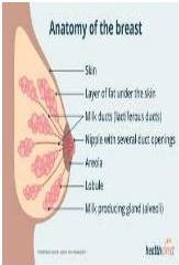
Objectives
- Discuss the features of different forms of benign non-neoplastic and neoplastic breast.
- List the benign breast diseases that increase a patient’s risk of developing breast cancer.
- Outline other risk factors predisposing to breast cancer & incidence/prevalence of breast cancer.
History
Part 2 opens its case series with the consultation approach:
-
A 50-year old female with a breast lump is in your clinic.
-
Q: What would you do?
-
Self introduction, privacy, chaperone, permission
-
History
- Name, age, nationality, gender
most common breast disease presentation ?
History of presenting complaint (lump)
- When noticed (duration)?
- How noticed?
- any other lumps?
- use of contraceptive pills?
- Any change since first noticed?
- progression
- gradual change
- Relationship with menstrual cycle?
- Any history of trauma?
5 cases necrosis and mimics malignancy
- Any pain (cyclical, non-cyclical), discharge (colour, uni/bilateral)
Painful more with benign Painless could be benign or malignant
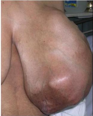
Past history
- Breast problem, mammogram, breast biopsy
- Exposure to radiation (face, chest) — risk factor
- Menstrual & pregnancy history
- early menarche
- Family history
- Other systems inquiry, medical/surgical history
Examination
Inspection of breasts
- Position: Semi-recumbent (45°), Supine, sitting
- Expose upper half (both breasts, chest wall, neck & arms)
- Arms by the sides / raised above head
- Stand in front of the patient
- 4 quadrants of breasts
Breast examination (positions)
Positions include the patient seated or standing (A) with arms at sides; (B) with arms raised over the head, elevating the pectoral fascia and breasts; (C) with hands pressed firmly against hips; or (D) with palms pressed together in front of the forehead, contracting the pectoral muscles. (E) Palpation of axilla; arm supported as shown, relaxing the pectoral muscles. (F) Patient supine with pillow under the shoulder and with the arm raised above the head on the side being examined. (G) Palpation of breast in a circular pattern from the nipple outward.
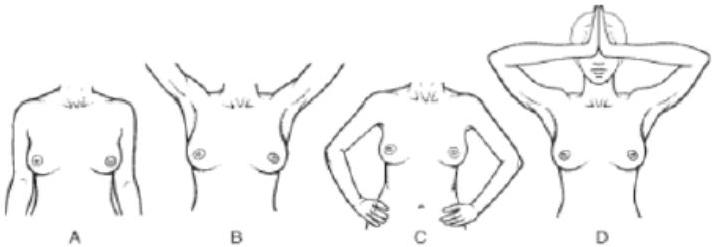

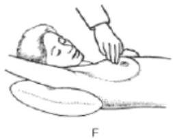
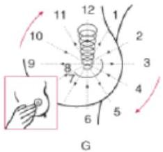
Figure 2: The Clinical Breast Exam [11].
Inspection
- Symmetry & size (underlying lump) — underdeveloped, enlarged
.size aprasmilly .nipples level .nipple - Arkin Complex
- Any obvious mass or lump?
- Site, size, shape
- Any skin change?
- Redness — infection, inflammatory carcinoma
- Oedema — peau d’orange
- Dimpling, ulceration — carcinoma
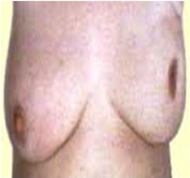
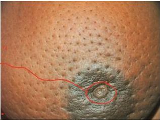
“Inflammatory Carcinoma is malignant the whole nipple is retracted
in benign there’s midline retraction is slit retraction
Inspection — nipple / areola
- Changes in the nipple/areola:
- raised level, retraction (carcinoma, duct ectasia)
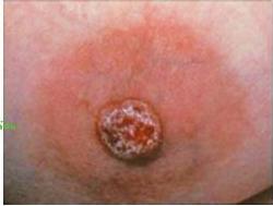
- ulceration (Paget’s disease)
unilateral malignancy dermatological disease usually Bilateral
- Discharge from the nipple:
- unilateral/bilateral
- spontaneous, colour
discharge duct ectasia young pit will come with
- Green discharge
- smoker with nipple retraction
- non lactating women
mildly discharge → Galacturhea Bloody discharge → duct papilloma Gill : morphology
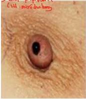
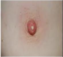
Raise arms above the head
- Both breasts & axillae — any change?
- (Exaggerates asymmetry & skin attachment)
- Supraclavicular area
Palpation
- Semi-recumbent / lying flat with arm under the head
- Ask for any painful area?
- Normal side first
- Palmer surface of the fingers/hand — searching for lump
- Use finger pulp/tips for characteristics of lump — what characteristics?
- Palpation of axillary/SC/IC lymph nodes
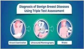
Triple assessment
Always remember
Triple Assessment
- Clinical — History & clinical examination
- Radiological — Breast imaging (mammogram, US, MRI)
- US — young (more fat tissue) + mammogram — 35, 40 — older +
- New older have mammogram + complementary US
- Pathological — Core biopsy (palpation / image guided) — core is better
How you investigate for PI with Breast lumbo? Diabetic diagnosis D
Pathological methods of diagnosis
- FNAC? True cut biopsy?
- Incisional biopsy
- Excisional biopsy
- Image-guided biopsy
Very small lump, not palpable — In situ
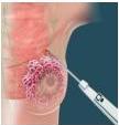
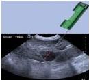
Investigations (breast lump workup)
- Hematology, Biochemical (U/E, LFTs)
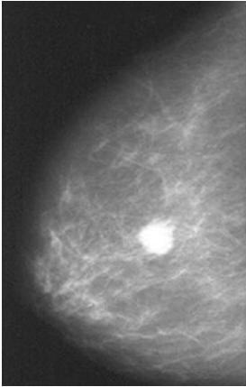
- Mammography (spiculated mass, clustered microcalcifications — linear/branching, architecture distortion)
- US, MRI
- Biopsy: Core needle biopsy by palpation / image guided
- Type, size, margin, grade, ER/PR status, Her2/neu
Staging workup
Done after histological confirmation. Aim: assess extent of the disease.
- CXR
- LFTs for liver metastasis
- Ultrasound
- CT
- Bone scan (if ALP, calcium raised)
- PET scan
2. Benign Breast Diseases
Acute mastitis & breast abscess
Theory
- Mostly diagnosed clinically
- Commonest form of mastitis ass. with breast-feeding
- Cracks/fissures in the nipples.
- Bacterial infection (esp. Staph. aureus)
- Usually unilateral — acute inflammation in the breast. Can lead to abscess formation I&D, aspirate and send for C/S.
Treatment = surgical drainage (often under general anesthesia) and antibiotics (according to C/S result) — empirical then shift.
Most common organism
- Staphylococcus
Su tts
- Cloxacillin
- Augmentin
Case 1 — Lactational mastitis (Part 1, “Case 1”)
A 30 years old lactating lady presented with Rt. breast pain fever for the last 2 days.
O/E Vitals pulse: 100, temp 38.5, BP 100/70
Local breast examination revealed swollen, red, tender to palpate Rt breast with white discharge.
- What is your differential diagnosis? — I’m to know if baby is teething or not
Case score
- Mastitis as signs of inflammation
- Can be breast cancer (but 2 days usually favor benign)
- If high WBC’s I might consider infection
Next step in management?
- Physical exam + then investigation
Complications?
- Abscess formation, chronic
Case 11 — Lactational abscess (Part 2, “Patient 7”)
A 30-year lactating woman presented with breast pain and swelling.
This abcess
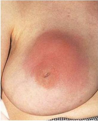
US — localize pus Aspiration / I&D, antibiotics, breast pump if small and early stage if big for lactation
Mammary duct ectasia
Case 2 — Green nipple discharge (Part 1, “Case 2”)
A 52 years old lady presented complaining of nipple discharge from her left breast which is green in colour, for the last 5 weeks, associated with swelling not tender soft on consistency ill defined edges, axilla free.
Green or bloody = malignancy or premalignancy
- What is your differential diagnosis? — duct ectasia
- Work up?
- Who to treat?
Theory
- 5th and 6th decades
- Affects mainly large ducts
- Periductal chronic inflammation
- Destruction and dilation of the ducts with fibrosis
- The underlying cause is unknown
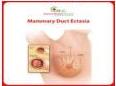
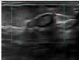
Retro areolar — No abnormal features — So benign but premalignancy
- Poorly defined periareolar mass; can be confused clinically/radiologically with carcinoma
- Can also present as a thick, cheesy or green nipple discharge +/- mass
- Periductal fibrosis → skin retraction
Treatment: Microductectomy for single duct and/or Hadfield’s operation for multiple ducts. Excision of all ducts involved with needle through nipple / wire, then see the duct around the probe.
Fat necrosis
- Uncommon lesion; may be a history of trauma, prior surgical intervention or radiation therapy
- Characterized by a central focus of necrotic fat cells with lipid-laden macrophages and neutrophils
- Chronic inflammation with lymphocytes and multinucleated giant cells
- Major clinical significance: its possible confusion with carcinoma (e.g. fibrosis → clinically palpable mass / calcification seen on mammography)
- Benign
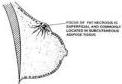
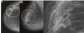
TRAUMATIC FAT NECROSIS
Non-proliferative (“fibrocystic”) changes
- Usually diagnosed 20 to 40 years
- Present as palpable lumps, nipple discharge or mammographic densities/calcifications — lumpy breast
- Often multifocal and bilateral → general “lumpiness”
- No if changes and benign
- No surgery
- Triple assessment to exclude malignancy
Benign tumours
Fibroadenoma
Theory
- Patients usually present < 30 years
- Classic presentation is that of a firm, mobile lump (“breast mouse”)
- Giant forms can occur, especially in younger patients
- Can be associated with proliferative changes in the adjacent breast tissue
- Approx. 20% of lesions are complex fibroadenomas — characterized by certain specific histologic features
Case 3 — Young lady with a mobile lump (Part 1, “Case 3”)
- A young lady of 18 years old presented with left breast lump 4×6 cm which is painless, moving in all directions from site to site, no other abnormality, right breast was normal.
- From this short scenario, what is the differential diagnosis? — Fibroadenoma
- What is your next step of management?
- How are you going to treat her? — Triple [assessment], might have FNAC
- Erty
- Or surgical if pt wants
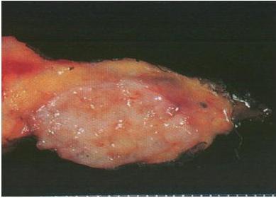
Case 7 — Painless lump in a 20-year-old (Part 2, “Patient 3”)
-
A 20-year old female with a painless breast lump.
-
Examination:
- Right: normal
- Left: well defined, mobile, 3×2 cm firm mass
- No LAP
-
What next?
-
U/S — smooth outline mass.
-
Core biopsy — Fibroadenoma
most Common Breast disease is a must
? Neoplasm, ? Aberration of development No
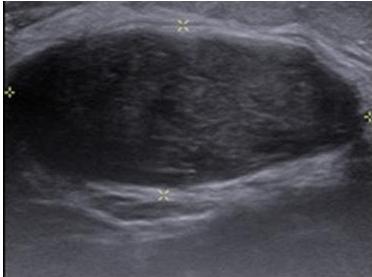
- Age group — < 30 years.
- Well-circumscribed, smooth, firm, mobile mass
- Multiple or bilateral
- Some increase in size. > 5 cm — giant fibroadenoma
- 1/3rd regress spontaneously
- Treatment: depend on size
- < 2 cm — reassurance & follow up
-
2 cm — excision, or less than 2 cm but pt is Anticus (garbled — likely “anxious”)
Duct papilloma
- Duct itself, not like fibroadenoma which is from the stroma
- Benign papillary epithelial tumour; occurs mainly in large ducts
- Papillae are fibrovascular stalks lined by layers of proliferating epithelial and myoepithelial cells
- Most patients present with a serous or bloody nipple discharge
Treatment
- Lumpectomy
- Excisional biopsy
- Wide local excision with free margin — Best
Case 9 — Bloody nipple discharge (Part 2, “Patient 5”)
- History: bloody nipple discharge
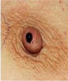
- Examination: 0.5×0.5 firm sub-areolar mass,
- bloody discharge from nipple on pressure
Differential:
- Intraductal papilloma
- Carcinoma
- Investigations: mammogram, U/S, ductogram
- to see which duct is involved
Duct papilloma
- Treatment: duct excision — microdochectomy.
Breast cyst
Case 8 — Premenopausal cyst (Part 2, “Patient 4”)
-
A 45-year female with a painless mass — left breast
- in premenopausal age ⇒ always think about cyst
-
Examination:
- A 6×6 cm mass, non-tender, firm, well defined with smooth surface
-
What next?
-
Mammogram, U/S — cyst
-
Distended involuted lobules.
-
Perimenopausal women.
-
Smooth discrete lump, usually painless.
-
Treatment: Observation (asymptomatic)
-
Aspiration (symptomatic): clear fluid & no residual mass
- Re-evaluate in 4–6 weeks then discharge patient.
-
Excision — hemorrhagic fluid / residual mass or relapsing cysts
so I will excise it to rule out malignancy
Phyllodes tumour
Case 10 — Large bosselated mass (Part 2, “Patient 6”)
- 40-year old female
- Large mass left breast, painless
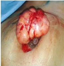
- Examination: large, bosselated mass, non-tender, no skin or deep attachment.
- No axillary LAP
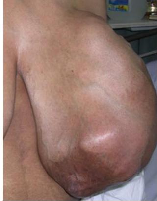
- Mammogram, U/S
- Core biopsy — Phyllodes tumour
- Fibroepithelial tumor, fibrous tissue
- Most benign, some malignant.
- Large, bosselated, no attachment.
- Malignant — metastasize by blood
- Treatment: Wide local excision.
- Mastectomy — very large tumours.
- No axillary lymph node clearance needed
3. Carcinoma of the Breast
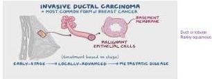
Case 4 — “Malignancy until proven otherwise” (Part 1, “Case 4”)
- A 46-years-old lady presented to her primary care physician with a complaint of a lump in her left breast, which she first noticed a few weeks ago. The lump is painless but has gradually increased in size. She also mentions occasional discomfort in the breast, particularly around her menstrual cycle. (size increased over 3 weeks) During palpation, firm, irregular mass of approximately 2 cm in diameter is palpable in the upper outer quadrant of the left breast. Axilla free.
- What is your differential diagnosis? — Malignancy until proven otherwise — Top
- How do you approach this case? — Triple assessment
Case 5 — Full carcinoma workup (Part 2, “Patient 1”)
A 50-year old female with a breast lump is in your clinic.
Patient 1 — history
- Age: 50 years
- Noticed the lump — 2 weeks back
- Painless, no discharge
- PMH & FH: unremarkable
- MH: menopause, menarche at 14
- Breast fed her 2 children
- No medication, allergies — nil
Patient 1 — examination
GE: unremarkable
Local Examination:
- Normal side — NAD
- Affected side:
- Inspection — NAD
- Palpation: Mass in UOQ, 2.5 cm, firm to hard,
Should be considered malignant until prove otherwise
- No skin or deep attachment
- Axilla: NAD both sides
- What next?
Provisional / working diagnosis
- ? Malignant ? Benign
- ? Most likely diagnosis
- Breast carcinoma
- ? What next
Patient 1 — biopsy result
- Patient 1:
- 2.5 cm, clear margin, low grade
- Invasive duct cell carcinoma,
- HR +ve (Hormone Receptor ER or PR)
good sign
- Her2/neu — negative
L3 if. Amitiv bad sign (garbled)
Epidemiology
- Commonest malignancy in women worldwide:
- Breast cancer is the number one cancer in women. There were 2,296,840 new cases of breast cancer in 2022 among women.
- From 1990 to 2021, KSA reported 19,440 deaths due to breast cancer, increasing from 201 cases in 1990 to 1,190 cases in 2021.
- Breast cancer & Lung cancer, Cervical cancer, Colonic cancer, Stomach cancer. (garbled list — likely “among the commonest cancers”)
Risk factors
Age
- Incidence of breast cancer ↑ increases with age
- Uncommon before age 25 years; incidence ↑ increases to the time of menopause and then slows
Family History
- At least two genes that predispose to breast cancer have been identified — BRCA 1 and BRCA 2
- Mutations in these tumour-suppressor genes also predispose affected women to ovarian cancer
- Prophylactic mastectomy
- Approx 10% of breast cancer is due to inherited genetic predisposition
- A woman whose mother or sister has had breast cancer is at ↑ relative risk 2 to 3 times compared to other women
Benign Breast Disease
Certain types of benign breast disease:
- Ductal ectasia
- Ductal papilloma
- Breast carcinoma in situ
History of Other Cancer
- A history of cancer in the other breast or a history of ovarian or endometrial cancer
Hormonal Factors
↑ levels of estrogen ↑ risk:
- Early age at menarche
- Late age at menopause
- Nulliparity
- Late age at first child-birth
- Obesity
Environmental Factors
- High fat intake
- Excess alcohol consumption
- Ionizing radiation
All risks should be covered in Hx
Histologic classification
Breast Cancer (Part 1 figures)
- Ductal
- DCIS (15%)
- IDC (75%)
- Lobular
- LCIS (5%)
- ILC (5%)
Invasive carcinoma (Part 2 figures)
Invasive Ductal Carcinoma
- Most common (80%)
Invasive Lobular Carcinoma
- 5 to 10%
- 30% bilateral, multicentric, multifocal, usually large mass
- Difficult to detect by mammogram
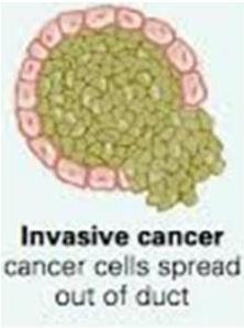
Origin: carcinoma develops from epithelium lining the terminal duct-lobular unit
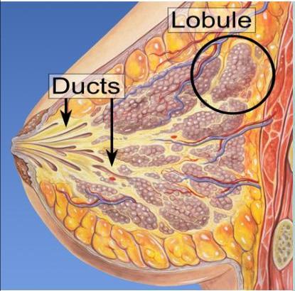
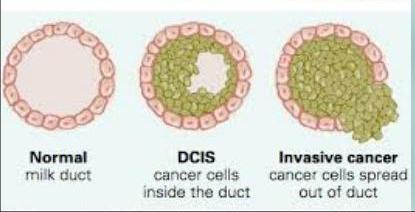
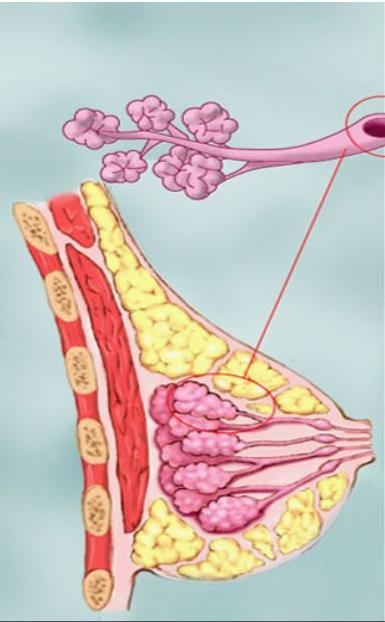
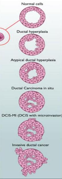
Carcinoma in situ — non-invasive, Stage 0
Malignant cells have not invaded the basement membrane
DCIS cancer cells inside the duct more common
- Ductal carcinoma in situ (DCIS) — most common.
- 3–4% of symptomatic, 25% of screen detected
- Lobular carcinoma in situ (LCIS) — marker of increased risk of invasive breast cancer (1%/year), familial disease
- multifocal (same quadrant), multicentric (different quadrants), bilateral
- DCIS to LCIS is 3:1
Management of DCIS (Stage 0)
- Localized & < 4 cm: wide local excision with 10 mm normal tissue + adjuvant radiotherapy
-
4 cm or multicentric: mastectomy
- No axillary lymph node dissection
- No adjuvant chemotherapy/hormone
Management of LCIS (Stage 0)
- Surveillance alone — mammography
if no family life and BRCA 1,2 are negative
- Surveillance + Tamoxifen
- if estrogen receptor +ve
- Bilateral prophylactic mastectomy
like Angelina Jolie
- (concerned of cancer risk, strong family history, mammographic dense breast)
- 20–30% lifetime risk of invasive ductal carcinoma
Staging
TNM classification (Part 2 table)
- The TNM system helps to establish the anatomic extent of the disease, and the combination of the 3 factors can define the overall tumor stage.
- This method allows for simplification, with cancers staged from I–IV, with stage IV being the most severe stage.
| TUMOURS | T0/ Tis | T1 | T2 | T3 | T4 |
|---|---|---|---|---|---|
| TUMOUR SIZE | T0- no tumour Tis- within duct/ lobule | 0-2 cm | 2-5 cm | > 5 cm | Any size with extension to skin/ chest wall Inflammatory breast cancer |
| LYMPH NODE | N0 No lymph node metastasis | N1 Cancer cell in 1-3 lymph nodes | N1 mi Lymph node tumour < 2mm | N2 Cancer cells in 4-9 Lymph nodes | N3 Cancer cells in infra/ supraclavicular LN or > 10 axillary LN |
| METASTASIS | M0 No evidence of metastasis | M1 Metastasis found in other area |
TNM classification (Part 1 table)
| Category | — | — | — | — |
|---|---|---|---|---|
| Tumor size (T) | Tumor size < 2 cm — T1 | Tumor size 2-5 cm — T2 | Tumor size > 5 cm — T3 | Tumor extends to size or chest wall — T4 (Early to deep dissolved orange) |
| Lymph Nodes (N) | N0 No lymph node metastasis | N1 Metastasis to ipilateral, movable, axillary LNs | N2 Metastasis to ipilateral fixed axillary, or IM LNs | N3 Metastasis to infraclavicular/supraclavicular LN, or to axillary and IM LNs |
| Metastasis (M) | M0 No distant metastasis | M1 Distant metastasis |
Original M-row fragment (repeated ”+ LN” collapsed to a single instance): N2 + IM + LN N3 + IM + LN
Stage groups
-
Staging systems includes TNM and the Manchester classification
-
Tumor size and axillary node status are important parameters
-
10-year survival rate for lymph node negative disease is 80% vs 35% for tumours with positive nodes
-
Stage 0 — Indicates carcinoma in situ. Tis, N0, M0.
-
Stage I — Localized cancer. T1-T2, N0, M0.
-
Stage II — Locally advanced cancer, early stages. T1-T2, N1, M0.
-
Stage III — Locally advanced cancer, late stages. T1-T4, N2-N3, M0. Neoadjuvant therapy then breast conservative surgery then radiation accordingly
-
Stage IV — Metastatic cancer. T1-T4, N1-N3, M1. Lung liver Bone brain — Supraclavicular Axillary Deep mediastinal — Blood, liver then brain
TIS depends: Early Locally advanced Late locally advanced
Prognosis
Tumour grade
- Different grading systems exist
- ↑ tumor grade = worse prognosis
Histologic subtypes
- Nottingham grading
Hormone receptors
- Estrogen receptors — ER/PR-positive → good prognosis & treatable with anti-estrogen therapy.
- Progesterone receptors
- =&HER2 receptors
- HER2-positive → bad prognosis, but treatable with trastuzumab.
Molecular markers
- Inc. c-erb-B2, c-myc and p53 — bad prognosis
- Brown medication, herceptin
- p53 and c-myc → bad prognosis due to genomic instability and aggressiveness.
Adverse tumour characteristics
- Positive LN
- Higher histological grade
- Triple negative
- Lymphovascular invasion
- High proliferation rate
- (>5% cells in S-phase, > 20% Ki-67)
Treatment options
Surgery
- Preserve fascia of the pectoralis sheath
- Mastectomy (simple & modified radical mastectomy, toilet mastectomy for palliative treatment)
- Breast conservation (lumpectomy, wide local excision with free margins)
- +/- Axillary dissection (sentinel lymph node biopsy & axillary sampling)
- Dye injected in the lump then see the drain, the first one, we remove and see histopathology
- If + we remove axillary lymph nodes = MRM usually
Surgery (breast + axilla) (Part 2):
- BCT + SLNB/ALND + Radiotherapy
- Mastectomy (MRM) + SLNB/ALND
modified Radical mastectomy “preserve the muscles”
- Multidisciplinary team: surgeon, radiotherapist, medical oncologist & nurse specialist
- Operable:
- Early (T1-T2, N0-N1, M0) — surgery + adjuvant therapy
- Locally advanced (T3-T4, N2, M0) — neoadj + surgery + systemic
(for debulking
Breast-conserving treatment (BCT)
- < 4 cm tumor — excision of tumor with 1 cm normal tissue margin
- LN: Sentinel node biopsy + node clearance
- Postoperative radiotherapy (RT)
- Contraindications:
- Widespread disease
- Unsuitable for radiotherapy
- (collagen disease, pregnancy — 1st & 2nd trimester)
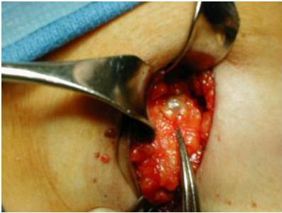
Modified radical mastectomy (MRM)
-
Mastectomy + SLNB ± clearance
- Larger tumor, widespread disease
- Choose this treatment if pt is anxious
-
Radiotherapy only if:
-
3 LN involvement,
- Lymphatic/vascular invasion,
- Grade 3 tumor,
-
4 cm tumor,
- Tumor attached to pectoral fascia or close surgical margin < 5 mm
-
-
Breast reconstruction — used selectively
Radiation therapy (local control)
- In local mets + breast conservative
Chemotherapy (neoadjuvant & adjuvant) (systemic control)
- Chemotherapy, hormone therapy, immunotherapy
- Adjuvant chemotherapy: given after surgery/RT.
- All patients — except tumor < 1 cm & grade 1
- Common regimens: FAC (5-fluouracil, adriamycin, cyclophosphamide) 6 cycles / 21 days.
- AC (adriamycin, cyclophosphamide), FEC (5-fluouracil, epirubicin, cyclophosphamide)
- Neoadjuvant chemotherapy (T3-T4 N2, M0):
- Before surgery/radiotherapy — shrinks larger tumors
Hormonal therapy (systemic control)
- In hormonal receptors +ve disease
- Tamoxifen (partial estrogen agonist): hormone receptor +ve tumours
- 20 mg / day for 5 years for pre & postmenopausal
- Luteinizing hormone-releasing hormone (LHRH) agonist (goserelin) —
- induces reversible ovarian suppression for premenopausal
- Aromatase inhibitors (blocks conversion of androgens to estrogen): letrozole, anastrozole, exemestane. Postmenopausal hormone receptor +ve tumors
- Oophorectomy: women < 50, ER +ve tumors,
- Metastatic disease (surgical or radiation)
Anti-HER2 therapy
- 15–20% tumors express HER2/neu
- Worse prognosis than HER2/neu negative tumors.
- Humanized monoclonal antibody:
- Trastuzumab (Herceptin)
Advanced disease
- Advanced = palliative surgery or care according to presentation (furgating + orange)
- Toilet mastectomy as palliative surgery / not curative
Mammary Paget disease
Case 6 (Part 2, “Patient 2”)
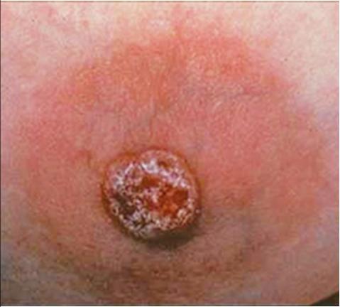
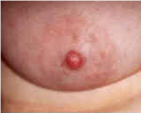
- 1–4% — breast cancers, predominantly unilateral
- Peak incidence — sixth decade
- Paget cells — large, pale epithelial cells, hyperchromatic, atypical nuclei
- In situ / invasive cancer
- Nipple-areola:
- Scaly, fissured, oozing, erythematous, itching, burning, or pain
- Retraction/ulceration
- Underlying palpable mass
what investigation you will order Mammographic, MRI
4. The Male Breast
Gynaecomastia
- True = the glandular tissue is enlarged
- Or pseudo gynecomastia = the fat is high but not the tissue
- Pathological or physiological
- Can be unilateral/bilateral; present as diffuse enlargement / defined mass
- Most important clinically as a marker of hyperestrinism
- Neoplasia needs to be excluded in certain cases
- Pseudo: no treatment, only counseling
- True: treat the cause, or retro-areolar mastectomy if other measures failed
Carcinoma
- Very rare occurrence; female cancer to male cancer ratio approx 100:1
- Pathology and behavior is similar to cancers seen in women although with less breast tissue, skin involvement is more frequent
