Common Pediatric & Neonatal Surgical Emergencies
Tariq Altokhais
Overview
| Neonatal | Pediatric |
|---|---|
| Malrotation/midgut volvulus | Malrotation/midgut volvulus |
| Pyloric stenosis | Intussusception |
| Hirschsprung’s disease | Hirschsprung’s disease |
| Esophageal atresia | |
| Intestinal atresia/obstruction/NEC | |
| Anorectal malformation (imperforate anus) | Acute appendicitis |
| Congenital Diaphragmatic Hernia | Acute scrotum |
1. Acute Appendicitis
Acute appendicitis → Same presentation and management as in adult
- Most common cause of acute surgical abdomen in children and adolescents
- Peak incidence 10-12 years
- Increased rate of perforation in children
2. Malrotation / Midgut Volvulus
If not treated, will be
Intestinal Rotation (Embryology)
- Normal rotation of the intestine requires transformation from a simple, straight alimentary tube into the mature fixed and folded configuration normally present at birth.
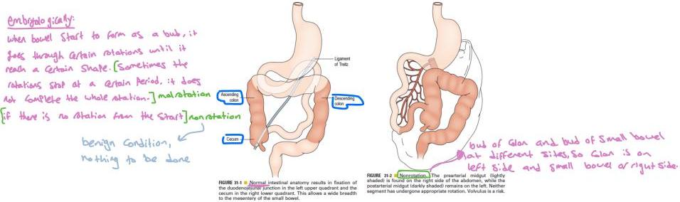
Normal Midgut Rotation
- Normal midgut rotation:
- 4-12 weeks of fetal life
- 270 counterclockwise movement of the midgut
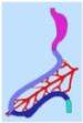
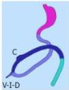
Rotation – Midgut (Hindgut Derivatives)
Also note hindgut derivatives
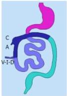
- C: caecum
- A: appendix
- V-I-D: vitellointestinal duct
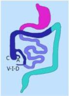
Malrotation (Incomplete Rotation)
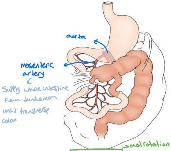
FIGURE 31-3 Incomplete rotation. Both the prearterial (lightly shaded) and postarterial (darkly shaded) segments have undergone partial, yet not complete, rotation. Ladd’s bands are seen attaching the cecum to the right posterior abdominal wall. The duodenum becomes compressed and possibly obstructed. Volvulus is a risk.
- Duodenum and small bowel go down, and cecum is up, and there is a band → this band normally attaches cecum to abdominal wall so it is fixed
- In malrotation, the band goes up, and when cecum expands, it pulls the bands which cause obstruction of duodenum
What Is the Problem with Malrotation?
- Ladd’s band obstructing the duodenum >> bilious vomiting
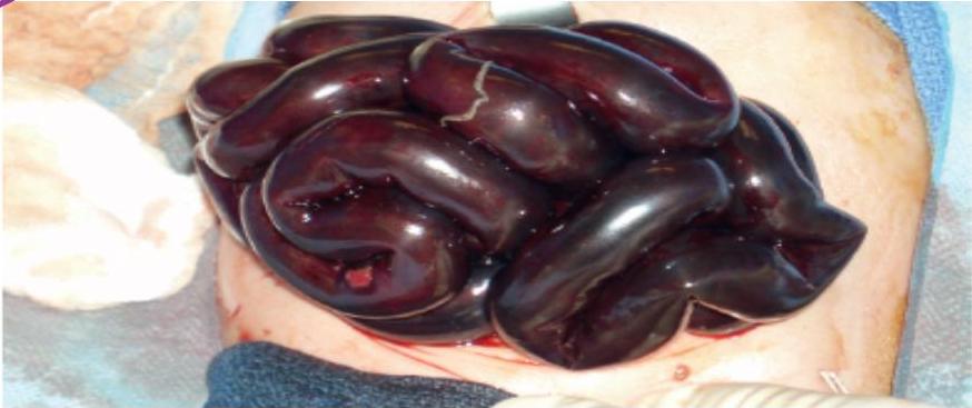
- Midgut volvulus (twisting) >> ischemia and bowel gangrene
- If whole bowel got twisted multiple times and there is delayed diagnosis (of duodenum till transverse colon)
- If not identified quickly >> patient will die due to septic shock
- If the patient does not die >> entire bowel loss (short bowel syndrome) – not compatible with life
Presentation
- 75% present in the first month of life
- 15% in the first year
Cardinal symptom:
- BILIOUS VOMITING → dark green (extremely important)
Other symptoms:
- Abdominal pain
- Scaphoid abdomen (initially)
- Distended abdomen (late)
- Rectal blood (not always)
The younger, the more common to get malrotation
- 30% present in the 1st week of life
- +30% → 60% present in the 1st month of life
- +30% → 90% present in the 1st year of life
- 10% present at any age
Physical Examination (P/E)
- Lethargic
- Dehydrated
- Tachycardia, hypotension, +/- fever
- Distended upper abdomen (stomach)
- Distended abdomen (late)
- Peritonitis (late)
- Abdominal wall erythema (late)
Investigations
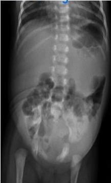
- Abdominal X-ray: → non-specific
- Can be totally normal
- Dilated stomach
- Upper GI contrast study (oral contrast):
- The most sensitive test to diagnose malrotation (gold standard)
- Takes time (should not be done if the patient has a picture of bowel ischemia)
- If not sure about the diagnosis → e.g. color of vomit is not dark or repeated vomiting led to bile reflux; do it if there is time and if the condition is stable enough
Upper GI Contrast Study – Duodenal Configurations
- C Shafes: Duodenum
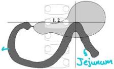
- Normal Position of Duodenum
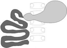
- Nonrotation of Duodenum
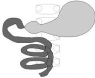
- Corkscrew Duodenum and Jejunum
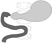
- Partial Duodenal Rotation with Jejunum in Right Upper Quadrant
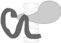
- Partial Duodenal Rotation with Duodenojejunal Junction over Right Pedicle
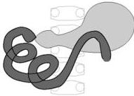
- Redundant Duodenum – Malrotation to Right of Spine
Contrast Study – Normal vs Malrotation
- Normal anatomy of the duodenum:
- Duodenal cap
- Duodenal loop
- C-shaped duodenum
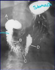
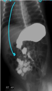
Anything different than this is a malrotation
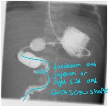
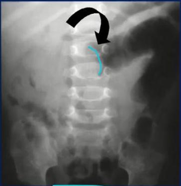
Abdominal X-ray Findings
- Duodenum and jejunum on right side
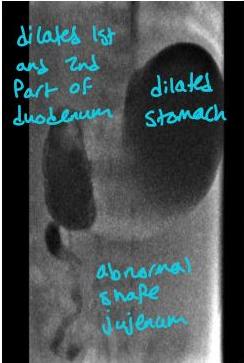
Treatment
- Intravenous fluid → rehydrate
- NPO
- NGT
- Broad spectrum antibiotics
- Immediate surgical consultation
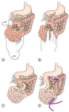
- Urgent Ladd’s procedure:
- Laparotomy:
- ① Untwist the bowel (derolate)
- Resect gangrenous intestines
- ② Divide Ladd’s band → so duodenum will be free
- ③ Place the bowel in non-rotation position (small intestine on the right, large intestine on the left of the abdomen)
- Do appendectomy at the same time → because if later on gets appendicitis, will have different presentation
- Laparotomy:
Ladd’s Procedure
Delayed Diagnosis / Management
- Bowel gangrene
- Short bowel syndrome → stay on TPN until they die
- Septic shock
- Multi-organ failure
- Death
- (Bowel transplantation is difficult to find donor)
- Prevention is key in these cases
3. Intussusception
In case of delayed presentation, child will present with ischemia, require resection and anastomosis
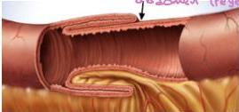
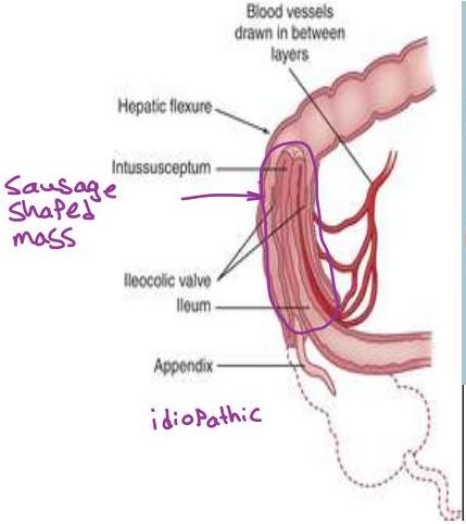
- Part of the intestine telescopes into the distal bowel by peristaltic activity
- Bowel is compressed > venous obstruction > bowel wall edema > arterial insufficiency > ischemia & bowel wall necrosis
Causes
- 3m-3y: idiopathic – 95% (Peyer’s Patch)
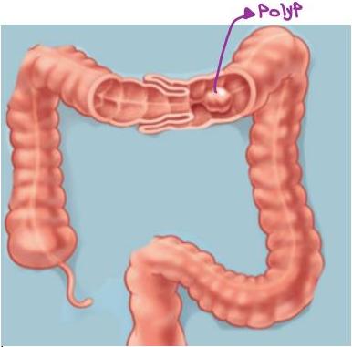
- >3y: pathologic lead point – 4% Meckel diverticulum
- Polyp
- Neoplasm
- Inverted appendix
- Celiac disease, CF, HSP
- Feeding tubes (GJ tube)
- Something leading bowel to go inside & any mass
- 1-2 weeks or even month before episode of intussusception
- Inflammation goes down to LN of the same lymphatic tissue of glodomen (Peyer’s patch)
- Swollen, so pulls the bowel, leading to intussusception
Presentation
- Typical age: 3 months-3 yrs
- Episodic colicky abdominal pain:
- Every 1-2 hours
- Every part of the child is flexed during the attack
- Child is well between the attacks
- Bilious vomiting
- Red currant jelly stool
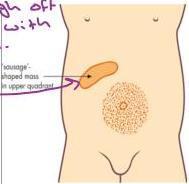
- Sausage-shaped abdominal mass in RUQ (65% of cases)
- Empty RLQ
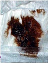

When bowel goes inside, part of bowel gets congested (venous congestion) so mucosa will slough off and go in stool; stool is mixed with mucosa.
Investigations
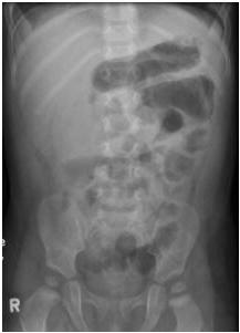
- ① AXR: RUQ mass, dilated bowel, air/fluid levels → not specific
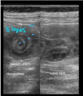
- ② US: gold standard, target sign
- Los Asueda kidney shape
Management
- NPO
- IVF
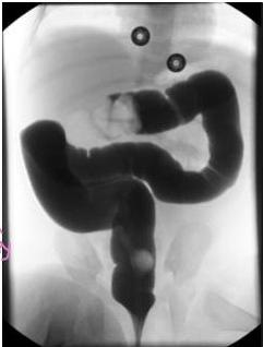
- Reduction enema: under fluoroscopic guidance (done by a radiologist)
- Successful in >75% of cases → if failed, do surgical reduction (depending on presentation and duration)
- By fluid, contrast, or by air, under controlled pressure
- Insert enema by Foley’s catheter or any catheter and see progression under x-ray fluoroscopy
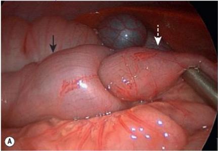
- Surgical reduction: if enema reduction is not successful or not available
- → Laparoscopy
Reduction (Enema Technique)
- Should continue contrast until completely reduced
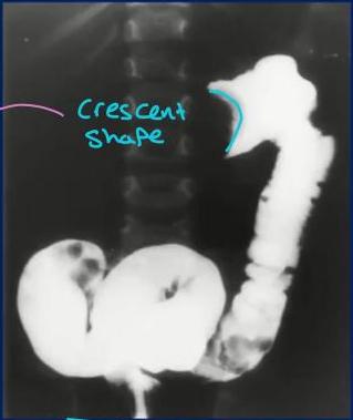
- Water-soluble contrast: → gastrografin ↓ barium
- Air contrast
4. Hypertrophic Pyloric Stenosis
- Gastric outlet obstruction due to hypertrophied pyloric muscle
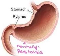
- The pylorus opens to let food out of the stomach into the small intestine
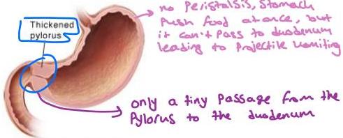
- With pyloric stenosis, thickened muscle around the pylorus prevents food from leaving the stomach
Classic Triad
- Term baby
- 2-8 weeks of life → unknown if congenital or acquired
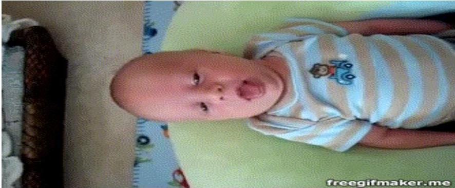
- Projectile, non-bilious vomiting (not as emergency as malrotation)
- May be progressive, that’s why it doesn’t present right after birth
- → Color is white due to milk
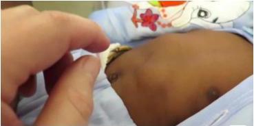
- Olive sign – can be felt by palpation

Barejo’s 2013
Projectile Vomiting
Clinical Features & Laboratory Findings
- Hungry & dehydrated baby
- Palpable pyloric muscle (Olive sign)
- Gastric peristalsis can sometimes be seen
Electrolytes + blood gas:
- Hypochloremia
- Hyponatremia
- Hypokalemia
- Metabolic alkalosis
Imaging
- ① X-ray → will not show anything
- ② US: gold standard
- Thick and elongated pyloric muscle
- Diagnosis depends on measurement of the thickness
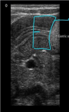
Thickened pylorus
Management
- NPO
- IVF
- Correct electrolytes and pH
- Surgical separation of pyloric muscles (pyloromyotomy) – no excision
Pyloromyotomy
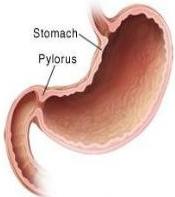
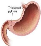
- The pylorus opens to let food out of the stomach into the small intestine
- With pyloric stenosis, thickened muscle around the pylorus prevents food from leaving the stomach
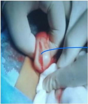
- Spread pyloric muscles to see → mucosa
5. Esophageal Atresia (Congenital; with or without Tracheoesophageal Fistula)
- Interruption in the continuity of the esophagus with or without fistula
- Caused by error in separation of esophagus & trachea (4th week gestation)
- 1:4500 live birth
- Male > female
- 50-60% associated anomalies
- VACTERL: Vertebral, Anal, Cardiac, TracheoEsophageal, Renal, Limb
Types of Esophageal Atresia
- Proximal pure esophageal atresia (no fistula): 5-10%
- Type 2/A
- Type 3a/B
- Type 3b/C: most common type (>85%); distal is altered to trachea (tracheoesophageal fistula)
- No gas = no fistula
- Type 4/E
Presentation
Prenatal:
- Polyhydramnios
- → Fetus doesn’t swallow the amniotic fluid, so it accumulates
Postnatal:
- Drooling
- Vomiting
- Respiratory distress
- Resistance upon insertion of NGT (coiled in the chest)
- Choking and aspiration (milk goes to trachea)
Part of routine neonatal exam: should rule out choanal atresia or obstruction by inserting NGT → do it before giving 1st milk; if there is resistance → x-ray
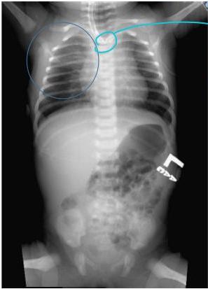
- → Coiled NGT ↓ esophageal atresia until proven otherwise
Management
- Urgent surgery → separate esophagus from trachea and connect the esophagus
- Right thoracotomy
- Division of fistula
- Repair esophagus
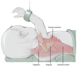
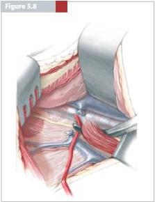
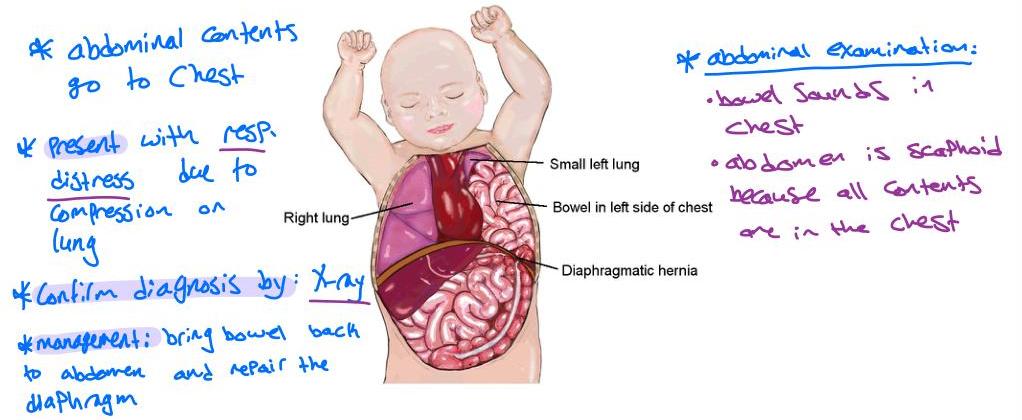
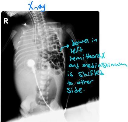
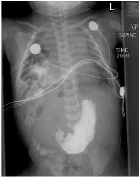
6. Congenital Diaphragmatic Hernia
Easy to diagnose and manage
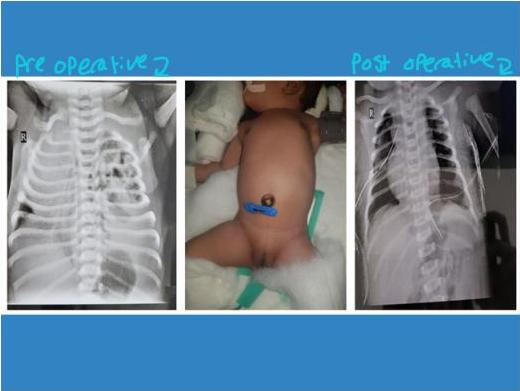
7. Imperforate Anus – Ano-Rectal Malformation (ARM)
- Various forms of anorectal anomaly
- Normal anus:
- Normal size
- Location at the center of the muscle sphincter
- 1:4000
- Male > female
- 65% has associated anomalies (VACTERL)
- Vertebral, anorectal, Cardiac, Tracheoesophageal, renal, limbs
- If you have one of these anomalies, you should look for the remaining other anomalies
- → Should look for other anomalies; types differ in male and female
Presentation
- Abdominal distension and vomiting (rectum is closed)
- But shouldn’t wait for this – in neonatal exam, should look for anus to make sure it’s in normal location and size
Types of Anorectal Malformation
| Sex | Type | Notes |
|---|---|---|
| Female | Perineal fistula | |
| Female | Vestibular fistula | |
| Female | Persistent cloaca | <3 cm common channel / >3 cm common channel |
| Female | Imperforate anus without fistula | |
| Female | Rectal atresia | |
| Male | Perineal fistula | |
| Male | Rectourethral fistula | Bulbar / Prostatic |
| Male | Rectovesical fistula | |
| Male | Imperforate anus without fistula | |
| Male | Rectal atresia |
Vestibular Fistula (or Rectovaginal Fistula)
- No complete obstruction like other parts
- Do anoplasty at age 6 weeks - 2 months (without colostomy)
- Rectum should open here, but here it opens just beside the vagina and the rectum shares a hole with the vagina (one hole for both) → seen by physical examination
- Called vestibular anus because anus opens in vestibule
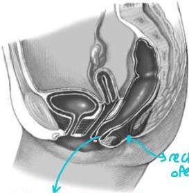
Seen by physical examination
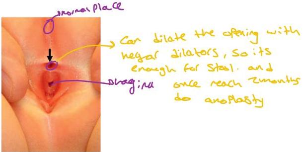
FIGURE 35-7 ■ (A) Schematic drawing of a rectovestibular fistula. (B) Female neonate with a rectovestibular fistula is in the prone position. The rectal fistula (arrow) is located in the posterior aspect of the vestibule.
Perineal Fistula
- Close to normal site → do anoplasty
- Anus should open here, in the perineum
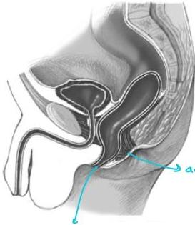
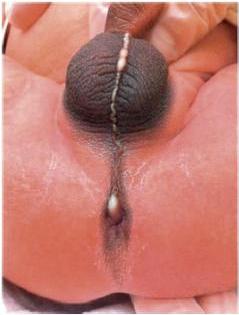
FIGURE 35-2 ■ This male infant has a rectoperineal fistula with a subepithelial tract filled with either mucus or meconium that extends into the scrotal raphe.
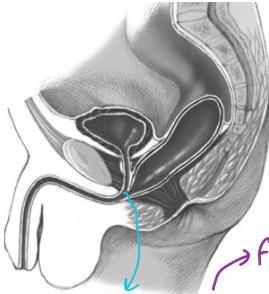
Rectourethral Fistula (Male)
- Most common in boys
- Presentation: stool in urine
- There is a single wall between rectum and urethra, and doing anoplasty immediately after birth can injure the urethra
- Opens in urethra, and there is no anus → far from normal site
- → Do colostomy and after 3-6 months do anoplasty
Surgical Options
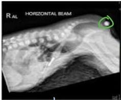
Primary Anoplasty
If rectum is close to skin
- Low type → better prognosis
- X-ray to decide which surgery to do: child in prone position, elevate pelvis, and do lateral X-ray while putting marker at site of anus, and see the gas of the rectum
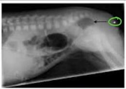
Colostomy with Delayed Anoplasty
If rectum is far from skin
- High type → usually have incontinence
- They don’t have enough fibers and muscles to contract – as part of the disease, not part of the surgery
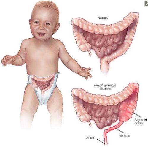
8. Hirschsprung’s Disease
→ Functional disease, not anatomical
- Absence of ganglion cells in the myenteric & submucosal plexus of the intestine
- → Absence of parasympathetic → overacting of sympathetic → bowel contracting all the time
- Arrest in ganglion cells migration from proximal to distal part of GI tract
- Transition zone (TZ): area between normal & aganglionated bowel
- Types:
- 80% - rectosigmoid TZ
- 10% - proximal colon TZ
- 5% - total colonic HD
- Rare - near total intestinal HD
Presentation
Neonatal:
- Delayed passage of meconium >24hr
- Bilious vomiting → picture of obstruction
- Abdominal distension
- Feeding intolerance
Delayed:
- Chronic refractory constipation
- Abdominal distension
- Failure to thrive
When doing PR exam (with cotton tip or temp probe): gush of stool and gas due to stimulation of the area
Diagnosis
- Rectal manometry: absent recto-anal inhibitory reflex
- Contrast study (enema)
Then, to confirm:
- Rectal biopsy:
- Gold standard
- → Bedside biopsy (suction biopsy) → no general anesthesia, it’s not painful
- → Look for ganglion cells; if no ganglion cells → reach diagnosis (histopathological diagnosis)
- Look for ganglion cells in the myenteric & submucosal plexus
- (This is the only disease where it’s asked for absence of cells)
Contrast Study Findings
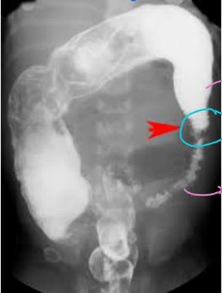
- → Dilated
- → Transition Zone
- → Contracted
Treatment
Excise the abnormal and bring the normal down
- Resection of aganglionic bowel
- Pull-through procedure
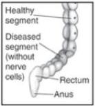
- Before pull-through surgery: the diseased segment doesn’t push stool
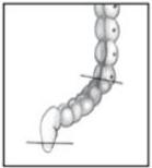
- Step 1: the diseased segment is removed
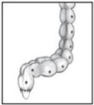
- Step 2: the healthy segment is attached to the remaining rectum
9. Intestinal Atresia
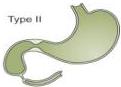
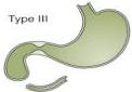
- Duodenal obstruction
- Jejunoileal atresia
- Colonic atresia
- Present with abdominal distension
- Rectal atresia
- Management: surgical
Duodenal Obstruction
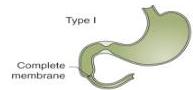
A
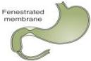
B
- Atresia usually in 2nd or 3rd part of duodenum
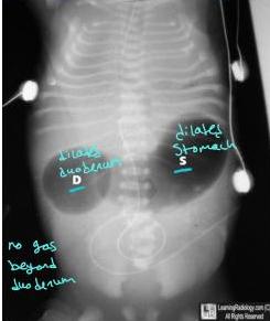
- On x-ray, duodenum is dilated because of long standing obstruction
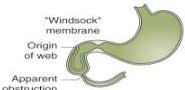
C
D
E
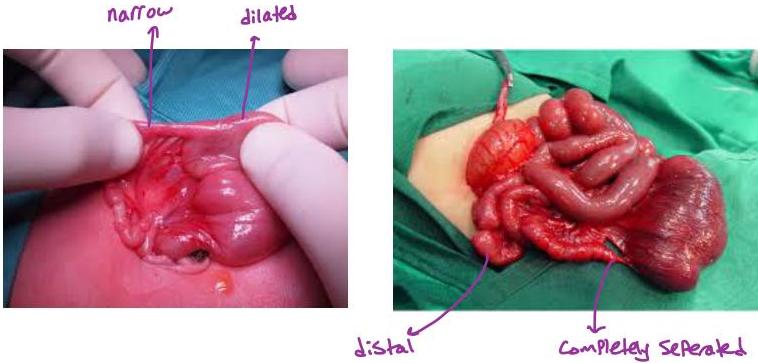
Jejunoileal Atresia
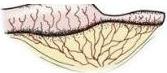
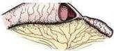
A: Stenosis
B: Type I
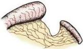
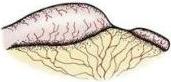
C: Type II
D: Type III (a)
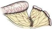
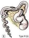
Type III (b)
F: Type IV
X-ray Findings – Bubble Signs
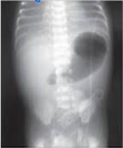
- Pyloric atresia (single bubble)
- Duodenal atresia (double bubble sign)
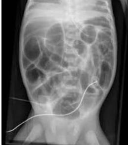
- Small bowel atresia → could be only stenosis or could be completely separated (multiple bubble)
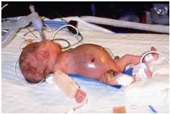
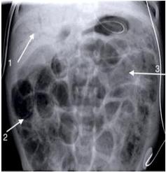
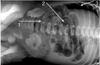
10. Necrotizing Enterocolitis (NEC) y
- Inflammation in small and large bowel
- Sometimes there is perforation (gas in bowel wall)
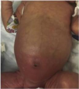
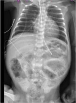
- Usually in premature babies esp. C/kg
- Presentation: abdominal distension
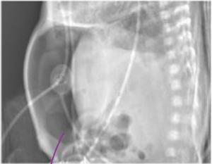
- Free air (perforation)
Management is conservative or surgical, depending on stage and condition of the child