OPERATION ROOM & SURGICAL TECHNIQUES
MOHAMMED ALSEBAVEL
LEARNING OBJECTIVES
- ❖ PRINCIPLES OF STERILIZATION, DISINFECTION, ANTISEPSIS, ASEPSIS
- ❖ PREPARATIONS FOR SURGERY
- ❖ HAEMOSTASIS IN SURGERY
- ❖ COMMON EQUIPMENTS: ELECTRO-SURGERY, DRAINS, SUTURES & INSTRUMENTS
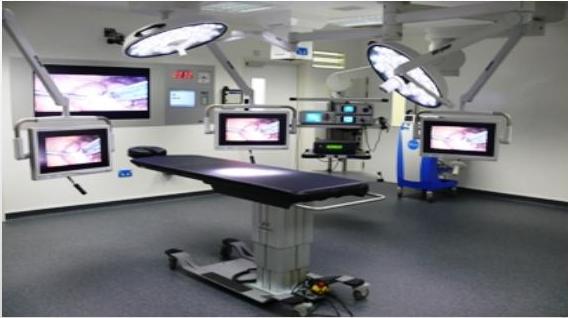
OPERATION ROOM (OR/ OT)
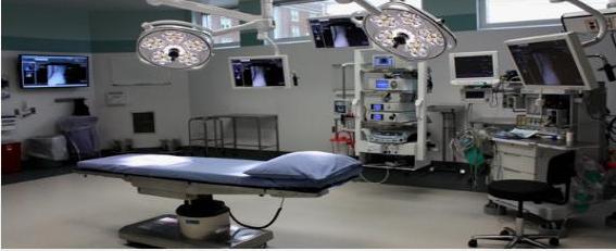
- UNFAMILIAR ENVIRONMENT
- NEW TERMS / TECHNIQUES / INSTRUMENTS
- HIGH STANDARD OF CLEANLINESS & INFECTION CONTROL
INFECTION CONTROL Z
Sterilization: Process of complete destruction of all micro-organisms including bacterial spores.
Disinfection: Process to reduces the number of viable organisms. Does not inactivates viruses and bacterial spores. Used on non-living surfaces e.g. instruments. (bleach, alcohol).
Antisepsis: Destroys organisms between wound & external environment. used on living tissues (chlorhexidine, povidone-iodine)
Asepsis: Process to have as few organisms in the immediate vicinity of the operating field (gloves, gowns, mask etc.)
STERILIZATION
- Two types: Heat & Cold
① Heat Sterilization (Autoclave)
→ machine produce high temp.
- Steam under pressure: increasing the atmospheric pressure raises water boiling point. instruments and drapes (pre-packed). 134°c & 30 lb./in² for 3 min. or 121°c & 15 lb./in² for 15 min.
- Dry heat: 160°c for 2 hrs. suitable for airtight containers & instruments for risk of corrosion.
② Cold Sterilization
- Irradiation: (gamma rays) - syringes, catheters, gauze, sutures.
- Ethylene oxide: highly penetrative gas used for delicate items - endoscopes, electrical equipments, single use plastic items.
- Glutaraldehyde (cidex): liquid chemical to clean lensed instruments
PATIENTS’ PREPARATION FOR SURGERY
- Shower
- Shaving vs clipping → done in the OR (to dec chance of wound infection) (cause more wound infection so instead do)
- Wide area of skin cleaned - povidone-iodine & alcohol
- Surgical field isolated with sterile drapes → to cover patient completely
- Prophylactic antibiotics: before skin incision (given once get the call to take Patient to OR to give the antibiotic as close as possible to the skin incision, to get highest level of antibiotic during skin incision)
PREPARATION OF SURGICAL TEAM
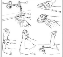
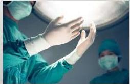
- Scrubbing: hand & fore-arm cleaning process
- All jewelry removed
- Fingers, hand & fore-arm cleaned - Chlorhexidine (Hibiscrub) 3-5 min.
- Drying of hands and forearm with sterile towel
- Gowns & gloves
- High-risk patients - HIV, hepatitis b & c → should do double gloving for these patients.
PATIENT POSITIONS
aim of Positioning is to make easier for surgeon for adequate exposure.
- Supine → in most abdominal surgery
- Trendelenburg: supine with 30° head down
- Reverse trendelenburg: head up
- Lithotomy: supine with knee & hip fully flexed & foot in stirrups → esp. for gynecological and rectal operations
- Lloyd-Davies: as trendelenburg - legs abducted, hips & knee slightly flexed and legs in rests. ↓ for AP resection (for cancer of lower rectum) (resection of whole rectum)
- Lateral: with upper arm raised above & in front of the head.
- ex. doing thoracotomy on right side, position the patient on left side during surgery
- ex. doing laP. splenectomy, position patient tilted slightly to right side (left side up, right side down) so by gravity spleen go down to help exposure
Trendelenburg & Reverse Trendelenburg


Lithotomy Position


Lloyd-Davies Position


Surgeon above and surgeon below
TEMPERATURE CONTROL
- → imp. esp. if long surgery
- unconscious patients - lose ability to control body temperature
- risk of hypothermia if ambient temp. < 21°C for 1-2 hours
- Prevention: → Control Temp of the room and Temp of the Patient
- minimize time patient left uncovered
- limiting exposure time of large area (chest, abdomen)
- use of heating mattress, warm-air heated blankets
- humidification of inspired anaesthetic gas
- warming devices for fluid & blood
- Cover patient with Bair hugger - plastic sheet to cover patient to control temp of patient
- IV fluid given to patient should be warm (but giving a lot of fluid can get patient into hypothermia)
SURGICAL TECHNIQUES
To minimize SCA/
- Incisions: adequate, along normal skin line (langer’s), avoid crossing joint crease
- in abd. Surgery most commonly done is midline incision advantage: not cut muscle only fascia from xiphisternum till umbilicus or below umbilicus → almost adequate for all operations of abdomen.
- Atraumatic handling of tissues: minimize necrosis
- Dissection in natural tissue planes
- beg. if dissecting peritoneum from fascia, go in plane between them, don’t go to peritoneum or fascia to not cut them.
- Good hemostasis: minimize blood loss/ hematoma formation
- Debridement of contaminated wounds → don’t leave dead tissue to avoid future infection
- Irrigation → continuously wash during surgery
SURGICAL HAEMOSTASIS
- Compression: at least for 5 min → pressure on bleeding area
- Packing: short/ longer duration e.g - liver laceration → put towel on bleeding area
- Ligation: absorbable or non-absorbable sutures, clips. → using sutures to tie vessels
- Vessel repair. → using sutures → if big vessels
- Topical haemostatic agents: oxidized regenerated cellulose, thrombin, gel foam, bone wax. → coagulation factors that are put in powders that can be put in bleeding area to stop the bleeding
(SURGICAL HAEMOSTASIS CONTD)
- Thermal coagulation: high frequency electric current diathermy/ cautery - most commonly used; bipolar or unipolar instruments; cutting diathermy can be cutting or blunt
- Ultrasound: coagulation/ cutting - harmonic scalpel and ligature. → generate heat which will stop the bleeding
- Laser. brandy used
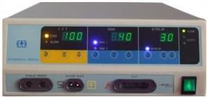
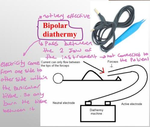
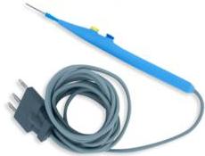
most commonly used
Unipolar Diathermy
- is pass through body of patient
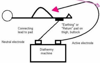
- go from generator to the patient, go through patient and come back to generator
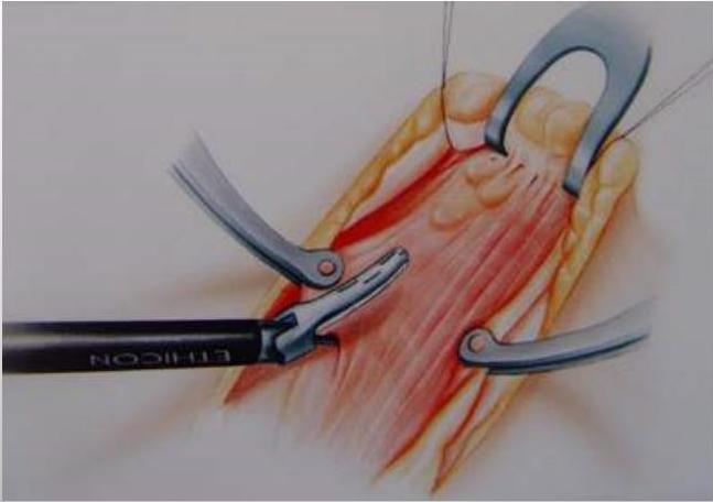
Ultrasonic Coagulation & Cutting (Harmonic Scalpel)
Surgical Haemostasis - Laser
X 6 not used
Argon beam coagulator (ABC): for parenchymatous organs, unipolar coagulation, non touch technique. and less depth of penetration 2-3 mm.
Other surgical lasers:
- Argon laser: ophthalmology, vascular anastomosis.
- CO2 laser: to cut tissue.
- Nd:yag laser: used for paranasal sinus and tracheobronchial tree.
- Er:yag laser: strongly absorbed by the water of tissue, can vaporize cartilage, fibrous tissue and bone.
SURGICAL NEEDLES z
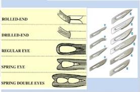
- Open french eye needle → not used any more
- Eyeless needles → ready made needles already attached to the thread (express needle)
- Straight or curved → mostly used
- Cross section: round, triangular or flattened
- Needle point: cutting (skin) , tapered or blunt (soft tissue/fascia) Z
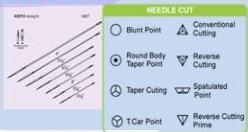
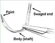
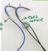
Figure 1: Example of Eyeless (Swaged) Needle (A) and Eyed Needle (B).
ABSORBABLE SUTURES
- Plain catgut: made from the intestine of cattle or sheep. absorption: about 10 days.
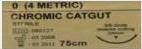
- Chromic catgut: treated by chromium salt, lasts up to 20 days.
- Polyglycolic acid (dexon): synthetic, braided, absorbable, higher tensile strength, reabsorption - 60 to 90 days.
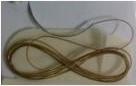
- Polyglyconate (maxon): synthetic monofilament.
- Polyglactic acid (vicryl): synthetic, braided, very high tensile strength absorbed in 60 days
- Polydioxanone (pds): monofilament absorbable
- These 3 used for: Soft tissue anastomosis eg: intestinal anastomosis, gastric anastomosis
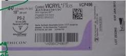
- synthetic - choose one of the 3 depending on preference of the Surgeon and kind of tissue dealing with
- Synthetic is used for than natural
NON-ABSORBABLE SUTURES
- SILK: protein filament from the silkworm larva. dyed, treated by polybutylene & braided. good tensile strength. natural → (not used most of the time)
- POLYESTER (DACRON): superior strength & durability.
- NYLON: synthetic polyamide polymer, monofilament and multifilament → non absorbable
- POLYPROPYLENE (PROLENE): monofilament, minimal tissue reaction → synthetic non absorbable
- STAINLESS STEEL: low carbon iron alloy, monofilament/multifilament. for bone suturing
| Suture Type | Product Description |
|---|---|
| MONOMID® | Monofilament Nylon Non-Absorbable |
| SILK | Braided Natural Silk Non-Absorbable |
| POLYPRO® | Monofilament Polypropylene Non-Absorbable |
| POLYBOND® | Braided Polyester Non-Absorbable |
| UHMWPE | Ultra-High Molecular Weight, Braided, Polyethylene Non-Absorbable |
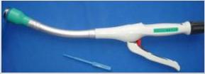
STAPLERS
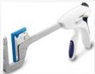
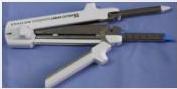
- TA instruments: linear everting double line, length 30, 55, 90 mm. Staple size 3.5 and 4.8 mm. 3.2 mm for vessel closure.
- GIA instruments: two double rows of staples - divide & anastomose. used for intestinal anastomosis
- EEAa instruments: end to end or end to side circular staplers
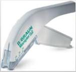
- Skin staples: skin closure.
DRAINS
- closed / open → not used anymore
- Dates back to Hippocrates (metal/ glass tubes, bone, gauze & rubber).
- Air vent suction by Heaton 1889.
- Prevents collection of serum, blood, pus/ drainage
- Passive - close or open
- Active - close & suction
- Prophylactic & therapeutic..
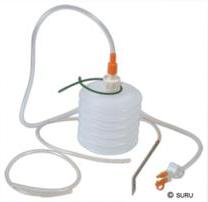
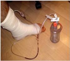
Closed Drains
- Active - suction drains:
- Multi-holed - polyvinyl chloride or silicone
- effective, drains soft tissue under large skin flaps
- lower infection rate
- clog & cease function
- Passive - non-suction close drains
Passive - Open Drains
- Leg. Corrugated drains → not used any more (no bag connected)
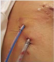
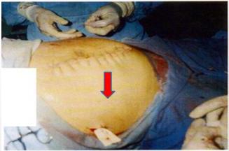
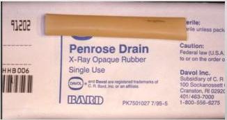
- Penrose drain (obsolete):
- Efficient, risk of infection.
- Soft, flexible latex rubber wick
- Non-active open drain - pus, blood or serum
- Brought out through a separate wound
- Anchored to the skin with a suture
Therapeutic Percutaneous Drainage Catheters
- To drain an abscess
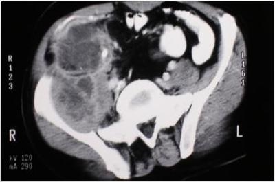
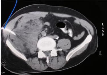
- CT or US guided - inserted by radiologists to drain accessible localized collection
Sump Drain
- used for pancreatic recosis or severe abdominal infections
- rarely used
- Tube within tube, so suction in inner tube, and air go in outer tube, which helps in suction.
- Large and bulky.
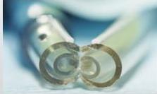
- Double/ triple lumen to allow irrigation & aspiration.
- Rely on continuous flow of air from outside.
- Predisposed to secondary infection.
- Less likely tissue occlusion.
- For high volume enteric fistula.
WOUND CLOSURE
- Primary closure: clean wounds.
- Delayed primary closure:
- contaminated wound
- left open for dressing,
- closure after 3-4 days
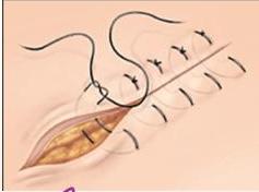
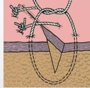
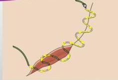
- Methods of suturing:
- simple interrupted suture
- mattress suture
- subcuticular suture - from under the skin
POST-OP WOUND CARE
❖ Dressing
- ❖ protects from mechanical trauma & bacterial invasion.
- ❖ sterile dressing applied before removal of drapes.
- ❖ infected wounds - dressing that absorb exudates.
❖ Immobilization
- ❖ reduce lymphatic flow & spread of wound flora
❖ Elevation
- ❖ reduces interstitial edema.
❖ Suture Removal
- ❖ proper timing for suture removal
MINIMAL ACCESS SURGERY
- Laparoscopic surgery - diagnosis/ therapy
- CO₂ pneumoperitoneum
- telescope & instruments - introduced through small trocars
- Common procedures: Cholecystectomy, GERD Bariatric, colon, splenectomy, Adrenalectomy, Pancreatic Tumour, Hernia repair
- thoracoscopic
- NOTS: natural orifice transluminal surgery
- Single port
Advantages
- less tissue trauma, less response to trauma.
- reduced pain
- fast post operative recovery,
- shorter hospital stay
- reduced wound complications - infection, dehiscence
- reduced risk of contact with patient blood
Disadvantages
- procedure generally slower
- special expertise necessary
- tactile feed back lost
- control of bleeding more difficult
- organ extraction some time difficult
PROPHYLACTIC USE OF ANTIBIOTICS
6 depends on what surgery is going to be done
Definition
- administration of antimicrobial(s) prior to surgical procedures to reduce the number of microbes that enter the tissue or body cavity.
- Antibiotics are selected according to microbes likely to be present at the surgical site.
SURGICAL WOUND CLASSIFICATION
MCO
- CLEAN WOUND (CLASS I) - THYROID SURGERY, BREAST BIOPSY (5%)
- both are class I
- → rate of infection is very low so no need for antibiotics
- CLASS 1D - GRAFT OR MESH USED (use of foreign body)
- → give antibiotic
- CLEAN-CONTAMINATED (CLASS II) - MINIMAL CONTAMINATION E.G., BILIARY, URINARY, GI TRACT SURGERY (11%)
- give antibiotic (1 dose of max for 2 doses)
- → Cholecystectomy for biliary colic (not acute cholecystitis)
- → There is minimal bacteria and chance of infection is low (10%)
- CONTAMINATED (CLASS III) - GROSS CONTAMINATION; E.G., DURING BOWEL SURGERY - (17%)
- give full course antibiotic (for 5 days)
- → There is bacteria in bowel. infection rate is 20%
- DIRTY (CLASS IV) - SURGERY THROUGH ESTABLISHED INFECTION E.G., PERITONITIS (> 27%)
- → (> 1/3 of patients will have infection)
- 5 days course of antibiotics
ANTIBIOTIC PROPHYLAXIS
- Prophylaxis in class id, ii, iii, iv
- Given just before surgery - (before GA)
- Duration: controversial. one dose/ 24 hour regimen / longer
- belonging on class