Spot Dx
Spot Diagnosis — Dr. Ahmed Khan
Name the clinical test? Diagnosis? Aetiology? — Cystic hygroma
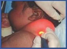
- Most common post triangle Tumor = cystic hygroma
- Clinical test: transillumination
- If infected, won’t transilluminate
Differential diagnosis? — Submandibular swelling
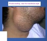
- FB skin swelling – near the mandibular angle
Bilateral submandibular lymph node is submandibular gland:
- Painful and malignant
- If solid → lymph node
- Cystic → Sublingual
- What causes it to swell
- The most anterior part of gland is palpable in the floor of gland
- In parotid — Pleomorphic Tumor — usually more benign → superficial parotidectomy
Differential diagnosis? — Thyroid isthmus nodule
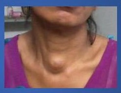
- Thyroid isthmus nodule
- Slowly, self, protruding on the tongue in swallowing. Normal thyroid. Tongue protrudes test
- If both are negative most likely dermoid
- Ttt: excision
Differential diagnosis? — Pendulous neck swelling
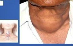
- Pendulous
Causes:
- Hypothyroidism
- Weight loss
- Hirsutism
- Menorrhagia
- Dry cold skin
- D- constipated
- By symptoms + signs and daily TSH = tx
Eye signs — Normal vs Lid retraction vs Exophthalmos
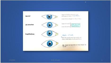
- Diagram of the upper lid relations of the eyeball: Normal / Lid retraction / Exophthalmos
Diagnosis & Other eye signs? — Toxic goiter
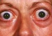
- Toxic goiter
- In primary hyperthyroidism — mostly eye manifestation
- In secondary — none
- Eye: ecchymosis conjunctivitis, lid lag or retraction, exophthalmos
Differential diagnosis? — Left neck (posterior triangle) cervical lymph nodes
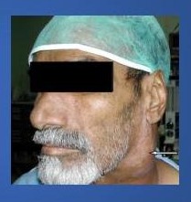
- Left neck
- Post triangle
- Cervical lymph nodes
- Pharyngeal pouches
- Dermoid cysts in kids
- Hx: fever, weight loss, hunger = TB
- Exd: acid hiselosis then penetrating then matting
- Ttt: ATD + excision biopsy
- When it exceeds through fascia and skin → forms a collar-stud abscess and later a chronic sinus
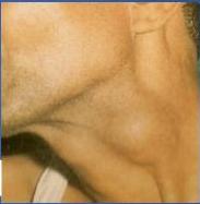
Location? — Carotid body tumor
- Blineath the sternodeidomastoid
- Deep to SCM, upper anterior triangle
Carotid body Tumor
- OK:
- Firm
- Pulsatile
- (1) — Transmitted
- Moves: horizontally / not vertically
Diagnosis?
- If small and asymptomatic = leave it
- If large compressing the vessels and might cause TIA = vascular surgery
- US, Duplex, CT angiography
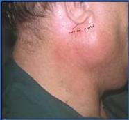
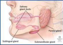
Differential diagnosis? — Parotid swelling
- Glateral = mumps
- Unilateral = parotid abscess
- Early: firm, solid swelling
- Late = fluctuation
- Ask for US = how many ml of pus, size of the cavity
- Effusion how to confirm? Intraoral examination of the duct/papila of the gland
- Ask the pt to maintain oral hygiene
Immunocompromised or pt kept NPO for long time = ascending infection
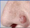
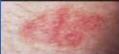
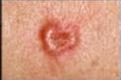
Differential diagnosis? — Basal cell carcinoma
Basal cell carcinoma / rodendant cell
- Roped edge
- In the nose
- Most common malignancy, rarely Meta but barrow adjacent structures
- Tis: wide excision
- Other (dix, sac, melanoma
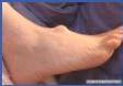
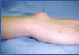
Differential diagnosis? — Ganglion cyst
Ganglion cyst
- How to confirm? Along the tendons
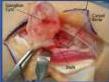
- Sometimes communicate w joints
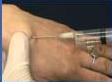
- Ttt: aspirate w big needle as thick fluid
- If recurrence or complicated we might excise it
Differential diagnosis? — Lipoma
- Any soft tissue tumor, lipoma
- By consistency
- Ask for US
- Confirmed by FNA
- Ttt: excision
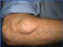
Differential diagnosis? — Neurofibromatosis
Neurofromatosis
- Reassure the pt + give symptomatic ttt
- As they are attached to the nerve, pt comes with pain + compression + cafe o late spots
- Van Recklinghausen disease
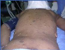
Differential diagnosis? — Squamous cell carcinoma
Squamous cell carcinoma
- Sun exposed area
- Reverted edge
- Less common than bcc but more aggressive
- Mets through lymphatic, if did = very poor prognosis
- Ttt: surgical excision + might give radiation
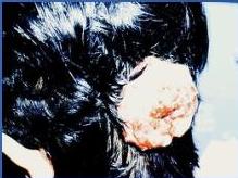
Differential diagnosis? — Hemangioma
Hemangioma
- Involuting type, superficial, go by 7 yr, reassure + leave it
- If complicated we treat + ILS (intralesional steroids. Photocoagulation
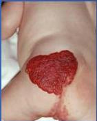
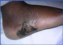
Differential diagnosis? — Gangrene (black spots)
- How to confirm the Dx?
- Black spots + pain + fever + systemic symptoms
- He: any signs of hemorrhage or petechial hemorrhage (low platelet), OM, trauma
- Investigations: fasting sugar, cbc, …
- How to confirm? Exclude others by cbc, , and biopsy will show gangrenous cells
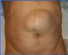
Differential diagnosis? — Incisional hernia
- The swelling appeared after abdominal surgery
- Incisions hernia
- If you reduce it you will feel the defect
- Investigations: defected abdominal wall
- Tilt repair + mesh, if 3–4 cm we might suture only, unless pt is not fit for the surgery
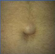
Differential diagnosis? — Umbilical / periumbilical hernia
- A partially reducible swelling
- Complications
- If from the center of umbilicus = umbilical
- If around > periumbilical
- Defect is smaller, more likely to have complications
- This is most likely omentum only, confirm by palpation
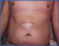
Differential diagnosis? — Incisional / Epigastric hernia
- Incisional hernia / Epigastric hernia
- What kind of surgery was done?
- Epigastric hernia — any hernia between xiphoid crescent + umbilicus, Detect in lines alba
Differential diagnosis? — Inguinoscrotal swelling
- How to confirm Dx?
- Inguinoscrotal swelling
- Indirect hernia due to patent vaginas processes
- Decreased in pit lying down
-
- hydrocele
- TC herniotomy
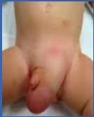
Name the clinical test
- Aetiology: Child / Adult
- In adults it is inflammatory, in kids no, more benign
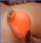
A groin swelling — Name the test?
- Ring occlusion test, at the midinguinal point occlude the internal deep ring
- Include their occlusion
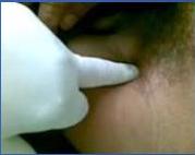
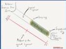
- Adults – mostly direct
- Kids indirect
- If butes Medial to my finger – direct
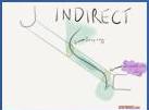
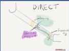
DIAGNOSIS & INDICATIONS? — Ileostomy
- Bleostomy
- Bulging + right side
- Why bulge? Digestive enzymes irritate the skin
- More likely to have electrolyte imbalance than colostomy
- Sympatory (toop) or permanent (end)
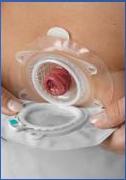
DIAGNOSIS & INDICATIONS? — Loop colostomy
- Loop colostomy
- Complications: bleeding, edema, ischemia / necrosis, retraction, prolapse, parasitomal hernia, stenosis, and skin irritation.
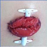
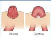
- Bleostomy as bulged
Name the procedure on left & its indications? — Hartmann
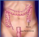
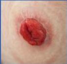
- hartmann
- Temporary but not a help, and! Exception
- Reansistomose later
Laparotomy for acute abdomen — Causes? Treatment?
- How to confirm? believe strangulation and check if color go back, or if bleeds/peristalsis = viable
- Return or resection + anastomosis fix A Same operation or stoma + later anastomose
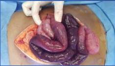
Laparotomy for acute abdomen
- Small bowel echemia
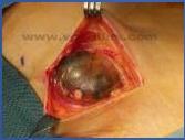
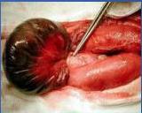
Laparotomy for liver mass — Hydatid cyst
- Terminal epithelium which grow to daughter cyst + laminated fibroses Wall — CT superior, US
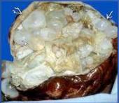
- Mydaloid cyst of liver
- Vague pain
- If bursts out = anaphylactic
- If calcified + away from portal v we can leave it
- Causative organism: Echromococcus granulosa
- Tic lap cystectomy and if big we replace the part w omentum, + albendazole
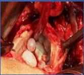
15-year girl — abdominal pain, vomiting & abdominal mass
- Microbiaceae (swallowed hair)
- Pt w psychiatric problem, hairs caused obstruction. Titred was it + refer to psychiatry
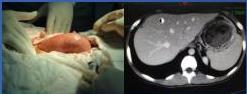
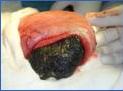

Diagnosis and management? — Perforated PU
- Perforated PU, anterior wall
- If in post, go breeding
- Laparotomy
- Closed by graham patch (smentum)
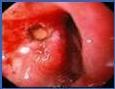
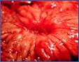
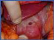
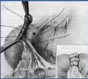
- Stitch gently or it will cut away + Guillotine effect 3-4 layers
Calot’s triangle — Why important?
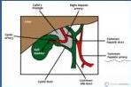
- Calot’s triangle
- Anatomy needs to be preserved (delineated?) during cholecystectomy to prevent bile duct injury
- Contains the cystic artery + cystic duct + lymph node of Lund
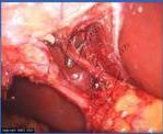
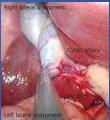
Abdominal pain — Laparoscopic appendectomy
- Laparoscopic appendicitomy
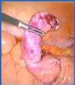
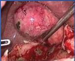
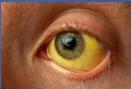
Differential diagnosis? — Jaundice
- Jaundice
- Mostly obstructive jaundice as surgical
- GGT + ALP
- US + dilated CBC
- Tilt depends on the cause
Differential diagnosis? — Erysipelas
- Etiology
- Non-necrotic
- Aeribiotic + raise
- Etiopyelas
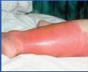
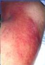
Differential diagnosis? — Furuncle
- Euroride
- Tilt I D + abs
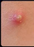
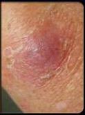
Inspection — Differential diagnosis? (red + inflamed swelling)
- Red + inflamed = abscess + complicated hernia
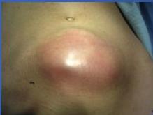
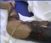
Differential diagnosis? — Management (necrotizing infection)
- Neo face
- Involving the subcutaneous
- Old age, immunocompromised
- If involved the whole circumference risk of compartment syndrome so fasciotomy
- Flt: urgent debridement + abx
- Former = common in dm
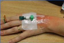
Pain at IV site — Differential diagnosis?
- Superficial thrombophlebitis, VTE
- Ricks: hyperosmolar fluid, trauma, endothelial injury, placing it for too long = inflammation + extravasation of fluid
- Cord like in palpation
- Tlt: remove the carruza + place it somewhere else
- Ddx: cellulitis - acute abscess, any inflammatory diseases, dermatological conditions
Post operative immobilized — Pain and swelling — Differential diagnosis?
- Hammam test
- Confirmed = Doyle’s
- DVT — usually unilateral
- Tlt: anticoagulation therapy. If not completely dissolve we put ivc filter to prevent PE
- Best is prevention by pharma (tmsh) + non pharma (stocking - intermittent pneumatic compression device)
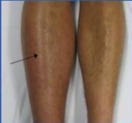
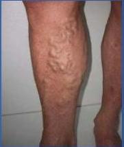
Inspection — Differential diagnosis? (varicose veins)
- Vancose vein, if late < medial ulcer
- Occupation, females, long standing jobs, congenital, familial
- Initial stage: telangiectasia — less than 1mm —, spider nives — 3,4 mm —, huncial varicose
- Duplex to see the flow, US
- Incompetent sepharofemoral junction
- Tlt: if initial = compression stocking - MIP laser ablation, Endovenous foam sclerotherapy
- If toe big < surgery to ligate the junction = perforators
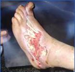
Inspection (large dorsal foot wounds)
- Large wounds extending to all over the dorsum
- Tlt: abx, debridement then graft
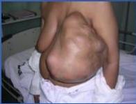
Differential diagnosis? — Phyllodes tumor
- Phyllodes tumor
- Tlt, depends on the tumor / breast ratio: if less than 1/3 median, if more mastectomy
Inspection — Differential diagnosis? (breast)
- Cancer
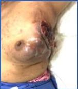
Inspection — Indication (breast)
- Inflammatory breast cancer
- Very rare
- Middle aged young women
- Biopsy
- Very aggressive
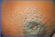
Inspection — Clean incised wound
- Clean incised wound
- Primary suture, if dead tissue we can use tertiary
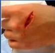
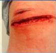
Inspection — Healed by secondary
- Healed by secondary
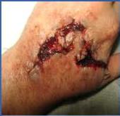
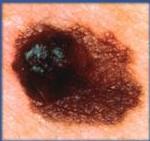
Differential diagnosis? — Melanoma
- Melanoma — most deadly
- White females
- Satellite
- Superficial spreading
- Sentinel lymph node biopsy
- Tlt. excie deeply Breslow depth
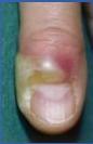
Differential diagnosis? — Paronychia
- paronychia
- I&D = Abs
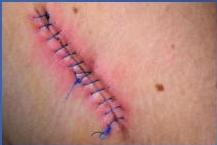
Inspection — Surgical site infection
- Surgical site infection
- If fluctuation is there = open and drain + send for cs
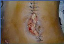
Inspection — Management (wound)
- from healing BSI
Inspection — Management (gangrene)
- Gangarounous
- Inc. dm
- Dry gangrene in dm (arterial), clear demarcation
- WM gangrene (vein) foul smelling = more element of infection
- Amputation
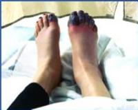
Differential diagnosis? — Transmetatarsal amputation
- Transmetatarsal amputation
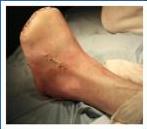
Inspection — Amputation level
- Deliver knee
- Disually below joint
- Complications: Phantom limb, as of the other lesion
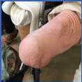
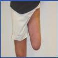
Differential diagnosis? — Sebaceous cyst
- Punctum + sebaceous cyst
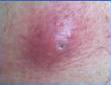
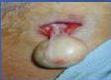
Pain and swelling — Differential diagnosis? — Pilonidal sinus
- Pilonidal sinus
- Mucopurulent discharge, hairy men, setting long
- Infection of the hair follicle in the natal cleft and above
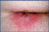
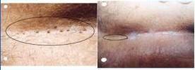
Differential diagnosis? — Pilonidal disease (pits)
- Many pit
- Initial = maintain hygiene + remove the hair
- Worse might I did
- Worst = surgical excision of the whole sinus if too big we might need graft
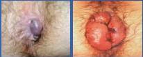
Differential diagnosis? — Hemorrhoids
- 3-7-11 typical sites
- Superficial thrombosed hemorrhoids
- Stapled Hemorrhoidectomy
- Grade I-II → Diet = fluids ± band ligation
- Grade III → Band or surgery
- Grade IV / complicated → Hemorrhoidectomy
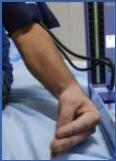
Inspection? — Carpopedal spasm
- Carpopedal spasm
- Hypocalcemia
- Post thyroid surgery
- IV calcium
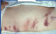
Inspection? — Pancreatitis (abdominal signs)
- Chronic hemorrhagic pancreatitis
- Grey tumor
- Cullen sign
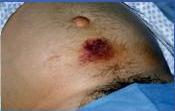
Differential diagnosis? — Pressure sores
- Bad / pressure sores
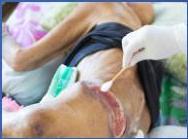
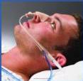
NG tube — Uses, placement, complications
- Uses — NOT
- Therapeutic + diagnostic
- How to place? Measure from earlobe to nostril
- Check correct position? Lubricant + insert = confirm by syringe + ph x ray
- Complications: In trachea, laceration
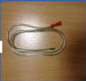
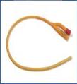
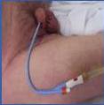
Catheters — Indications & complications
- Two entry points
- Foley
- Simple catheter — single opening
Differential diagnosis? — Pedicle flap
- Pedicle flap
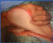
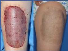
Differential diagnosis? — Partial thickness graft
- Partial thickness graft
- Meated to be enlarged
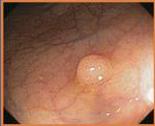
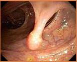
Colonoscopy findings — Pathology: Polyps
- Villous
- Tabasilosus
- Tabulos
- Pedunculated polyps less likely malignant