Mechanisms of Injury: Blunt Trauma/ Penetrating
Trauma Classified into 3:
1. Blunt Trauma
- No penetration of tissue
- Most common is MVA
2. Penetrating Trauma
- Leg. - Stab wound
- Gunshot
- bullet
- firearm
- There is a difference based on the harm they cause and the approach
- Injuries depend on depth, angle, device, location.
- Size can be misleading
- Object penetrates internal tissues causing injury
- Stabbings, firearms
3. Acceleration-Deceleration Injury
- Injuries caused by rapid change in velocity
- Deceleration injuries
- Body stops, tissues and organs continue to move forward
- MVAs, MCAs, falls, contact sports
- Body move fast then stop so there is difference in movement of different parts of tissue
- Leg injury to hepatic veins
- & acceleration deceleration lead to shearing force which will injur hepatic veins → bleeding
Case No. 1: Chest Trauma — Stab Wound
Pre-Hospital Data
- A 17-year-old male brought to A tertiary care center in AL Naseem District, when he became a victim of a stabbing.
- This all began when the victim confronted an old friend about a personal conflict which occurred between them years ago.
- What started out as a verbal argument eventually resulted in physical violence.
- The victim sustained a single stab wound to the left chest in the mid axillary line, just below the level of the nipple.
- He was transported to ER at National Guard Hospital by red crescent.
- He was noted to be awake and alert throughout the entire transport.
Key findings to look for (Tension Pneumothorax):
- lung: Pleura → Tension Pneumothorax → different movement of chest
- Palpable for detailed trachea
- dec air entry by auscultation
- recrepitus palpable with subcutaneous emphysema
History…Continue
- Past Medical/Surgical History: Asthma
- Family History: Non-significant
- Medications: Inhalers as needed
- Allergy: No Known Drug Allergy (NKDA)
Trauma Room Assessment
- The patient was moved from the stretcher onto the examination table, and the only complaint obtained from the patient was shortness of breath
Nursing Care
- Cardiac monitors, blood pressure-cuff and oxygen saturation probes were then placed on the patient.
- Obtain Vital signs:
- Heart rate — 90/min
- Blood Pressure — 130/70
- Respiratory rate 25 → Tachypnic
- Temperature — 37 C
What Next?
Primary Survey

What is Primary Survey
- The primary survey is the initial assessment and management of a trauma patient.
- It is conducted to detect and treat actual or imminent life threats and prevent complications from these injuries.
- A systematic approach using ABCDE is used.
- If a group of clinicians is assembled to perform the initial assessment, there will be multiple simultaneous activities occurring and resuscitation does not always proceed in a purely linear, sequential fashion.
- On arrival to the emergency department, the patient should have full non-invasive monitoring applied.
② 1st radiological investigation to be done: Chest X-ray
Immediate Nursing and Monitoring
- ECG monitoring
- Non-invasive blood pressure cuff
- Oxygen saturation probe
- The patient’s temperature should be recorded
- Removal of the patient’ clothes is encouraged so that they can be fully assessed → to exclude any other injury
- Oxygen should be applied to achieve saturation of 94-98%
The primary survey of a trauma patient
In single responder settings these may need to be addressed in a linear or sequential fashion; however, when a team is assembled, these elements may be addressed simultaneously. The term ‘survey’ is somewhat misleading in that it implies that only assessment is occurring; however, each phase requires simultaneous assessment for, and management of, any life threats detected.
- Airway – with cervical spine control
- Breathing
- Circulation including control of exsanguinating external haemorrhage
- Disability
- Exposure
② 1st radiological investigation to be done: Chest X-ray
Trauma Team
- K Trauma team is led by one person.
- This person will decide on priorities of the patient:
- breathing
- circulation (hemorrhage)
- of. Should be diagnosing and treating at the same time

Control of Exsanguinating External Hemorrhage
- Failure to recognise and control large-volume external haemorrhage has been found to be a frequent occurrence in trauma resuscitation.
- Obvious large volume external blood loss must be managed as an immediate priority in the field and on arrival of the patient into the emergency department, with the aim being to control life-threatening external haemorrhag.
Possible Interventions during the Golden Hour
defend on country and facility it differ
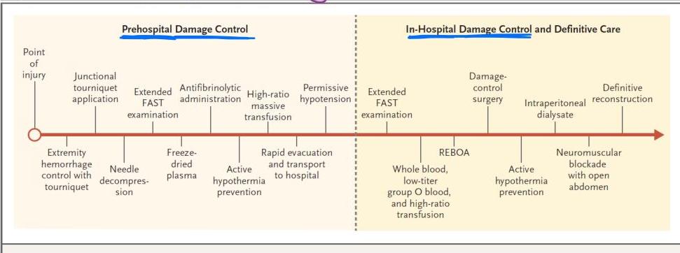
The primary purpose of the golden hour concept is to achieve early hemorrhage control. Prehospital and in-hospital maneuvers toward this goal include initial care, triage, rapid evacuation, and resuscitation. FAST denotes focused abdominal sonography for trauma, and REBOA resuscitative endovascular balloon occlusion of the aorta.
Airway with cervical spine protection

- An assessment of airway patency and stability should be performed during the primary survey and a plan for airway management instituted if required.
- Unless the patient is in cardiac arrest, immediate securing of the airway with endotracheal intubation is rarely required upon arrival of a major trauma patient.
Airway Life Threats
The following airway life threats must be assessed and managed:
- ■ Airway obstruction — Vomit, blood, dislodged teeth. Remove with suction.
- ■ Blunt or penetrating neck injury → Should be careful about how we deal with airway
- Reduced conscious state → This can lead to hypoventilation and/or airway obstruction and hypoxia
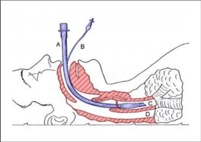
Consider early endotracheal intubation if neck wounds or hematomas are causing the airway obstruction or if there is an airway disruption.
Caution: Rapid-sequence intubation in patients with blunt or penetrating neck injuries carries the risk of total loss of airway patency upon administration of sedative and/or muscle relaxant medication.
Clinical example: If patient has big neck hematoma and try to intubate and have difficulty, and 4 mins already passed from giving muscle relaxant, leading patient to have anoxic brain damage.
Rapid Sequence Intubation (RSI)
Primary Survey — Case Application
- Airway and Breathing:
- Airway — patent airway as demonstrated by his ability to talk.
- Breathing — decreased breath sounds at the left base.
- Oxygen mask with 100% FiO2 was placed; & an oxygen saturation of 100% was obtained
- Circulation: no active external bleeding
- Disability or Deficits: neurological exam grossly intact
- Exposure: the patient’s clothes were removed to thoroughly examine for other injuries
- Secondary survey:
Secondary Survey
The secondary survey is performed once the patient has been resuscitated and stabilized.
- It involves a more thorough head-to-toe examination
- Aim is to detect other significant but not immediately life-threatening injuries.
- If during the examination any deterioration is detected, go back and reassess the primary survey.
1. History
Taking an adequate history from the patient, bystanders or emergency personnel of the events surrounding the injury can assist with understanding the extent of the injury and any possible other injuries.
Use the AMPLE acronym to assist with gathering pertinent information:
- Allergies
- Medication
- Past medical history including tetanus status
- Last meal
- Events leading to injury
② Head-to-Toe Examination
During this examination, any injuries detected should be accurately documented and any required treatment should occur, such as covering wounds, managing non-life-threatening bleeding and splinting of fractures
Head and Face
- Inspect face and scalp. Look for any lacerations or bruising, including mastoid or periorbital bruising which may be indicative of a base of skull fracture.
- Gently palpate for any depressions or irregularities in the skull.
- Look in the eyes for any foreign body, subconjunctival hemorrhage, hyphemia, irregular iris, penetrating injury or contact lenses.
- Assess the ears for any signs of cerebrospinal fluid leak, bleeding or blood behind the tympanic membrane.
- Check the nose for any deformities, bleeding, nasal septal hematoma or cerebrospinal fluid leak.
- Look in the mouth for any lacerations to the gums, lips, tongue or palate. Note any swelling which may indicate inhalation injury.
- Inspect the teeth, noting if any are loose, fractured or missing.
- Test eye movements, pupillary reflexes, vision and hearing.
- Palpate the bony margins of the orbit, maxilla, nose and jaw. Inspect the jaw for any pain or trismus.
Chest
- The chest should be palpated for rib tenderness and subcutaneous emphysema.
- The entire thorax must be palpated including the supraclavicular fossae, right and left ribs and both axillae.
- A hand can be slid posteriorly along a supine patient to check for occult blood loss; however, a formal examination of the back of the chest occurs when the patient is log rolled.
- Auscultate the lung fields; note any percussion abnormality, lack of breath sounds, wheezing or crepitations.
- Check the heart sounds: apex beat and presence and quality of heart sounds
Neck
- A plain lateral neck x-ray may be indicated when assessing a poly-trauma patient but if normal does not clear the C spine. This is particularly relevant in the intubated patient.
- To examine the neck the cervical collar should be opened and the head supported with manual in-line stabilization throughout the exam. Three staff members are normally required to conduct a neck examination safely.
- Gently palpate the cervical vertebrae. Note any cervical spine pain, tenderness or deformity.
- Check the soft tissues for bruising, pain and tenderness.
- Trachea (midline or deviated): The trachea may deviate away from the side of a tension pneumothorax
- Wounds: blunt or penetrating injuries, size and depth.
- Subcutaneous emphysema: The presence indicates an airway disruption such as a laryngeal fracture or pneumothorax.
- Larynx: Laryngeal tenderness or crepitus; this may indicate an underlying laryngeal fracture. Caution: firm palpation may disrupt a fractured larynx leading to total airway obstruction.
- Veins: Look for distension – neck vein distension may be seen in tension pneumothorax or pericardial tamponade (a late and peri-arrest sign).
- Esophagus: To assess the esophagus, ask the patient to swallow. An esophageal injury may be suspected if the patient has pain or difficulty swallowing
Abdomen
- Inspect the abdomen. Palpate for areas of tenderness especially over the liver, spleen, kidneys and bladder. Look for any bruising, lacerations or penetrating injuries. Document seat belt marks.
- Check the pelvis. Perform a pelvic x-ray if there is any suspicion of injury. Gently palpate for any tenderness. Do not spring the pelvis. Any additional manipulation may exacerbate haemorrhage.13 Apply a binder if a pelvic fracture is suspected even if low clinical suspicion.
- Auscultate bowel sounds.
- Inspect the perineum and external genitalia for bruising or hemorrhage
Limbs
- Inspect all the limbs and joints, palpate for bony and soft-tissue tenderness and check joint movements, stability and muscular power. Note any bruising, lacerations, muscle, nerve or tendon damage. Look for any deformities, penetrating injuries or open fractures.
- Examine sensory and motor function of any nerve roots or peripheral nerves that may have been injured.
- Assess distal colour, warmth, movement, sensation and capillary refill
Buttocks and Perineum
- Look for any soft-tissue injury such as bruising or lacerations.
- Genitalia: Inspect for soft-tissue injuries such as bruising, lacerations or burns. Note any priapism that may indicate a spinal injury.
- The priorities for further investigation and treatment may now be considered and a plan for definitive care established
Back, and buttock and Perineum
- Blood in rectum indicate there is injury inside
- Log roll the patient. Try to do this once. It can be achieved at the time of patient transfer when you have the most personnel to do it safely.
- Maintain in-line stabilization throughout.
- Inspect the entire length of the back noting any deformity, bruising and lacerations.
- Palpate the spine for any tenderness or steps between the vertebrae. Include a cervical examination at this stage.
- A digital rectal examination should be performed only if a spinal injury suspected
Secondary Survey — Case Findings
- HEENT: no lacerations, no hematomas, no fractures palpated
- Neck: midline trachea, no JVD, no crepitus
- Chest: clear on right, single stab wound to the left chest in the mid-axillary line in the 4th intercostal space, no crepitus, no bleeding, decreased breath sounds at the left base
- Cardiac: regular rate and rhythm (RRR), normal S1 and S2
- Abdomen: present bowel sounds, soft, non-tender, non-distended
- Extremities: warm, present distal pulses
- Neuro: awake, GCS 15, no focal deficits
What Next?

Radiological Survey
Chest X-ray: left sided hemopneumothorax → Place chest tube
- An upright CXR was obtained. ??sit the patient up (isolated penetrating injury to the chest) and the mechanism of injury did not warrant spinal precautions.
- A pelvis and lateral C-spine films were also not obtained because of the isolated nature of the injury.
- Transthoracic Echocardiogram: no pericardial effusion
- Because the weapon can be aimed at any direction (medially, superiorly, inferiorly), the heart can be potentially injured.
- A pericardial tamponade is lethal unless discovered and treated quickly
Pneumothorax management notes:
- urgency of dealing with Pneumothorax depends on the amount and symptoms of the patient.
- little amount → no need for chest tube
- When inserting central line, can injure pleura → pneumothorax
- So after doing Central line, do chest X-ray; if the amount of pneumothorax is only 10% and the patient is asymptomatic → no need for Chest tube
- Tension Pneumothorax need urgent action → emergency insertion of chest tube or at least a needle, to get air out
- → shift mediastinum → dec venous return → hypotension
Blood work ordered:
- type and screen
- Coagulation panel
- Complete blood count (CBC)
- Arterial blood gas
- Toxicology screen
Next…
- Chest tube insertion: 300 CC of Blood.
- The patient at this time began complaining of a new subscapular pain, or pain between the shoulder blades. This was alarming to the trauma team for the following reasons.
- Patients with diaphragmatic injuries and irritation from the blood frequently exhibit referred pain in this distribution.
- Surgery
Case No. 2: Abdominal Trauma — Stab Wound
H&P
- Red crescent bringing “stab wound” to abdomen, 2 am “25 cm knife”
- 25 y/o male stabbed left lower flank and left upper quadrant, intoxicated.
- GCS 14, verbalizing, not cooperative.
- VS: 90, 140/80, 26, 95% RA
- Physical exam: 2 lacerations, 4-5 cm each, oozing
- mid axillary line, 9-10th intercostal space
- right left upper quadrant
Stab wound may not show direction of injury. So injury may not only be below site of injury. Could be below, above, right, left. So should think about surrounding organs.
2014-2015
H&P… (Continued)
- Vitals stable after primary, secondary survey, airway intact.

H&P… (Continued): Next Step
- Vitals stable after primary, secondary survey, airway intact.
- Next step: FAST
- Labs: Bicarbonate 21, glucose 108, creatinine 1.3, EtOH 148, HGB 12.6, UA neg, drug screen neg. INR normal.
- Portable chest x-ray: small pneumothorax on right with small pleural effusion
- CT scan chest/abdomen/pelvis: 9th left rib fracture, hemothorax and 20% right pneumothorax, perforation of colon, pneumoperitoneum, hemoperitoneum
- Decision to proceed to OR
Note: Put chest tube to Stabilize Patient before taking to OR (if there is a patient with fractured ribs, but not have hemo or pneumothorax, should put chest tube because the fractured ribs can cause pneumothorax at any moment)
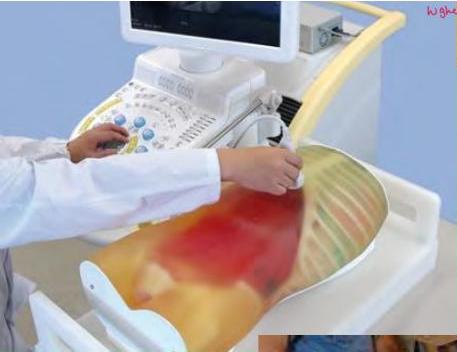
Image notes (verbatim): Collection of blood, fluids, or fluid occurring: 1-Sulphuric, 2-heptagonal, 3-Revis (in absence)
FAST (Focused Assessment with Sonography for Trauma)
- quick easy way to look for bleeding
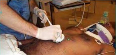
Image notes (verbatim): Because when putting ETT and mechanical ventilation in air, the inflation of the lung can be higher than normal, and there is such a need to be certain can have pneumothorax — barry supply.
Diagram labels (verbatim): longitudinal cut — Liver — < blood — hepato renal pouch → separated if there is: black, fluid, or — Kidney — black medulla (fluid — Lung — black — diarrhea is recurrent on us shaft, shiny — ERPocketBooks.com — Focus Assessment Sonography for Trauma
OR Management
Case outcome:
- Left chest tube placement for hemo/pneumothorax
- Exploratory laparotomy
- Stab wound through transverse colon — partial colectomy with anastamosis
- Control of mesenteric vascular bleeding
- Repair of left hemidiaphragm laceration
- Patient stabilized
Post operative complication:
- Infection in pleural fluid (empyema)
- Leaking from colon anastomosis
- Back to OR for ileostomy
- Requiring TPN
- Massive transfusion
OR management:
- Right chest tube → for hemo-pneumothorax, before patient go to OR
- Take patient for explorative laparotomy → might not know what I will find exactly in the abdomen, so need good exposure and cast access to abdomen. So that determines the incision (midline incision) from xiphisternum to umbilicus or down to pubis, in mid line
- In the mid line there is linea alba → fibrous tissue, no muscle, so its easy to go in → can reach abdomen in 15-30 seconds
- Also the plane is avascular → so no bleeding from abdominal wall
- For stab wound of transverse colon: excise area of injury and do [Primary anastomosis] → wrong (shouldn’t be done)
- Control of mesenteric vascular bleeding
- repair of lacerated right diaphragm
- Patient is stabilized
Options to treat in case of injury to colon:
- If small injury (1 cm hole) in small bowel or stomach: → suture
- If large injury (5cm hole) in small bowel: → resect and anastomose
- If injury in colon:
- Don’t stitch only and don’t do primary anastomosis (colon is dirty, risk of stool spillage)
- Put Stoma → if injured part is visible, bring that part to outside or close injured part
- do it before doing primary anastomosis, to protect anastomosis from contamination
- if do primary anastomosis, Post-op complications can occur:
- Patient already has diaphragmatic laceration and there is spillage of stool in peritoneal cavity → So infection went from abdomen into the chest
- Doing anastomosis in a dirty field, the anastomosis will open up after 3-4 days
- So to deal with this complication: take patient back to OR, do diverting ileostomy
- divert content of intestine to outside of body so it doesn’t go to area inhaled → keep it until the infection has been controlled
- (trying to minimize contamination by bringing content of intestine to the outside by putting temporary ileostomy)
Which part of colon can be used for colostomy → mobile part of colon:
- beg. Transverse colon, Sigmoid
- Do loop colostomy
- Do end colostomy
ATLS: Stab Wound of Abdomen
Most commonly affected organs (defending or Site of organ):
- Liver (40%) → most common, because its more exposed
- Small bowel (30%)
- Diaphragm (30%)
- Colon (15%)
Most commonly affected organs with GSW to abdomen:
-
Small bowel (50%) → taking more surface area
-
Colon (40%)
-
Liver (30%)
-
Abdominal vascular structures (25%)
-
Bullet: defend on which organs it passed through (from entrance to exit)
Does every patient with stab wound need OR?
- depend on weather it passed through Peritoneum
- 6 know clinically: OR or No OR…
- Passed through Peritoneum: visible bowel or omentum
- do exploration of wound in the OR. (Put local anesthesia) Check if Peritoneum is intact or not
- do FAST. if show bleeding, take to OR
Decision aids:
- History: knife length?
- Exam: signs of peritonitis?
- Vitals: Hypotensive?
- FAST exam positive?
- DPL (diagnostic peritoneal lavage) positive?
- Stable patient, CT findings?
- Stabbing to back/flank less likely to have to go to OR due to deep muscle presence → replaced with CT scan → better diagnostic and non-invasive
DPL (diagnostic peritoneal lavage):
- Put needle below umbilicus and infuse 1L of NS in the Peritoneal Cavity, done under local anesthesia. Then drop glass into the floor. 150 fluid will come back to the bottle, and look at the color. If color is red, its blood. So take to OR (can do CBC for that fluid)
Key statistics:
- 98% of GSW to abdomen require laparotomy → no need for investigations
- 60% of stab wounds that penetrate anterior peritoneum have hypotension, peritonitis, or evisceration of omentum and/or small bowel that would necessitate going to OR for exploratory laparotomy.
Abdominal trauma: OR guidelines
- Blunt abdominal trauma with hypotension and positive FAST with evidence of intraperitoneal bleeding
- Blunt or penetrating abdominal trauma with positive DPL
- Hypotension with penetrating abdominal wound
- GSW transecting peritoneal cavity or visceral/vascular retroperitoneum
- Evisceration
- Bleeding from stomach, rectum or GU tract from penetrating trauma
- Peritonitis
- Free air, rupture of hemidiaphragm
- CT positive for ruptured GI tract, intraperitoneal bladder injury, renal pedicle injury, or severe solid organ injuryding from stomach, rectum or GU tract from penetrating trauma.
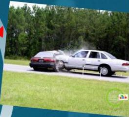
Case No. 3: MVA or MVC or RTA
Overview

- 23 yo (year old) wm (white male), unrestrained driver in high speed t-bone MVC (motor vehicle collision) on drivers side
- Unresponsive initially at the scene, then increase to GCS (Glasgow Coma Scale) 13
- Arrived in trauma center: GCS 13, HR 120, BP 110/55, SaO2 (oxygen saturation) 94% on face mask, moaning loudly, somewhat combative
T bical: small temporal epidural hematoma — spoke to hematoma (SHR) in correlation of blood between the skull and the stripped off distal hematoma. — LRS is a small SHR, then it can be removed without surgery. If surgery is needed, then remove the clot to lower pressure on the brain and stopping bleeding to prevent the hematoma from retaining.
- Maintain by reducing supportive therapy. Always control and blood pressure support are the most important issues. Establish IV access, administer oxygen, and monitor and maintain the brain blood pressure.
Physical Exam Reveals
- Facial abrasions and lacerations, tympanic membranes (eardrum) clear and pupils round and reactive
- Neck & back unevaluable (unable to be evaluated because he is on a board)
- Chest tenderness on left with decreased breath sounds
- Abdomen slightly distended and tender left greater than right; Pelvis stable.
- Obvious deformity Left thigh with good distal pulses
TRAUMA MANAGEMENT
- What are the management priorities? (Hint: ABCDE) — (airway, breathing, circulation, disability, and exposure)
- What injuries are you looking for / concerned for? (Hint: assess head, chest, abdomen, neck / back, extremities) → its multiple trauma so assess everything
- What additional studies do you want to obtain? (Hint: xrays, CTs, labs)
Chest: Look for hemo/Pneumothorax
Case Scenario
if not act urgently, Patient can go into cardiac arrest
- While doing the secondary survey it was observed that the patient becoming hypotensive with systolic blood pressure in the 80s
- The heart sounds were muffled
- JVP is raised → exclude hypovolemia
- What do you suspect and what is the next step?
- ✗ Hypovolemia ??? → can thing of this but raised JVP go against it
- Cardiac tamponade ???
- What test you want to do ???
- What is your next step?
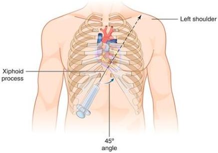
Pericardiocentesis
- identify xithisternum, put long needle and 2S-SOCC syringe, and insert needle angled at 45° and point to left shoulder
- To confirm diagnosis by echo
- (while doing FAST, when Probe is on liver, angle the Probe superiorly to the heart and detect cardiac Tamponade)
- Then go in and will going in, aspirate fluid, and will immediately save improvement of patient
- Even small amount of blood in pericardium can cause tamponade
Pericardiocentesis — 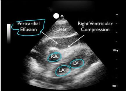
Case Scenario (Continued)
- You obtained 300 cc of blood and patient blood pressure start to improve and JVP went down.
- After 10 minutes the patient’s blood pressure started to deteriorate going down to 80s systolic.
- Condition of Trauma Patient Can change, so should observe closely
- This patient went from hypotension due to tamponade, to hypotension due to hypovolemia
- So next step → do eFAST to see source of bleeding if its in abdomen
- Bleeding source could be from extremities so should monitor distal pulses
Case Scenario (Continued): What Next?
- You obtained 300 cc of blood and patient blood pressure start to improve and JVP went down.
- After 10 minutes the patient’s blood pressure started to deteriorate going down to 80s systolic.
- What to do next: FAST
- What next. OK???
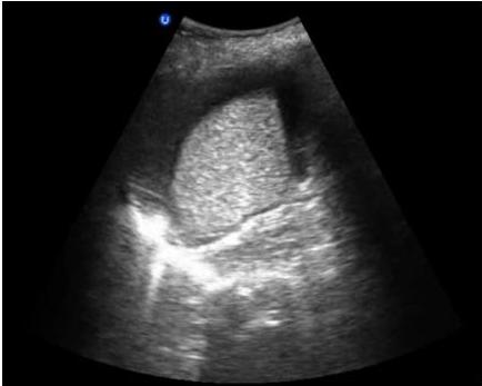
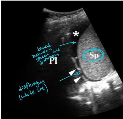
FAST Show bleeding in abdomen
What Next?
- FAST Show bleeding in abdomen
- Patient should be observed carefully: ICU or in high dependency unit
- Correct hypotension
- After that decide: if stable → no need for OR
- OR
- Or
- No OR
- So presence of blood in abdomen does not mean patient needs OR
- Can do serial CT scans to see if hematoma is increasing inside.

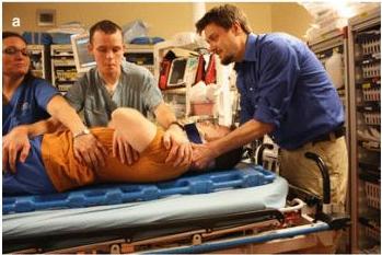
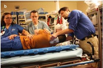
LOGROLLING (for spinal injury)
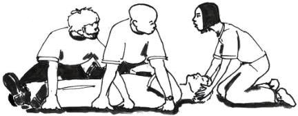
Care for Cervical Spine
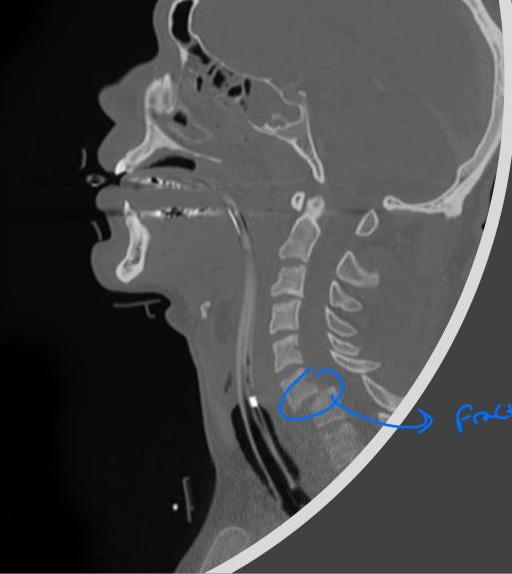
Care for cervical spine — Fractured
Rules for Cervical Collar (When to Remove the Collar)
- do X-ray, CT → and get report from radiologist that there is no cervical injury
Meets all low-risk criteria? (NEXUS)
- No posterior midline cervical-spine tenderness
- No evidence of intoxication
- A normal level of alertness
- No local neurologic deficit
- No painful distracting injuries
- Yes → No Radiography
- NO → Radiography
Fail nexus or obtunded
- The patient has shown gross motor function of all four extremities.
- The patient has no paresthesia’s or neurologic symptoms.
- The patient is able to range the neck.
- The attending radiologist has dictated a final report of a high quality CT scan of the cervical spine and it shows no cervical spine fracture or acute abnormality (e.g. widening)
deits imp to guard against cervical bone injury or else can get quadriplegia
Leave the cervical collar
- The patient has shown gross motor function of all four extremities.
- The patient has no paresthesias or neurologic symptoms.
- The patient is able to range the neck.
- The attending radiologist has dictated a final report of a high quality CT scan of the cervical spine and it shows no cervical spine fracture or acute abnormality (e.g. widening)
- Any questionable findings on CT cervical spine should be discussed with responsible attending
- Obtunded patient = GCS <15 or ≤11 if intubated
Glasgow Coma Scale (GCS)
| Behaviour | Response |
|---|---|
 Eye Opening Response Eye Opening Response | 4. Spontaneously 3. To speech 2. To pain 1. No response |
 Verbal Response Verbal Response | 5. Oriented to time, person and place 4. Confused 3. Inappropriate words 2. Incomprehensible sounds 1. No response |
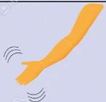 Motor Response Motor Response | 6. Obeys command 5. Moves to localised pain 4. Flex to withdraw from pain 3. Abnormal flexion 2. Abnormal extension 1. No response |
In trauma, a Glasgow Coma Scale score (GCS) of 8 or less indicates a need for endotracheal intubation