ORTHO
Degenerative Spine Disorders
Causes
- Recurrent disc prolapse attacks
- Aging leading to loss of disc hydration
- Spinal instability
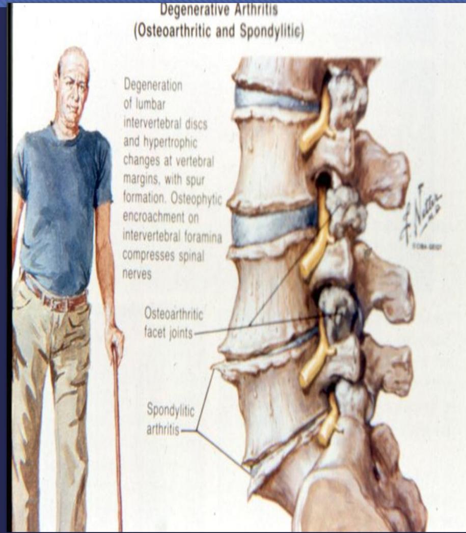
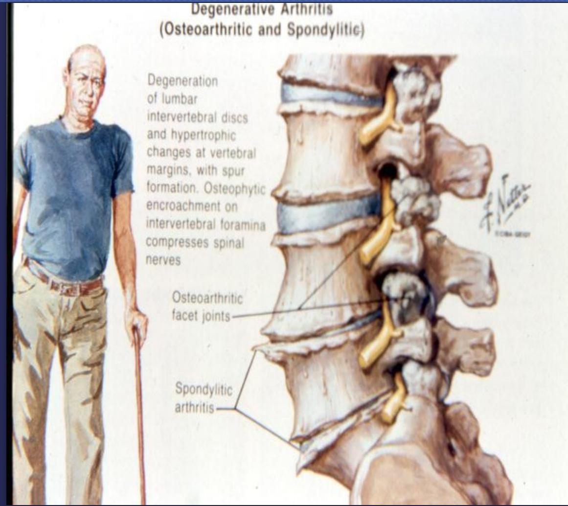
Pathology
- Decreased disc height
- Osteophytes of vertebral margins
- Degenerative facet joint changes
Clinical Features
- Recurrent back pain attacks
- Catching sign (locking)
Imaging
X-ray Findings
- Narrowing of disc space
- Osteophytes
- Osteoarthritic changes in facet joints
Treatment
- Conservative measures
Spinal Canal Stenosis
Definition
Narrowing of the spinal canal
Causes
- Degenerative changes of bone and soft tissue
Clinical Features
- Neurogenic claudication after prolonged standing or walking
- Relief with sitting or squatting (spine flexion)
Imaging
- X-ray: May show degenerative spondylolisthesis or degenerative changes
- MRI: Essential to show stenosis extent
Treatment
- Mild cases: Conservative management
- Severe cases: Surgical intervention
THANK YOU
IMAGING
Plain X-rays:
Flexion-extension views are useful for identifying spondylolisthesis and spinal instability.
Supportive findings (on AP and lateral views):
- Disk space narrowing.
- Vertebra body osteophytes.
- Endplate and facet sclerosis.
Disk protrusion: protrusion of the vertebral disk nucleus pulposus through the annulus fibrosus
Disk herniation (disk extrusion or disk prolapse): complete extrusion of the nucleus pulposus through a tear in the annulus fibrosus
Disk sequestration: extrusion of the nucleus pulposus and separation of a fragment of the disk.
MRI spine without IV contrast
Preferred initial imaging modality for suspected radiculopathy or myelopathy.
Supportive findings
Disk degeneration: Dehydrated disk that appears hypointense on T2-weighted images.
Disk prolapse/herniation: herniation of disk tissue with surrounding edema.
Evidence of impingement/compression of a spinal nerve or the spinal cord: may be visible, e.g.:
- Focal narrowing of the spinal canal
- Compression of the thecal sac.
- Edema of the spinal cord: Appears hyperintense on T2-weighted images)
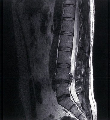
Degenerative disk disease with disk extrusion
MRI lumbar spine (T2-weighted; sagittal plane)
Hypointense degenerated disks at L4–5 and L5–S1 are accompanied by disk space narrowing. A disk extrusion at L4–5 has migrated superiorly behind the L4 vertebral body and narrows the thecal sac.
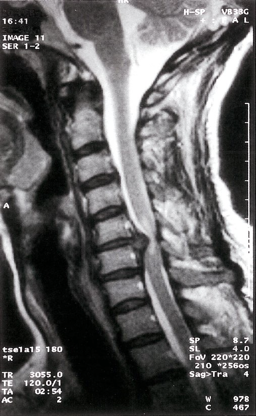
Cervical disk herniation
MRI cervical spine (T2-weighted; sagittal plane) of a patient with symptoms of cervical myelopathy A herniated disk at C5–6 effaces the dural sac and compresses the spinal cord. Hyperintense compression-induced edema is seen within the cord.