SURGERY-2
Case No. 3
- A 54 year old male presents to your ER with left lower abdominal pain, diarrhea, nausea and vomiting.
- He states that he has a decreased appetite along with a low-grade fever and chills.
- His past medical history includes a diagnosis of hypertension and obesity. He is a smoker.
- Upon arrival to the ER his vital signs are normal aside from a pule of 109 and a (temperature of 38.7.) → inflammation
- On exam you note the patient is visibly uncomfortable. When you examine his abdomen, you note tenderness and guarding to the (left lower quadrant.) → Sigmoid Colon
Diverticulitis
More questions to ask patients
- lower GI bleeding
Support diverticular Disease
- diarrhea
Support ulcerative colitis or Crohn’s
Investigations:
- Although you suspect diverticulitis,
- CT scan of the patient’s abdomen and pelvis → imp to diagnose diverticular disease or abscess or colon cancer or mesenteric ischemia
- Basic labs including a CBC, electrolytes creatinine and a urinalysis to rule out other causes of his symptoms. → suspect leukocytosis → low Hb if there is bleeding
The patient’s labs are grossly normal aside from a WBC count of 12,400.
Results of the CT scan confirm a diagnosis of diverticulitis
- 3 view abd. x-ray → if nothing obvious on abd. x-ray → Then do CT scan
- erect chest x-ray
*Treatment of diverticular disease
most of the time its conservative even during acute attack
but if its complicated (Perforation) → resect colon, end colostomy
Presentations Requiring Immediate /Urgent Surgery
(Temporary) Staple upper rectum then reconnect after 6 weeks - 3 months (hastman procedure)
- The most serious cause of intra-abdominal bleeding is a Ruptured Abdominal Aortic Aneurysm (AAA) which requires swift referral to the vascular team and immediate surgical intervention.
- Other common causes usually involve a slower rate of bleeding, but with urgent surgery still required, include Ruptured Ectopic Pregnancy, bleeding Peptic Ulcer and Trauma.
- These patients will typically go into hypovolemic shock. Clinical features include tachycardia and hypotension, pale and clammy on inspection, and cool to touch with a thread pulse.
Can get infected → abscess
Can perforate → peritonitis

Sigmoid colon
Diverticuli
*most common cause of perforation is duodenum


CT scan for varying degrees of diverticular disease (1) diverticulum in the sigmoid colon (2) degree of diverticulitis present (3) abscess formation, secondary to ongoing diverticulitis
IMAGING
Abdominal pain and diarrhea
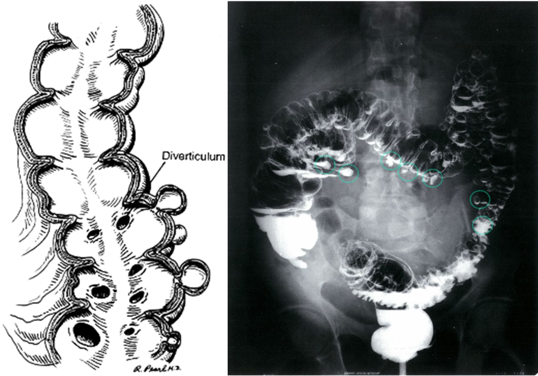
Diverticular disease. Small mucosal out pouching occur from the side large intestine.
Colonoscopy: diagnostic modality of choice for suspected symptomatic diverticulosis
Double-contrast barium enema: highly sensitive test to detect diverticulosis but not commonly performed
Diverticulosis
- Double-contrast barium enema of the colon
- Multiple diverticula in the colon can be seen (examples circled in green).