Table of content
Vesiculobullous Skin Diseases
Dr. Sami Fatehi
MD, MSc, PhD
Vesiculobullous Skin Diseases
- Bullous diseases are skin conditions characterized by blister formation.
- Bullae (blisters) are an accumulation between the epidermis or upper dermis.
Causes of Blisters
- Genetic since birth or in early life
- Physical (heat or friction)
- Inflammatory (eczema)
- Immunologic (autoimmune reaction)
- Non-immunologic (drug reaction)
How Are Keratinocytes Held Together?
- There are two levels holding:
- Between keratinocytes: ⇒ desmosomes
- Between basal keratinocytes and basement (dermis): ⇒ hemidesmosomes
Structure of the Epidermis and Sites of Target Antigens/Cleavage in Bullous Diseases
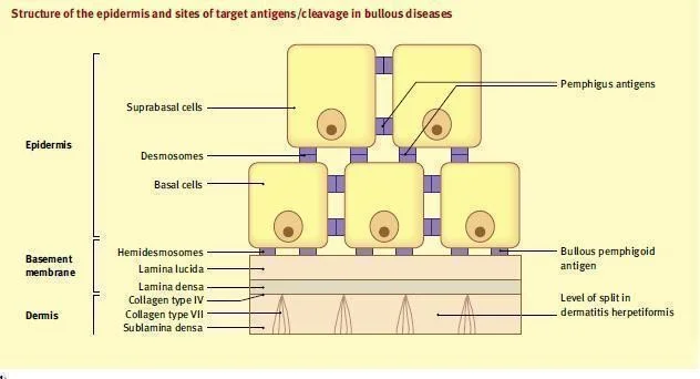
Keratinocytes and Desmosomes
- Keratinocytes in the epidermis are tightly bound together by desmosomes and intercellular substance for a barrier with high tensile strength and stability.
- Basal keratinocytes bind to hemidesmosomes.
Basement Membrane Zone (BMZ)
-
BMZ lies beneath the epidermis as specialized area of cell- extracellular matrix adhesion.
-
Collagens & laminins traversing the zone forming hemidesmosomes and anchor the epidermis to the dermis
BMZ Structure
- BMZ is divided into:
- Lamina lucida
- Lamina densa
- Sub lamina densa
- The BMZ is vulnerable to damage or malformation and is a common site of blister formation, particularly the lamina lucida.
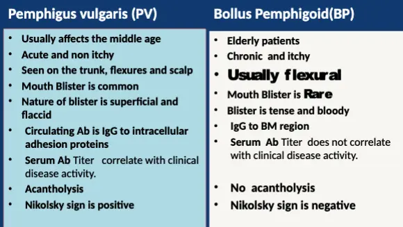
Classification and Pathophysiology of Bullous Diseases
- Intra-epidermal bulla → the base and the roof of the bulla are epidermal cells → bulla is flaccid and easy to be ruptured
- Sub-epidermal bulla → the base is the dermis and the roof is the epidermis → bulla is tense and stable
Detailed Classification and Pathophysiology of Bullous Diseases
-
Intraepidermal bullae:
- A. Acantholytic bullae: This is due to the breakdown of desmosomes
- B. Nonacantholytic bullae: It is due to the death of the cells or physical destruction
-
Subepidermal bullae: Lesions formed between the epidermis and the lamina propria of the dermoepidermal junction like:
- Bullous Pemphigoid
- Epidermolysis Bullosa