FM
Thyroid Gland
- The thyroid is a small, butterfly-shaped gland located at the base of the neck just below the cricoid.
Thyroid Gland Function
-
The thyroid’s main role in the endocrine system is to regulate the metabolism in the body by producing hormones.
-
The two main thyroid hormones are:
- T3
- T4
Physiology
- Hypothalamus
- TRH
- Pituitary
- TSH
- Thyroid gland
- T3
- T4
- Thyroid gland
- TSH
- Pituitary
- TRH
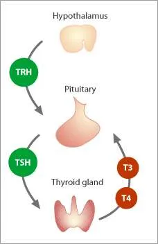
Thyroid Axis
- Thyrotropin-releasing hormone (TRH) from hypothalamus stimulates the pituitary to release TSH.
- TSH in turn stimulates the thyroid to secrete the pro-hormone thyroxine (T4) and to a lesser extent the receptor active hormone tri-iodothyronine (T3).
- The majority of circulating T3 is generated by peripheral conversion of T4 by the intracellular iodothyronine deiodinases.
- Thyroid hormone (TH) is transported over the cell membrane by specific TH transporters. After transport and metabolisation in the cell,
- T3 can interact with nuclear TH receptors and activate or inactivate TH responsive genes.
ENT
Anatomy
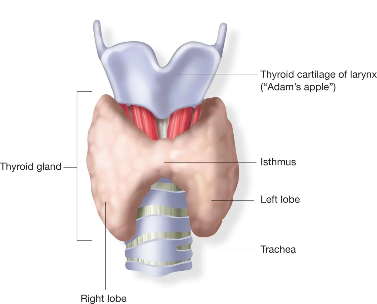
 The thyroid gland is a butterfly-shaped organ located at the front of the neck.
-
It consists of two lobes (left and right) connected by a narrow thyroid isthmus.
-
A small pyramidal lobe may project upward from the isthmus, often to the left of the midline.
-
Positioned near the front of the neck, it lies against and around the front of the larynx and trachea.
-
The thyroid cartilage and cricoid cartilage are situated just above the gland.
-
The isthmus extends from the second to third rings of the trachea, with the uppermost part of the lobes extending to the thyroid cartilage, and the lowermost to sixth tracheal rings.
-
The thyroid gland is covered by a thin fibrous capsule, which has an inner and an outer layer.
-
The outer layer is continuous with the pretracheal fascia, attaching the gland to the cricoid and thyroid cartilages, via a thickening of the fascia to form the posterior suspensory ligament of thyroid gland also known as Berry’s ligament. This causes the thyroid to move up and down with swallowing.
-
Two parathyroid glands usually lie on each side between the two layers of the capsule, at the back of the thyroid lobes.
-
The infrahyoid muscles lie in front of the gland and the sternocleidomastoid muscle to the side.
-
Behind the outer wings of the thyroid lie the two carotid arteries.
-
The trachea, laryngx, lower pharynx and esophagus all lie behind the thyroid.
Blood Supply
- The thyroid receives arterial blood from the superior thyroid artery and the inferior thyroid artery (a branch of the thyrocervical trunk).
- Occasionally, the thyroid ima artery, with a variable origin, also supplies blood.
- Venous blood is drained by the superior and middle thyroid veins into the internal jugular vein, and the inferior thyroid veins into the left and right brachiocephalic veins.
Lymphatic Drainage
- Lymphatic drainage typically passes to the prelaryngeal lymph nodes (located just above the isthmus), and the pretracheal and paratracheal lymph nodes.
Nerve Supply
- The gland receives sympathetic nerve supply from the superior, middle, and inferior cervical ganglion of the sympathetic trunk.
- Parasympathetic nerve supply is provided by the superior laryngeal nerve and the recurrent laryngeal nerve.