Osteoarthritis
Approach to Patient with Osteoarthritis
03/10/2024

Background
-
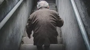
Osteoarthritis is the most common type of joint disease, affecting more than 30 million individuals in the United States alone. It is the leading cause of chronic disability in older adults, costing the US greater than $185 billion annually.
-
Osteoarthritis predominantly involves the weight-bearing joints, including:
- Knees
- Hips
- Cervical and lumbosacral spine
- Feet
-
Other commonly affected joints include:
- Distal interphalangeal (DIP)
- Proximal interphalangeal (PIP)
- Carpometacarpal (CMC) joints
-
Traditionally, osteoarthritis was thought to affect primarily the articular cartilage of synovial joints; however, pathophysiologic changes are also known to occur in:
- Synovial fluid
- Underlying (subchondral) bone
- Overlying joint capsule
- Other joint tissues
Table of Contents
- Risk Factors for Osteoarthritis
- Osteoarthritis Signs and Symptoms
- Osteoarthritis of the Hand
- Pain Mechanisms in Osteoarthritis
- Osteoarthritis Diagnosis
- Imaging Studies
- Differential Diagnoses
- Osteoarthritis Management
- Non-Pharmacologic Interventions
- Pharmacologic Therapy
- Hand Osteoarthritis
- Knee Osteoarthritis
- Hip Osteoarthritis
- Surgical Therapy
Examination
Imaging
Radiological signs of osteoarthritis
-
Irregular joint space narrowing
-
Subchondral sclerosis: a dense area of bone (visible on x-ray) just below the cartilage zone of a joint.
-
Osteophytes (bone spurs): spurs or densifications that develop on the edges of the joint.
-
Subchondral cyst: a fluid-filled cyst that develops on the surface of a joint

-
Lateral compartment osteoarthritis of the knee X-ray right knee (AP view) Marked lateral compartment narrowing is accompanied by subchondral sclerosis and osteophyte formation. Tibial spine osteophytes are also visible.
-
Degenerative disease of spine with osteophyte formation and vacuum disc phenomenon.