Medical Ophthalmology
Case Presentation
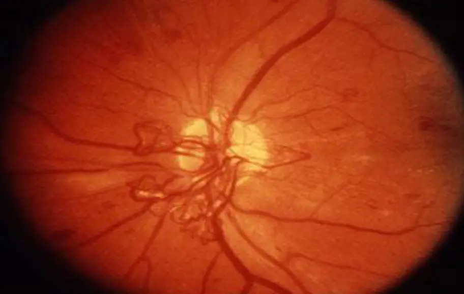
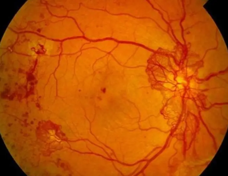
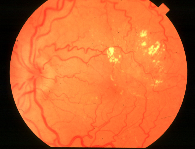
A 40-year-old woman had diabetes for 20 years. On her annual ophthalmic visit, hard exudates were noted together with retinal edema adjacent to the left macula. Her vision was 6/6 in the right eye and 6/9 in the left eye. —Laser treatment was recommended.—
Uncontrolled high a1c
PDR

NPDR
1- What is diagnosis?
PDR; proliferative diabetic retinopathy insulin depedent HBA1c is high, or NPDR; non proliferative diabetic retinopathy
2- Differential diagnosis
-
CRVO (Central Retinal Vein Occlusion)
-
BRVO (Branch Retinal Vein Occlusion)
-
Hypertensive retinopathy
-
Radiation retinopathy
-
Cataract
-
Macular degeneration or dystrophy.
-
Retinitis pigmentosa
-
Error of refraction
-
Chronic glaucoma
3- Clinical Examination & Investigations
Clinical Exam
- Fundus examination (Visual field – retinal and pupil exam. )
Investigations
- Lab investigation for diabetes & hyperlipidemia (glucose, lipid profile, HbA!C every 3m to asses DM control)
- Florescence Angiography (FA)
- OCT
- Fundus photo
4- Treatments
NB: NPDR
- General control of all risk factors(DM,HTN, hyperlipidemia)
- Argon laser Photocoagulation: focal or Grid pattern.
- Intravitreal injection of steroids or AVEGFs.
- Surgical treatment in resistant cases.
NB: PDR
- General control of all risk factors(DM,HTN, hyperlipidemia)
- Panretinal laser photocoagulation: PRP.
- Intravitreal injection of steroids or AVEGFs.
- Surgical treatment in resistant cases.
DD of Other Retinopathy
- CRVO
- BRVO
- Hypertensive retinopathy
- Radiation retinopathy
DD of Chronic Visual Loss
- Cataract
- Error of refraction
- Chronic glaucoma
- Retinitis pigmentosa
- Macular degeneration or dystrophy.
Clinical Features Y
History
Retinal Examination Visual Acuity