Professor of Ophthalmology Dr. Amani Badawi
Table of Contents
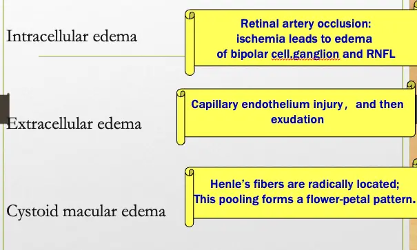
- Retinal Edema and Exudates
-
Intracellular Edema
-
Extracellular Edema
-
Exudates
-
- Preretinal Hemorrhage
- Subretinal Hemorrhage
- Deep Hemorrhage
- Superficial Hemorrhage
-
Blood Vessel Change
-
Classification of Retinal Diseases
-
Vascular diseases
-
Macular diseases
-
Retinal detachment
-
Retinal degeneration
-
Retinal tumor
-
Ocular manifestation of general diseases
-
Retinal Vascular Disease
-
Retinal artery occlusion
-
Retinal venous occlusion
-
Diabetic retinopathy
-
Vasculitis
-
Coats disease
-
Central Retinal Artery Occlusion (CRAO)
- Clinical Manifestation
- Comparison of Eye Fundus
- FFA of CRAO
- Treatment
- Prognosis
-
-
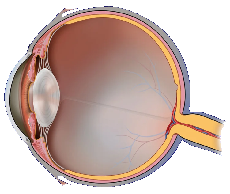
Anatomy and Physiology of Retina
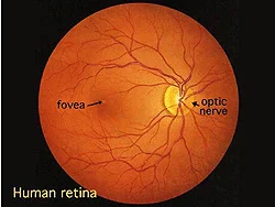
- Human Retina
- Macula Lutea
- Histology of Retina
- Neuroconduction of Retina
- Vasculature of Retina
- Retina Barrier
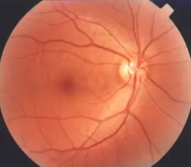
Human Retina
- Fovea
- Optic nerve
Normal Right Eye
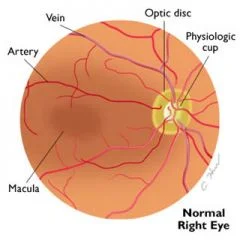
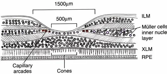
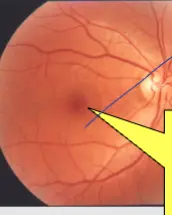
Macula Lutea Y
-
Located 3mm temporally to the optic papilla, right on the visual axis.
-
A concave central retinal depression is called Fovea Centralis.
-
Macula lutea contains only cones; 1 cone synapses to 1 bipolar cell, which synapses to 1 ganglion cell, leading to the most sensitive vision. In peripheral retina, 600 rods connect to 1 ganglion.
Histology of Retina Y
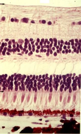
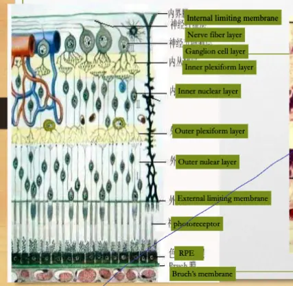
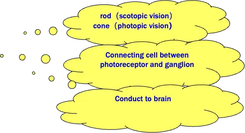
Neuroconduction of retina
3 neurons:
Photoreceptor
Bipolar
Ganglion cell
Supporting tissue: Müller cell
Vasculature of retina
-
inner layer→ central retinal vascular system
-
outer layer→ choroid(ciliary vascular system)
-
macula lutea→ choriocapillaries
Retina barrier
Inner barrier(blood–retina barrier) dense connection of retinal capillary endothelium
Outer barrier(choroid-retina barrier) zonula occludens between the RPE
RPE- Bruch’s membrane- choriocapillaries complex
Symptoms
-
Visual impairment - Related to lesion site
-
Metamorphopsia
-
Flickering - Vitreous traction to the retina
-
Macropsia
-
Micropsia - Retina edema→ fewer cones stimulated →micropsia