Retina
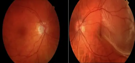
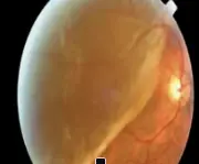
A 38-year-old healthy female presented for a routine eye exam because she was running on contacts lenses, had not been seen in a year and she needed new lenses. She had an ocular history of high myopia but was a successful user of daily wear disposable contact lenses. After change of the lens she noted no any improvement in her left eye. Left VA was 3/60 while OD VA was 6/9.She said that she began to notice vision loss inferiorly (sup detach) one week prior, which progressed to central vision loss two days prior to exam. Upon a dilated fundus examination, there was a retinal lesion shown in the photo. Both clear corneas, clear lenses, Normal IOP and reactive pupils
1- What is diagnosis?
Retinal detachment
2- DD acute painless VL
- RD
- V hge
- CRAO
- CRVO
3- Clinical Examination & Investigations
Clinical Exam Full opthalmic examination (VA, Pupil, Retinal examination)
Investigations
- Ocular US in some cases
- OCT
4- Treatments RD
-
Horseshoe retinal tear: Close the tears: by photocoagulation, condensation, electric coagulation
-
If total: Surgery: Scleral buckling: Retinopexy & Virectomy
Clinical features Y
- History, Retina, Visual acuity, Pupil